
Recomendados
Mais conteúdo relacionado
Mais procurados
Mais procurados (20)
Semelhante a Otitis media syanthika medsurg
Semelhante a Otitis media syanthika medsurg (20)
Mais de SYANTHIKADUTTA
Último
Último (20)
Otitis media syanthika medsurg
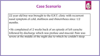
- 1. Case Scenario 22 year old boy was brought to the E.N.T. clinic with recurrent nasal symptom of cold, stuffiness and rhinorrhoea since 1/2 months. • He complained of 2 weeks back of an episode of left earache followed by discharge which was profuse and mucoid. Pain was severe at the middle of the night due to which he couldn’t sleep.
- 3. by- Syanthika Dutta B. Sc Nursing 3rd year CON, MCH Presentation on Care of a patient with OTITIS MEDIA
- 5. Definition Otitis Media is the inflammation of part or all of the lining mucosa of the middle ear cleft i.e the eustachian tube, tympanic cavity, attic, aditus, antrum and mastoid air cells
- 7. Acute OTITIS MEDIA Acute OTITIS MEDIA without effusion Acute OTITIS MEDIA with effusion Aero OTITIS MEDIA Chronic OTITIS MEDIA Chronic Suppurative Otitis media Tubercular otitis media Syphilitic otitis media CLASSIFICATION OF OTITIS MEDIA
- 8. ACUTE OTITIS MEDIA (without effusion) • It is the acute inflammation of the mucosal lining of the middle ear cleft. • Basically affects the infants and the younger age group. Normal AOM
- 9. Predisposing factors • Younger age group • Recurrent attacks of common cold. • URI • Tonsillitis • Adenoids • Chronic exposure to secondhand smoking. • Chronic rhinitis & sinusitis • Nasal Allergy • Tumours of nasopharynx • Cleft palate • Eustachian tube dysfunction • Medical conditions like Down syndrome, cystic fibrosis.
- 10. Aetiology • Extension of Infection from nasopharynx (upper respiratory tract infections)via eustachion tube • Through Perforation or Grommet • Often starts with Viral infection • (RSV,Rhinovirus,Parainfluenza virus, Influenza virus) followed by • Bacterial infection • (Strep.Pneumoniae, H.Influenza,M.catarrhalis & Staph. Aureus) • Inflammation of surrounding structures (rhinosinusitis) • Allergic reaction ( allergic rhinitis)
- 12. 1. Tubal Occlusion 2. Pre- suppuration 3. Suppuration 4. Resolution STAGES
- 13. Clinical Manifestations 1. Pain 2. Decrease in hearing 3. Discharge 4. Fever 5. Malaise 6. Headache 7. Tympanic membrane bulging out, appears erythematous.
- 14. Acute OTITIS MEDIA (with effusion) Accumulation of non-purulent/ sterile fluild in the middle ear cleft resulting in hearing impairment.
- 15. Aetiology 1. Allergy 2. Eustachian tube dysfunction 3. Palatal abnormalities 4. Viral infections 5. Nasopharyngeal tumors 6. Environmental factors-. Bottle feeding, passive smoking.
- 16. Pathogenesis Eustachian tube dysfunction Negative Middle ear pressure Goblet cell hyplasia and hypersecretion Decrease in mobility of tympanic membrane Hearing impairment
- 17. Clinical Presentations 1. Conductive deafness 2. Otalgia 3. Delayed and defective speech 4. Nasal Obstruction 5. Tympanic membrane appears bulging with erythema. 6. Fever.
- 18. Aero OTITIS MEDIA • Also known as Otitis Barotrauma • It is a non-suppurative condition owning to the failure of eustachian tube to maintain middle ear pressure at ambient atmospheric level.
- 19. Clinical features 1. Severe Ear ache 2. Tinnitus 3. Tymphanic membrane retracted and congested 4. Hearing loss
- 20. Chronic Suppurative Otitis Media It is the chronic (>3 months) inflammation of the periosteal lining of the middle ear cleft.
- 21. Risk factors for CSOM 1. History of multiple episodes of acute OTITIS MEDIA 2. Living in crowded places 3. Member of a large family
- 22. Etiology for CSOM 1. Traumatic injury 2. Iatrogenic causes 3. Pathogens- Pseudomonas aeuroginosa, Staphylococcus aureus, Aspergillus, Candida etc
- 23. Chronic Suppurative OTITIS MEDIA Classification of CSOM Tubotymphanic (Mucosal disease) Atticoantral (Cholesteatoma) Active Inactive
- 24. Profuse, Mucoid, Odourless Scanty, Purulent, Foul smelling Central Attic or marginal Pale Red and fleshy Absent Present Rare Common Audiogram Mild to moderate conductive deafness Conductive or mixed deafness Criterion Tubotymphanic Atticoantral Common Discharge Perforation Granulation Polyp Cholesteatoma Complications Uncommon
- 25. Chronic Suppurative OTITIS MEDIA- Tubotymphanic Types- 1.Active- when the ear is discharging. 2. Inactive- when the ear is dry. Characteristics- 1. Perforation is almost always central. 2.Mucosa is inflamed and edematous. 3. Polyps are seen. 4. Ossicular chain is usually intact.
- 26. Chronic Suppurative OTITIS MEDIA- Atticoantral This type of C.S.O.M. is considered as dangerous / unsafe type. This type of CSOM is associated with formation of Cholesteatoma, granulation tissues & occasionally cholesterol granulomas. If left untreated, it leads to serious complications involving the temporal bone and cranial cavity & brain.
- 27. What is Cholesteatoma? Cholesteatoma is simply “skin in wrong place”. Aural cholesteatoma is a bag of stratifiedsquamous epithelium within the middle ear which contains desquamated epithelial debris (mainly keratin) and germs. (This term is a misnomer: it is neither a tumor nor it contains cholesterol crystals) The correct term: Keratoma
- 28. Cholesteatoma
- 29. Pathophysiology of CSOM Irritation and inflammation of the middle ear mucosa Mucosal edema Mucosal ulceration Breakdown of the mucosal epithelium Granulation tissue formation Destruction of the margins of the bone margins CSOM
- 30. Clinical features of CSOM 1. Otorrhoea 2. Otalgia 3. Fever 4. Malaise 5. Vertigo 6. Hearing loss in the affected ear 7. Perforation of tympanic membrane 8. Facial paralysis 9. Headache
- 31. Tubercular otitis media Secondary to pulmonary Tb Clinical features :- Slow onset of disease- --Painless condition --Discharge is thin, scanty and odourless --Tympanic membrane is pale yellow to rosy pink in colour. --Perforations in the membrane are usually multiple and may be associated with pale granulations. --Hearing loss is disproportionate to other symptoms.
- 33. Syphylitic OTITIS MEDIA Rare condition caused by Spirochetes reaching middle ear cleft through eustachian tube or blood borne. Clinical features- --Sensorineural hearing loss --Tinnitus --Vertigo --Bone necrosis --Sequestrum formation
- 34. Diagnostics and Investigations 1. Medical history of previous Otitis Media 2. Otoscopic Examination 3. CT scan – Temporal bone CT scanning may reveal osseous erosion of the scutum, ossicles, ear canal, mastoid, otic capsule, fallopian canal, or tegmen, which typically suggests cholesteatoma. Also includes subperiosteal abscess or intracranial abscess, and, in some cases, lateral sinus thrombosis. 4.MRI scan- reveal dural inflammation, sigmoid sinus thrombosis, labyrinthitis, and extradural and intracranial abscesses.
- 35. Diagnostics and Investigations… .contd 5. Audiometry and Audiogram- Conductive hearing loss is expected, but mixed or sensorineural hearing loss may indicate more extensive disease and should alert the treating physician of impending complications, including labyrinthitis. 6. Mastoid X-ray 7. Tympanogram 8. Middle ear Endoscopy
- 37. X-ray view of Otitis Media
- 38. CT scan image of Otitis Media
- 39. MRI scan for Otitis Media
- 40. Endoscopic view of Acute OTITIS MEDIA of right middle ear
- 41. Treatment and Management Patients with OTITIS MEDIA are frequently subjected to topical therapy as compared to systemic therapy.
- 42. Dimensions of management Pharmacological Management Non-pharmacological management Surgical management Nursing management
- 46. Non-pharmacological management 1. Application of hot compression 2. Aural toilet 3. Drainage suction from the ear
- 47. Surgical management General indications for surgery- 1. Patients who donot respond to topical therapy 2. Perforation that persists beyond 6 weeks 3. Otorrhea that persists for longer than 6 weeks despite antibiotic use 4. Cholesteatoma formation 5. Radiographic evidence of chronic mastoiditis, such as coalescent mastoiditis 6. Conductive hearing loss.
- 48. A. Myringoplasty It is the operation specifically designed to close tympanic membrane defects. The approach to the ear can be transcanal, endaural, or retroauricular. -The transcanal approach requires less surgical exposure and leads to faster healing. -The endaural approach can improve exposure in ears with a lateral soft tissue or cartilage overgrowth, but again, it tends to limit the surgical view. -The retroauricular approach allows for maximal exposure but requires an external skin incision.
- 50. B. Tympanoplasty This surgical approach contains tow techniques- 1.Underlay technique involves placing the graft material underneath (or medial to) the eardrum. 2. Overlay technique involves grafting lateral to the eardrum.
- 51. C. Ossiculoplasty Ossiculoplasty refers to the surgical process of restoring the hearing system between the tympanic membrane and oval window. It involves the use of grafts and prosthesis.
- 53. D. Mastoidectomy It consists of the removal of the outer wall of the mastoid cortex and the exteriorization of all the mastoid air cells.
- 54. Surgical complications The complications of myringoplasty and tympanoplasty are- - Infection - Hematoma - Taste disturbance owning to damage of chordae tympani nerve - Ear numbess - Conductive hearing loss - Senironeural hearing loss - Vertigo - Facial paralysis
- 55. Surgical complications… contd The complications of mastoidectomy are- 1. CNS leakage 2. Intracranial complications-. abscess, meningitis.
- 56. Nursing management Nursing Assessment Assessment of patient with otitis media include the following: History Assess if there is a history of trauma to the ears, affected siblings, a history of cranial/facial defects or any familial history of otitis media. Physical examination The patient’s ear is examined with an otoscope by pulling the ear upwards and back to straighten the ear canal. If the patient is suffering from hearing loss, the nurse should be alert for the following- Speech deterioration, Fatigue, Insecurity, Loneliness, Social Withdrawal.
- 57. Subjective Data Objective data 1. Pain in the ear 2. Ear discharge 3. Nasal congestion 4. Sleepless night due to ear pain. 5. Fever for 2 weeks. 1. Facial grimacing, irritable 2. Pain rate -7/10 3. Vital signs- -Pulse rate- 120beats/min -Temperature-100 °F -Respiratory rate- 26breaths/min 4.Tymphanic membrane appears red with bilateral bulge. 5. Pharynx slightly red without exudate. 6. Discharge appears mucoid and purulent.
- 58. Nursing Diagnoses 1.Acute pain related to the inflammation of the middle ear as evidenced by facial grimacing and verbalisation of the patient. Interventions- * Assess the intensity, duration of the pain. * Assess the possible causes responsible for pain. * To rate the intensity of pain according to scale. * To record and monitor vital signs closely. * To assist with divertional therapy. * To administer with hot compression, as per order. * To administer analgesics, as per order. Pre-operative Nursing management
- 59. 2.Impaired verbal communication related to effects of hearing loss as evidenced by neurological assessment. Interventions- * Assess reasons for impaired verbal communication. * To make atmosphere conducive to communication. * To assist with pen and paper to enable patient with communication. * To observe for non-verbal communication and gestures. * To speak in low and attentive manner. * To maintain a specific routine for speech practice sessions. * To arrange for speech and audio rehabilitation measures.
- 60. 3.Disturbed sensory perception related to obstruction, infection of the middle ear, or auditory nerve damage as altered sensorium of patient. Interventions- * Assess for possible signs of obstruction in middle ear. * Assess signs for auditory nerve damage. * To assess with diagnostic procedures- Ctscan, MRI. * To assess for hearing ability. * To assess for any visible discharge from ear. * To arrange for calm and quiet environment. * To observe for non- verbal communication and gestures.
- 61. 4.Anxiety related to health status and impending surgery as evidenced by increased apprehension of health deterioration. Interventions- * Assess the level and reasons for anxiety. * Prepare the patient for surgery. * Provide bed and sleep. * Provide complete information such as possible outcome, side-effects of the surgery. * Encourage patient and family members to ventilate concerns about the health status. * To arrange discussion sessions to ventilate grievances. * To introduce patients with the similar diagnosis and with better benefits and outcomes. * Reassessment.
- 62. 5. Risk for injury related to hearing loss and loss of balance. Interventions- * Maintain a safe environment. * Assist the patient with ADLs. * Assist with locomotion and movement.
- 63. 6. Risk of infection related to presence of pathogens. Interventions- * To monitor and record vital signs. * To perform hand wash during patient handling. * To avoid invasive procedures. * To maintain aseptic measures during invasive procedures. * To assess the discharge from ear and send for culture sensitivity.
- 64. Post-operative Nursing management 1. Acute pain related to surgical procedure as evidenced by patient’s verbalisation. Interventions- * Assess duration, intensity and reason for pain. * Rate the pain. * Monitor vital signs, signs of inflammation. * To elevate head at 30 to 45°. * To administer hot compression, as per order. * To assess the site of incision, any signs of discharge. * To assist with divertional therapy. * To administer analgesics, as per order.
- 65. 2. Impaired verbal communication related to hearing loss secondary to surgery as evidenced by edema, accumulation of tissue fluid in the middle ear. Interventions- * Assess the causes of impaired verbal communication. * Elevation of head at 30-45° to relieve edema. * To maintain a calm and quiet environment. * To speak in clear and soft language with the patient. * To observe for non-verbal clues. * To educate family members about effective ways of communication.
- 66. 3. Impaired tissue integrity related to surgical incision as evidenced by assessment of incision site. Interventions- * Assess the site of incision and the surrounding tissue. * Assess any signs of edema and inflammation. * Assess the discharge, the colour, amount of drainage. * Elevation of head at 30-45°. * Planning of diet rich in proteins, vitamins. * Administration of antiseptics, as per order.
- 67. 4. Deficient knowledge about postoperative care as evidenced by patient’s assessment of knowledge. Interventions- * Inform patient and family members about the expected effects and potential side effects of medication. * Instruct about aseptic measures during dressing. * Avoid getting water in the operative ear for 2weeks after surgery. * Any signs of increased drainage and inflammation should be immediately brought into notice of the primary care giver. * Follow up care.
- 68. 5.Risk for infection related to placement of grafts, prosthesis secondary to ossiculoplasty. Interventions- * To monitor and record vital signs for every 2hrs. * To perform hand hygiene while handling with patient. * To maintain strict aseptic measures during dressing. * To inspect the condition of wound. * To observe for any signs of elevated temperature and purulent drainage. * To maintain a clean environment. * Administration of prophylactic antibiotics , as per order.
- 69. 6. Risk for injury related to vertigo due to dislodgment of prosthesis. Interventions- * Instruct patient to avoid heavy lifting, straining and nose blowing for 2-3weeks. * Observe for any signs of difficulty in nerve injury such as – dropping of mouth, slurred speech, decreased sensation and difficulty in swallowing. * Administer antiemetics, as per order.
- 70. Prognosis Death from AOM is rare in the era of modern medicine. With effective antibiotic therapy, the systemic signs of fever and lethargy should begin to dissipate, along with the localized pain, within 48 hours. Children with fewer than 3 episodes are 3 times more likely to resolve with a single course of antibiotics, as are children who develop AOM in nonwinter months. Typically, patients eventually recover the conductive hearing loss associated with AOM.
- 71. THANKYOU