5. Filling of Bladder
Stimulation of stretch receptors
Afferent impulses via Pelvic nerve
Sacral segments of Spinal cord
Efferent impulses via Pelvic nerve
Urethral relaxation & contraction of detrusor
Flow of Urine into urethra and stimulation of stretch receptors
Efferent impulses via Pelvic nerve
Inhibition of pudendal nerve & relaxation of external sphincter
Micturition Reflex
6. HIGHER CENTRES FOR MICTURITION:
• Periaqueductal gray area(PAG) of midbrain-receive afferents in
pelvic nerves via spinal cord.
• Pontine Micturition Centre(PMC) of brainstem- essential centre in
coordinating micturition process.
• Suprapontine areas of brain-frontal cortex,hypothalamus,para-central
lobule,limbic system and cingulate gyrus.
1) Important in conscious and unconscious control of PMC.
2) Role in delaying micturition ,inhibiting premature detrusor
contractions and in initiating voiding at appropriate time.
7. PHYSIOLOGY OF MICTURITION:
STORAGE PHASE: 99% of the time
Fills at a rate of 0.5- 5 ml/min.
Compliance of bladder: receptive relaxation of bladder-accomodating
the increase in volume without increase in intravesical pressure.
Under sympathetic control predominantly
Factors contributing to Compliance:
1. Passive elastic properties of tissues of bladder wall
2. Proximal urethral musculature.
3. Neural reflexes controlling detrusor tension during filling.
4. Spinal centres inhibiting cholinergic system.
5. External sphincter.
8. Drop in intraurethral pressure, sympathetic blockade, obliteration of post
urethrovesical angle
Detrusor contracts
Funneling of bladder neck and proximal urethra
Urine flows into upper urethra
EUS opens & Voiding occurs
Distal end of urethra closes, milking back last drop into bladde
Process of Micturition
10. Measurement of intravesical bladder pressure during bladder
filling ( measures volume-pressure relationships).
Simultaneous measurement of bladder pressure and voiding
function-allowing the site of dysfunction to be localised
specifically to either bladder or bladder outlet/urethra
Principal aim: to reproduce patients symptoms and correlate
symptoms with UDS findings.
Can be used to define the behaviour of bladder and urethra
during both phases.
Used to assess bladder compliance, sensation, capacity, flow
rate and detrusor activity.
CYSTOMETRY
11. CYSTOMETRY TECHNIQUES
Simple Cystometry- only intra-vesical pressure is measured,
so inaccurate.
Subtraction cystometry:
• measure both Pves and Pabd simultaneously.
• Pves –Pabd gives us the Pdet.
• Accurate determination of Pdet and do not involve any
radiation of pressure.
VCMG: combines subtraction cystometry with contrast
media bladder filling and radiological screening, visualise
lower tract during storage and voiding phases.Hence is a
GOLD STANDARD urodynamic inv.
AUM: allow bladder to fill naturally.
12. COMPLICATIONS
Discomfort during the procedure
Discomfort and dysuria following the procedure
Transient bleeding post procedure
UTI- in 2-4% patients , prophylactic antibiotic for at risk
patients
Radiation exposure during videoCMG
Failure- urdynamic question may remain unanswered.
13. EQUIPMENT SETUP
Transducers to measure pressures
Fluid filled catheters to transmit intravesical &
intraabdominal pressures to the transducers
Second intravesical catheter to fill bladder with fluid.
Infusion pump
Flowmeter to measure flow rate.
14. Catheter placement:
Intra-vesical : per urethrally or suprapubic route.
Intra-abdominal : inserting catheter in rectum.(stoma or upper
vagina).
Catheter has a balloon at the rectal end, it should be filled only
by 10-20%.
Rectal catheter inserted 10 cm above the anal verge and secured
Transducers: 3 are in common use
External fluid charged pressure transducers-recommended by
ICS.
Catheter mounted transducers-no ref height,no movt artefacts.
Air charged, pressure sensing technology- newest
15. Filling the bladder
CATHETER TYPES:
• Dual lumen –recommended by ICS. Can be used for both
intra-vesical pressure measurement and also for bladder
filling.Thinnest possible is used to limit the artefacts.8 Fr
preferred.
• Single lumen- requires 2 separate catheters.
FILLING FLUID:
• Sterile water
• Normal saline
• Radiological contrast-VCUG
16. FLUID TEMPERATURE:
• Ideal- at body temperature
• More practical to use fluid at room temp, doesnot appear to affect
results.
• Colder fluids – may irritate the bladder –ppt detrusor overactivity.
QUALITY CONTROL:
• Setting zero pressure- zeroed either to the surrounding atm pressure or
the internal pressure. 3-way taps are used to zero.
ICS recommends surrounding atm pressure-as it standardises.
• Setting reference height(level at which transducers must be placed )
i. External fluid filled systems: superior edge of symphysis pubis.
ii. Microtip transducers: transducer itself
iii. Air filled transducers: position of the internal balloon.
17.
18. Resting pressures :
• Pdet should be < 6 cm water and ideally as close to zero as
possible .
Dampening :poor transmission of pressure to the transducer.
• Cough before and after the investigation
• Assessed through out the procedure –cough every 1 min, cough on
changing position,voids.
Medications:
Drugs affecting LUTS should be discontinued.
One week prior to study normally.
19.
20. Cystometry stages
Urethral and bladder function evaluated in both phases.
1. Storage phase /filling cystometry : pump on to maximum
tolerated capacity ( permission to void)
2. Voiding phase / voiding cystometry : permission to void to
complete voiding.
Liquid cystometry is more physiologic.
Bladder filling either by diuresis or through a catheter.
21. FILL RATES –
o Slow ( upto 10ml/min )-more physiological, used in neurogenic patients
o Medium ( 10 to 100 ml/min ) most frequently used
o Rapid ( > 100 ml/min )
ICS recommends 50ml/min in non-neurogenic patients and 20 ml/min in
neurogenic patients.
PATIENT POSITION :
Standing position is ideal atleast for some part of filling phase.
Supine position – helpful as catheters are placed in supine position.
Patient should be brought to standing as soon as possible.
Supine may be the only position in patients with neurological dysfunction or
very small children.
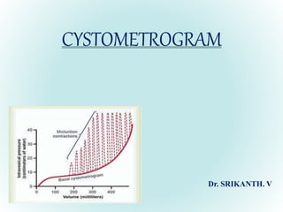
22. NORMAL CMG
Capacity 350 – 600 ml
First desire to void between
150- 200 mi
Constant low pressure that
does not reach more than 6
– 10 cm H2O above
baseline at the end of
filling.
Provocative maneuvers
(cough,fast fill etc.,) should
not provoke a bladder
contraction normally
Absence of systolic
detrusor contractions.
No leakage on coughing.
A voiding detrusor
Pressure rise of < 70 cm
H2O with a peak flow
rate of >15 ml / s for a
volume > 150 ml.
Residual urine of < 50 ml.
23. CMG PARAMETERS
Intravesical pressure (Pves ) : Total pressure within the bladder.
Abdominal pressure (Pabd ) : Pressure surrounding the bladder,
currently estimated from rectal, vaginal or extraperitoneal pressure or
a bowel stoma.
Detrusor pressure ( Pdet) : Component of intravesical pressure
created by forces on the bladder wall, both passive and active.
True detrusor pressure = Intravesical pressure – intraabdominal
pressure. (Pdet = Pves – Pabd )
24. First sensation of bladder filling : Volume at which patient first
becomes aware of bladder filling.
First desire to void : Feeling during filling cystometry that would
lead the patient to pass urine at the next convenient moment.
Strong desire to void : Persistent desire to void without fear of
leakage.
27. Detrusor function during storage phase:
• Normal – little or no change in pdet during storage phase. Any detrusor
activity prior to voiding phase is Involuntary detrusor activity.
• DO-Detrusor Overactivity –
• Characterised by involuntary detrusor contractions during storage phase.
• Spontaneous or provoked.
• Idiopathic DO-OAB
• Neurogenic DO
28. Compliance :
Intrinsic ability of bladder to change in volume without significant
alteration of detrusor pressure.
Relationship between change in bladder volume and change in Pdet (
∆volume/ ∆pressure ) : measured in ml/cm H2O.
Normal bladder is highly compliant, and can hold large volumes at low
pressure.Normal compliance is >30-40 cm H2O
Normal pressure rise during the course of CMG in normal bladder will be
only 6-10 cm H2O.
Decreased compliance < 20 ml/cm H2O, poorly distensible bladder.
29. Impaired Compliance is seen in :
Neurologic conditions : Spinal cord injury/lesion, spina bifida,
usually results from increased outlet resistance ( e.g., detrusor
external sphincter dyssnergia [ DESD ] or decentralization in case of
lower motor neuron lesions.
Long term BOO ( e.g., from benign prostatic obstruction)
Structural changes : Radiation cystitis or tuberculosis.
Impaired compliance with prolonged elevated storage pressures is a
urodynamic risk factor and needs treatment to prevent renal damage
30.
31. Urgency : A sudden compelling desire to void.
Normal detrusor function : Allows bladder filling with little or no
change in pressure, no involuntary contractions.
Detrusor overactivity : Involuntary detrusor contractions during the
filling phase , spontaneous or provoked.
Storage greater than 40 cm H2O is associated with harmful effects on
the upper tract.
Overactive bladder : Storage symptoms of urgency with or without
urgency incontinence , usually with frequency and nocturia.
Neurogenic detrusor overactivity : Overactivity accompanied by a
neurologic condition , also k/a detrusor hyperreflexia.
32. Abdominal leak point pressure (ALPP): Intravesical pressure at
which urine leakage occurs because of increased abdominal pressure
in the absence of a detrusor contraction.
ALPP is a measure of sphincteric strength or ability of the sphincter to
resist changes in Pabd.
Applicable to stress incontinence ; ALPP can be demonstrated only in
a patient with SUI.
There is no normal ALPP , because patients without stress
incontinence will not leak at any physiologic Pabd.
Lower the ALPP, weaker is the sphincter.
33. ALPP< 60 cm H2O : significant ISD
ALPP 60-90 cm H2O : equivocal
ALPP> 90 cm H2O : urethral hypermobility, little or no ISD.
34. Detrusor leak point pressure ( DLPP ) : Lowest detrusor pressure at
which urine leakage occurs in the absence of either a detrusor
contraction or increased abdominal pressure (risk with > 40 cm
H2O).
It’s a measure of Pdet in a patient with decreased bladder compliance.
Higher the urethral resistance, higher the DLPP, the more likely is
upper tract damage as intravesical pressure is transferred to the
kidneys.