
Recomendados
Recomendados
Mais conteúdo relacionado
Mais procurados
Mais procurados (20)
Destaque
Destaque (18)
Semelhante a Tempel, et al IAI
Semelhante a Tempel, et al IAI (20)
Tempel, et al IAI
- 1. INFECTION AND IMMUNITY, Sept. 2006, p. 5095–5105 Vol. 74, No. 9 0019-9567/06/$08.00ϩ0 doi:10.1128/IAI.00598-06 Copyright © 2006, American Society for Microbiology. All Rights Reserved. Attenuated Francisella novicida Transposon Mutants Protect Mice against Wild-Type Challenge Rebecca Tempel,†* Xin-He Lai,† Lidia Crosa, Briana Kozlowicz, and Fred Heffron Department of Molecular Microbiology and Immunology, Oregon Health & Science University, Portland, Oregon Received 12 April 2006/Returned for modification 17 May 2006/Accepted 5 June 2006 Francisella tularensis is the bacterial pathogen that causes tularemia in humans and a number of animals. To date, there is no approved vaccine for this widespread and life-threatening disease. The goal of this study was to identify F. tularensis mutants that can be used in the development of a live attenuated vaccine. We screened F. novicida transposon mutants to identify mutants that exhibited reduced growth in mouse macro- phages, as these cells are the preferred host cells of Francisella and an essential component of the innate immune system. This approach yielded 16 F. novicida mutants that were 100-fold more attenuated for virulence in a mouse model than the wild-type parental strain. These mutants were then tested to determine their abilities to protect mice against challenge with high doses of wild-type bacteria. Five of the 16 attenuated mutants (with mutations corresponding to dsbB, FTT0742, pdpB, fumA, and carB in the F. tularensis SCHU S4 strain) provided mice with protection against challenge with high doses (>8 ؋ 105 CFU) of wild-type F. novicida. We believe that these findings will be of use in the design of a vaccine against tularemia. Francisella tularensis is a gram-negative, facultative intracel- lular pathogen that causes tularemia, a debilitating and poten- tially fatal disease that affects humans and a wide range of animals. Infections can be acquired through bites from an arthropod vector, skin lesions, ingestion of contaminated food or water, and, most dangerously, inhalation of as few as 10 bacteria (10). The low dose required to cause tularemia by the aerosol route resulted in the development of F. tularensis for use as a biological weapon by several national weapons pro- grams. This has led the U.S. Centers for Disease Control and Prevention to classify F. tularensis as a category A bioterrorism agent; members of this category are considered the organisms that pose the most serious risk to national security (http://www .bt.cdc.gov/Agent/Agentlist.asp). There is currently no ap- proved vaccine available in the United States or Europe. Thus, the development of a vaccine against F. tularensis has become an international research priority. Although the molecular mechanisms of F. tularensis patho- genesis remain relatively obscure, it has been established that replication in human and animal macrophages is central to this organism’s ability to cause tularemia (14). Several F. tularensis genes associated with intracellular growth have been identified, including iglB, iglC, mglA, pdpD, and a clpB homolog (2, 18, 20, 31, 33). Additionally, it is thought that many of the genes in the recently described F. tularensis pathogenicity island (FPI) con- tribute to the survival and growth of this organism in macro- phages (32, 35). Of these, only iglC has been studied as the basis for a potential vaccine strain. Pammit et al. recently reported that intranasal vaccination with an F. novicida strain carrying an iglC deletion resulted in Ͼ50% protection against challenges with the wild-type organism (38). However, the capacity of mutant derivative strains with mutations in other FPI genes to confer protection against challenge with wild-type bacteria has not been studied. Three main subspecies of F. tularensis are commonly recog- nized: F. tularensis subsp. tularensis (type A), F. tularensis subsp. holarctica (type B), and F. tularensis subsp. mediasiatica. All of these biotypes along with F. novicida exhibit more than 95% DNA sequence identity (3). Although type A and type B strains are highly infectious, only type A strains cause signifi- cant mortality in humans. The current live vaccine strain (LVS) is an attenuated type B strain that provides different levels of protection against challenge with type A F. tularensis strains depending on the route of immunization, the route of chal- lenge, and the genetic background of the host (4–6, 21, 43, 49). Because the molecular basis for LVS attenuation is not known, this strain is not licensed as a tularemia vaccine. F. novicida U112 provides an ideal model for studying Fran- cisella pathogenesis for several reasons. While F. novicida is not considered a human pathogen, it exhibits a degree of virulence in mice similar to that of F. tularensis subspecies (27, 42). Moreover, F. novicida is easier and less dangerous to manipulate genetically than F. tularensis. In addition to the considerable genomic similarity (Ͼ95%), the close relationship between F. novicida and F. tularensis is further highlighted by their nearly identical 16S rRNA gene sequences (13). The degree of genetic identity suggests that the two organisms utilize similar virulence genes and that F. novicida is thus an apt platform for the development of a tularemia vaccine. In this study, we used transposon mutagenesis to identify F. novicida genes required for intracellular growth. The resulting mutant strains were screened for attenuation in macrophages and mice and tested for the ability to provide protection against a wild-type challenge in mice. Five F. novicida mutant strains were found to protect mice against challenge with Ͼ8 ϫ 105 CFU of wild-type F. novicida. These results will be used in the future for construction of a Francisella vaccine. * Corresponding author. Mailing address: 6543 Basic Sciences Ad- dition/CROET Building, Department of Molecular Microbiology and Immunology, Oregon Health & Science University, 3181 SW Sam Jackson Park Rd., Portland, OR 97239. Phone: (503) 494-6841. Fax: (503) 494-6862. E-mail: tempelr@ohsu.edu. † R.T. and X.-H.L. contributed equally to this work. 5095
- 2. MATERIALS AND METHODS Bacterial strains and culture. F. novicida strain U112 was a kind gift from Fran Nano (University of Victoria). All Francisella strains were cultured at 37°C in tryptic soy broth supplemented with 0.1% cysteine (TSBC) (Becton, Dickinson and Company, Sparks, MD) or on cysteine heart agar (CHA) (Difco/Becton, Dickinson and Company) plates. Kanamycin was added to a final concentration of 20 g/ml to TSBC and to CHA (CHA/Kan20) for selection of U112 strains carrying the transposon. Escherichia coli Genehogs (Invitrogen, Carlsbad, CA) that were used in subcloning for sequencing were transformed according to the manufacturer’s directions. Colonies containing the transposon were selected by growth at 37°C on Luria-Bertani (LB) agar plates containing 60 g/ml kanamy- cin. The Salmonella strain used for Southern blotting was grown in LB broth and on LB agar plates at 37°C. Generation of bacterial transposon mutant strains. A library of F. novicida transposon insertion mutants was created by electroporating mini-Tn5 transposon- transposase complexes into appropriately prepared F. novicida. Although inde- pendently developed, our technique was similar to that of Kawula et al. (26, 45). The mini-Tn5 cycler transposon was constructed as previously described (17). The transposon-transposase complex was prepared as described by Goryshin et al. (19). F. novicida U112 was grown to confluence on CHA plates at 37°C and resuspended with 5 ml of ice-cold 10% glycerol–500 mM sucrose buffer. Aliquots (1 ml) were transferred to 1.5-ml microcentrifuge tubes, pelleted by centrifuga- tion at 12,000 ϫ g for 5 min at 4°C, and resuspended in 1 ml of buffer. This wash step was repeated until a total of four washes had been performed. After the final wash, each aliquot was resuspended in 100 l buffer. One microliter of transposon- transposase complex was added to each tube, and the samples were electropo- rated in 1-mm-gap cuvettes at 1.5 to 1.7 kV, 200 ⍀, and 25 F. The bacteria were recovered in 1 ml TSBC in glass tubes for 4 h in a 37°C rotator and plated on CHA/Kan20 plates. The frequency of isolation of transposon insertion mutants was rather low (about 10 to 100 insertions per 109 cells following electropo- ration). Culture and infection of cell lines and primary macrophages. The J774A.1 and RAW264.7 murine macrophage cell lines (American Type Culture Collection, Manassas, VA) were cultured in Dulbecco’s modified Eagle’s medium (DMEM) (Gibco-BRL, Rockville, MD) supplemented with 10% fetal bovine serum (FBS) (Gibco-BRL), 1 mM nonessential amino acids (Gibco-BRL), and 0.2 mM so- dium pyruvate (Gibco-BRL) at 37°C in the presence of 5% CO2. Bone marrow- derived macrophages (BMDM) were collected by flushing the femurs of BALB/c mice with serum-free DMEM and were cultured in DMEM supplemented with 20% L929 and an antibiotic cocktail of penicillin (10,000 U/ml) and streptomycin (10,000 g/ml). For infection, bacteria were added to 50% confluent cells in 24- or 96-well culture dishes (Corning, Corning, NY) or four-chamber microscope plates (Nalge Nunc, Naperville, IL) at the multiplicities of infection (MOI) indicated below, and the cells were centrifuged at 1,000 ϫ g for 5 min at room temperature and incubated at 37°C in the presence of 5% CO2. One hour after infection, the cells were washed twice with phosphate-buffered saline (PBS), and DMEM containing 100 g/ml of gentamicin was added to prevent the growth of any extracellular bacteria (29). Two hours after infection, the cells were washed twice with PBS and either lysed or incubated in the presence of 10 g/ml gentamicin for an additional 22 h. Cells were lysed with TSBC containing 0.5% saponin (Sigma) for 30 min at 37°C in the presence of 5% CO2. Screening for reduced growth in macrophages. RAW macrophages were seeded to obtain 50% confluence in 96-well tissue culture plates and infected (MOI of ϳ1,000) with overnight cultures of F. novicida mutant strains that were grown in stationary 96-well tissue culture plates, as described above. At 24 h postinfection (p.i.), the macrophages were washed and lysed as described above. Three percent of each lysate was plated onto CHA/Kan20 plates and incubated overnight at 37°C. Mutants that exhibited growth defects were identified visually. To eliminate false positives, the potentially attenuated mutants were subjected to another round of selection by infecting RAW macrophages in 24-well plates, as described above, using an input MOI of 100, which corresponded to about one bacterium per macrophage. After lysis, 50 l of each lysate was plated onto CHA plates and incubated overnight at 37°C. F. novicida mutants compromised for growth in macrophages were identified visually by comparison to wild-type U112 infection lysates; the attenuated mutants yielded individual colonies, while the wild-type bacteria grew to confluence. Sequencing of mini-Tn5 insertion sites and sequence analysis. The method described by Geddes et al. was used for sequencing mini-Tn5 insertion sites and sequence analysis (17). Briefly, chromosomal DNA from F. novicida mutants exhibiting reduced growth in macrophages was prepared (1), digested with EcoRI, and subcloned into pACYC184. Ligation reaction mixtures were elec- troporated into GeneHogs E. coli cells (Invitrogen) and selected for growth on LB medium containing 60 g/ml kanamycin. Plasmids from kanamycin-resistant colonies were purified using a QIAprep Spin miniprep kit (QIAGEN, Valencia, CA) according to the manufacturer’s instructions. The DNA sequence of the fusion junction was obtained using a primer complementary to bp 166 to 190 of the 5Ј end of mini-Tn5 cycler (5Ј GTTGACCAGGCGGAACATCAATGTG 3Ј). Sequence analysis was performed using the MacVector 7.2.3 software and the NCBI BLAST server at http://www.ncbi.nlm.nih.gov/BLAST/. Mouse studies. Six- to 8-week old female BALB/c mice were purchased from the Jackson Laboratory (Bar Harbor, ME). The animals were fed autoclaved food and water ad libitum. All experiments were performed in accordance with Animal Care and Use Committee guidelines. For vaccination and challenge studies, mice were inoculated intraperitoneally (i.p.) with bacteria in 150 l (total volume) of PBS. Mice were vaccinated with the number of CFU indicated below. Surviving mice were challenged 28 days later with the doses indicated below. Dissemination and clearance of the bacteria were determined by harvesting the lungs, liver, and spleen on the days postinfection indicated below, homogenizing the organs with a stomacher, and plating serial dilutions. The 50% lethal doses (LD50) were calculated by the method of Reed and Muench (39). Mice were checked for signs of illness or death twice each day following infection. Bacterial growth in liquid media. Overnight cultures of F. novicida were diluted into 10 ml of TSBC to obtain an optical density at 600 nm (OD600) of 0.1. Optical densities were then recorded at the times specified below. Note that the cultures were diluted 1:10 to obtain OD600 of Ͼ1 for accuracy. We previously determined by plating that an OD600 of 1 was equivalent to approximately 4 ϫ 109 bacteria/ml. Quantification of bacterial entry and growth in macrophages. J774 and RAW cells and BMDM were seeded in triplicate to obtain 50% confluence in 24-well tissue culture plates and were infected as described above with F. novicida mutant strains at an input MOI of 100. Cells were lysed at 2 or 24 h p.i. Serial dilutions of the lysates were plated onto CHA/Kan20 or CHA (wild-type and mock infection controls) plates. After overnight incubation at 37°C, the colonies on each plate were counted. Means and standard deviations were calculated using Microsoft Excel X for Mac. The 24-h data were statistically analyzed by paired two-tailed t tests using Microsoft Excel X for Mac. Southern blot analysis. F. novicida chromosomal DNA was prepared using the cetyltrimethylammonium bromide method (1), and 250 ng of each preparation was digested to completion with HindIII. Digested DNA was electrophoresed on a 0.8% agarose gel for 2 h at 90 kV and then transferred to a positively charged nylon membrane (Roche) using a standard capillary transfer method (1). DNA was cross-linked to the membrane at 120,000 J/cm2 using a Stratalinker 1800 UV cross-linker (Stratagene, La Jolla, CA). The digested bacterial DNA was probed with a digoxigenin-labeled probe using a DIG High Prime II DNA labeling and detection starter kit (Roche, Indianapolis, IN), and the membrane was exposed to film (Kodak, Rochester, NY) for 2 or 8 min as described below. By using a DNA probe that spans a HindIII site in the transposon and hybridizes to two separate locations of the HindIII-digested chromosomal DNA, we were able to determine the number of transposon inserts in each strain. Cytotoxicity assay. A cytotoxicity assay was conducted as described by van der Velden et al. (48). Briefly, J774 cells seeded in 96-well culture plates were infected in triplicate with either the transposon mutants or wild-type F. novicida U112 at an input MOI of 100. After 48 h, the supernatants were removed and assayed for released lactate dehydrogenase (LDH) using the CytoTox 96 non- radioactive cytotoxicity assay (Promega, Madison, WI). Cytotoxicity was deter- mined for each mutant strain by calculating the amount of LDH released as a percentage of the maximal amount released from macrophages infected with wild-type strain U112. Microscopy. J774 cells were infected at an input MOI of 100, as previously described, in four-well chamber plates (Nalge Nunc). After 24 h, the cells were washed twice with PBS, fixed for 1 h with 4% paraformaldehyde, and stored in PBS at 4°C. After three washes for 10 min in PBS, the cells were permeabilized with 0.5% Triton X-100 (Sigma Chemical) in PBS for 20 min at room temper- ature, blocked with 5% FBS in PBS for 30 min, and incubated for 1 h at 4°C with a polyclonal antibody against F. tularensis (Becton, Dickinson and Company). After three washes for 10 min in PBS, the cells were again blocked with 5% FBS. A goat anti-rabbit antibody conjugated to Alexa 488 (Molecular Probes, Eugene, OR) was applied to the cells overnight at 4°C. The cells were again washed three times for 10 min in PBS and incubated with a 1:1,000 dilution of FM 4-64 membrane stain (Molecular Probes) and 1:1,000 dilution of Draq5 DNA stain in PBS (Alexis Biochemicals, San Diego, CA) for 10 min at room temperature. The cells were washed twice with PBS and mounted in Fluormount-G antifade solu- tion (Southern Biotechnology, Birmingham, AL), and images were obtained with an Applied Precision DeltaVision deconvolution microscope system (Advanced Precision Instruments, Issaquah, WA). All images were taken obtained a ϫ60 5096 TEMPEL ET AL. INFECT. IMMUN.
- 3. objective. Stacks of 10 z-plane images that were 1 m apart were captured at 1024 ϫ 1024 pixels and deconvolved for seven iterations. Selected images were saved in TIFF format and imported into Adobe Photoshop to be formatted. Complementation of disrupted genes. Plasmid pKK202 (30) was modified to include unique NotI, SfiI, and XhoI restriction sites by digestion with ClaI and XbaI, followed by ligation with a DNA fragment. Oligonucleotides CGGCGG CCGCTTGGCCTCGAGGGCC and CTAGGGCCCTCGAGGCCAAGCGG CCGC were annealed to obtain a double-stranded product encoding the new restriction sites. Using SCHU S4 DNA as a template, full-length genes were amplified by PCR. The dsbB gene was cloned using primers GCGGCCGCCTT CTTAACGTCCACAGTTTTGTCC and GGCCCTCGAGGCCCTTTCTGAT GGTTTGTCATTTCTCC, FTT0742 was cloned using primers GCGGCCGCG CAGCATTACCTGGAATTACAAG and GGCCCTCGAGGCCCAAACAGC AAATAAATATACAACACC, and fumA was cloned using primers GCGGCC GCTAGTGATAAAATTAGCGAGG and GGCCCTCGAGGCCATTAACTA TAATGCCGAG. The modified pKK202 vector and the PCR products were digested with NotI and XhoI and ligated. pKK202-dsbB was electroporated into the dsbB mutant F. novicida strain and used to infect J774 and RAW cells, primary macrophages, and mice as indicated below. pKK202-FTT0742 and pKK202-fumA were similarly tested with RAW cells. RESULTS Transposon mutagenesis and identification of disrupted loci. An ongoing challenge to the establishment of a suitable set of genetic tools for F. tularensis is the difficulty of creating stable mutations in the genome. To address this issue, we developed a transposon mutagenesis technique independent of phages and shuttle vectors that yielded as many as 150 trans- poson mutants from a single electroporation procedure (see Materials and Methods). Our method, although independently developed, is quite similar to the technique used by the Kawula laboratory with LVS (26, 45). The bacteria were electropo- rated with a transposase-transposon complex that completed the transposition event once it was inside the bacteria. This approach yielded the library of 779 Francisella transposon mu- tants used in this study. Macrophages are the primary host cell type for Francisella in both humans and animals (14). It follows that one approach for developing a tularemia vaccine would be to discern which Francisella genes are necessary for growth in macrophages. Thus, we screened our F. novicida transposon mutant library for mutants that had a reduced ability to grow in macrophages, as described in Materials and Methods. Of the more than 700 F. novicida transposon mutants screened, 34 exhibited reduced growth in RAW macrophages. We obtained sequences for 28 of these mutant strains and identified the disrupted open read- ing frames (ORFs) by comparison to the SCHU S4 sequence (Table 1). Sixteen F. novicida mutants exhibited attenuation in mice. To narrow our study to the mutant strains that had an atten- uated phenotype in an animal model, we infected wild-type BALB/c mice with the 28 F. novicida mutants that were atten- uated for growth in macrophages. Mice were inoculated intra- peritoneally with 6 ϫ 103 bacteria in 150 l of PBS, a dose which is about 100-fold greater than the wild-type F. novicida TABLE 1. F. novicida transposon mutant strains generated in this study Mutant Corresponding SCHU S4 FTT no. Gene disrupted % Identity to SCHU S4a Nucleotide location of transpson insertb % Survivalc 1 FTT0107c dsbB 100 114151 100 2 FTT0145 rpoC 99 163108 0 3 FTT0203c purH 99 222340 100 4 FTT0334 rpsQ 100 342324 0 5 FTT0356 htpG 95 356504 100 6 FTT0504c sucC 97 524250 0 7 FTT0583 fopA 98 599781 0 8 FTT0742 Hypotheticald 89 765155 100 9 FTT0893 purM 98 901556 100 10 FTT0893 purM 97 901647 100 11 FTT0893 purM 98 901848 100 12 FTT0894 purCD 99 904045 100 13 FTT0894 purCD 99 904160 100 14 FTT0917 maeA 98 926193 33 15 FTT1165c aspC2 99 1179264 0 16 FTT1222 dedA2 98 1240288 33 17 FTT1241 glyA 97 11261475 67 18 FTT1269c dnaK 100 1291446 100 19 FTT1345/1700 pdpBe 98 1384141/1777485 100 20 FTT1369c tktA 97 1416905 100 21 FTT1535c ocd 99 1597434 0 22 FTT1535c ocd 97 1597841 0 23 FTT1600c fumA 98 1667516 100 24 FTT1629c Hypotheticalf 99 1692570 0 25 FTT1664 carB 99 1730805 100 26 FTT1720c purL 98 1804171 100 27 FTT1720c purL 97 1805882 100 28 FTT1769c clpB 96 1858564 0 a Level of identity when our fragment sequence was used. b Corresponding to SCHU S4 location. c Data for groups of three mice at 28 days after infection with 6 ϫ 103 CFU. d Lipoprotein gene. e SCHU S4 contains two copies of pdpB. f Membrane protein gene. VOL. 74, 2006 ATTENUATED AND PROTECTIVE FRANCISELLA MUTANTS 5097
- 4. LD50 for mice. At 28 days postinfection, the survival rate for 16 of the 28 groups of mice was 100%, indicating that 16 of the insertion mutants were highly attenuated in this animal infec- tion model (Table 1). Five F. novicida mutants protected mice against challenge. The ideal living vaccine strain produces an asymptomatic in- fection that provides complete protection against subsequent exposure to the wild-type organism. To determine if any of our 16 attenuated F. novicida transposon mutants could confer protection against wild-type infection, we next challenged the surviving vaccinated mice with the wild-type parental strain. Four weeks after infection with mutant F. novicida strains, surviving mice were intraperitoneally challenged with 8 ϫ 105 CFU F. novicida U112. We considered this to be a very strin- gent challenge, as the dose was more than 10,000 times the LD50 observed for wild-type infection. At 28 days after the challenge, 5 of the 16 mutants exhibited 100% protection after a single vaccination; these mutants had mutations in dsbB, the ORF corresponding to FTT0742 (referred to as FTT0742 be- low), pdpB, fumA, and carB (Table 2). In the same experi- ments, all wild-type control infections resulted in 0% survival. LD50s for F. novicida mutants. Additional infections of mice with the F. novicida transposon mutants were conducted to determine the LD50s of the five protective strains. In our ex- periments, the F. novicida U112 parental strain was observed to have an LD50 of 66.25 CFU (Table 3). Our carB mutant exhibited the least attenuation and had an LD50 of 6.75 ϫ 103 CFU. The LD50s for our dsbB and fumA mutants were 6.625 ϫ 105 CFU and 6.17 ϫ 105 CFU, respectively. The mutant strains with the highest levels of attenuation in an animal infection model were the FTT0742 and pdpB mutants, both of which were observed to have LD50s of Ͼ6 ϫ 107 CFU. Taken to- gether, these results showed that our five F. novicida transpo- son mutants were significantly attenuated in a mouse infection model compared to the wild-type parental strain. F. novicida mutants were highly attenuated for growth in mouse macrophage cell lines. Wild-type strain F. novicida U112 and the five mutants that conferred protection were examined in order to quantify entry and attenuation in mac- rophage cell lines and primary mouse macrophages (BMDM). RAW and J774 macrophage-like cells and BMDM were in- fected in triplicate wells in duplicate plates with an input MOI of 100, as described in Materials and Methods. The contents of one plate containing each cell type were lysed at 2 h after infection to determine the abilities of the transposon mutants to enter host cells, and lysis of the contents of the second plates at 24 h allowed us to quantify the intracellular growth of the mutant strains. In J774 mouse macrophage-like cells, the levels of entry were quite similar for the mutant and wild-type strains, except for the pdpB mutant (Fig. 1A). The dsbB mutant exhibited the highest level of attenuation in J774 cells, and for the FTT0742 mutant derivative there was also a decrease in the number of CFU after 24 h. Although fewer pdpB mutant bacteria than bacteria of the other strains entered J774 cells, this mutant still displayed an attenuation phenotype. For the fumA mutant there was very little difference between the level of entry and the level of replication at 24 h, indicating that there was no death, no replication, or a balance between the two. Interest- ingly, the carB mutant was able to replicate within J774 cells, albeit at a lower rate than the wild-type control. Each of the mutants exhibited statistically significant attenuation in J774 cells at 24 h p.i. (P Ͻ 0.005). The levels of entry for the transposon mutants and the wild- type bacteria were similar when RAW mouse macrophage-like cells were used (Fig. 1B). Again, the dsbB and pdpB mutant derivatives exhibited the strongest attenuation phenotypes. In- triguingly, the FTT0742, fumA, and carB mutant strains all were able to replicate in RAW cells, whereas only the wild type and the carB mutant replicated in the J774 cell line. Compared to the wild type, each of the mutants strains was significantly impaired for replication inside RAW cells at 24 h p.i. (P Ͻ 0.05). As observed for the J774 and RAW cells, the dsbB and pdpB mutants were the most attenuated strains in primary murine BMDM (Fig. 1C). Despite its high LD50 for mice, the ability of the FTT0742 mutant derivative to enter or replicate within BMDM was not impaired, and both the fumA and carB strains displayed only slight attenuation. This apparent disparity un- derlies the differences in infection among various cell popula- tions. It should also be noted that the BMDM were not stim- ulated prior to infection and were probably less microbicidal than tissue-resident macrophages in the mouse model. In BMDM, only the dsbB, pdpB, and carB strains were signifi- cantly attenuated for growth at 24 h p.i. (P Ͻ 0.01). To visually assess the replication of F. novicida transposon mutants in macrophages, fluorescence microscopy was per- formed. J774 macrophages were infected and prepared for microscopy as described in Materials and Methods. As ex- TABLE 2. F. novicida transposon mutants attenuated in mice F. novicida mutant % Survivala dsbB.............................................................................................100 purH ............................................................................................ 0 htpG ............................................................................................ 0 FTT0742 .....................................................................................100 purM............................................................................................ 0 purM............................................................................................ 0 purM............................................................................................ 33 purCD.......................................................................................... 0 purCD.......................................................................................... 0 dnaK............................................................................................ 0 pdpB............................................................................................100 tktA.............................................................................................. 66 fumA ...........................................................................................100 carB.............................................................................................100 purL............................................................................................. 0 purL............................................................................................. 33 a Levels of survival after challenge with 8 ϫ 105 CFU of wild-type strain U112. Groups of three mice were inoculated intraperitoneally. TABLE 3. LD50s for protective F. novicida mutants Strain LD50 (CFU)a Wild-type U112........................................................................ 66.25 dsbB...........................................................................................6.625 ϫ 105 FTT0742 ................................................................................... Ͼ6 ϫ 107 pdpB .......................................................................................... Ͼ6 ϫ 107 fumA.......................................................................................... 6.17 ϫ 105 carB ........................................................................................... 6.75 ϫ 103 a LD50 when mice were inoculated intraperitoneally. 5098 TEMPEL ET AL. INFECT. IMMUN.
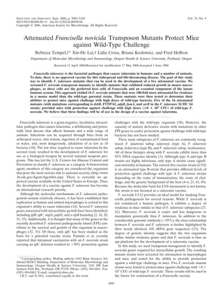
- 5. pected, macrophages infected with F. novicida U112 contained more bacteria than cells infected with our mutants contained (Fig. 2). Although several bacteria were observed inside host cells infected with the fumA mutant (Fig. 2F), infections with the dsbB, FTT0742, pdpB, and carB mutant strains resulted in only one or two intracellular bacteria at 24 h p.i. These findings corroborated our finding that the F. novicida transposon mu- tants were defective for replication or survival inside macro- phages. Furthermore, although the macrophages were initially seeded at the same concentration, fewer cells remained in the wells after infection with wild-type strain U112 than after in- fection with the mutants and in uninfected controls. This ob- servation indicated that host cell death occurred during the course of the wild-type infection but not during infection with attenuated mutant strains. FIG. 1. Five F. novicida transposon mutants are attenuated for growth in macrophages. J774 (A) and RAW (B) cell lines and mouse BMDM (C) were infected with the five F. novicida mutants and wild- type strain U112 at an MOI of 100 for 2 h and 24 h. Cells were lysed, and serial dilutions of the lysates were plated onto CHA/Kan20 (mu- tants) or CHA (U112 and mock infection controls). Colonies were counted, and the numbers of CFU/ml were calculated and converted to a log scale. Each column shows the average for three individual infec- tions. No bacteria were detected in J774 or RAW cells infected with the pdpB mutant after 24 h. Asterisks indicate the statistically signifi- cant results for 24 h (J774 cells, P Ͻ 0.005; RAW cells, P Ͻ 0.05; BMDM, P Ͻ 0.01). FIG. 2. Fluorescence microscopy of macrophage cell line infec- tions. J774 macrophages were infected in four-chamber microscope plates for 24 h at an MOI of 100. The cells were fixed in 4% paraform- aldehyde, probed with a polyclonal antibody against Francisella (Alexa 488) (green), and stained with FM 4-64 (membrane not shown) and Draq 5 (DNA) (blue). Cells were imaged with an Applied Precision DeltaVision deconvolution microscope system using a ϫ60 objective. The arrows indicate individual bacteria. wt, wild type. VOL. 74, 2006 ATTENUATED AND PROTECTIVE FRANCISELLA MUTANTS 5099
- 6. To address the possibility that the attenuation phenotypes of our F. novicida mutant strains could have been due to overall defects in replication, a simple growth curve was determined for each mutant. While the dsbB, FTT0742, and pdpB mutants replicated at levels similar to that of wild-type strain U112, both fumA and carB mutants exhibited defects in replication after 4 h of growth (data not shown). At 24-h after inoculation, the OD600 of cultures of these two strains were approximately 1.5, and the OD600 of cultures of the other strains ranged from 2.5 to 3.3. Interestingly, this phenotype was not rescued by supplementing the media with malate (fumA) or arginine (carB) (data not shown). It is possible that the addition of exogenous substrates could not complement the defects be- cause the enzymes are part of multienzyme complexes from which additional intermediates are excluded. Nonetheless, the fumA and carB mutants remained potential vaccine can- didates because they protected mice against wild-type chal- lenge (Table 2). Infection with F. novicida mutants did not reduce host cell integrity. The observed attenuation phenotypes could have been a result of increased host cell killing, which would have yielded fewer live infected macrophages and thus fewer bac- teria, as they would have been killed by the gentamicin in the extracellular media (29). One method for determining the de- gree of cytotoxicity that results from bacterial infection is to measure cell lysis by quantifying the release of the stable cy- tosolic enzyme LDH. J774 macrophages were infected with either wild-type strain U112 bacteria or one of the five mutant strains for 48 h at an input MOI of 100. The levels of LDH in the supernatants were then recorded. As shown in Fig. 3, the abilities of the five mutant strains to cause cell lysis were significantly impaired compared with the ability of wild-type F. novicida. With the levels of LDH released during wild-type infection normalized to 100%, the amounts of LDH released during infection with the five attenuated mutants ranged from 9.75% (FTT0742) to 24.52% (fumA). These results indicated that the attenuation phenotypes were not due to increased killing of host cells by the transposon mutants and that the intracellular replication of these strains was indeed compro- mised. Each F. novicida mutant harbored a single transposon in- sertion. Transposon mutagenesis has the potential to produce strains with more than one transposon insertion and thus mul- tiple causes for an observed phenotype. Each of our five pro- tective F. novicida transposon mutants was subjected to South- ern blot analysis to ensure that the attenuation phenotypes of the mutant strains were the result of a single transposition event. To determine the number of inserts, a DNA probe that spanned a unique HindIII site in the transposon was designed so that digestion of chromosomal DNA harboring a single transposon insert would yield two targets for this probe. Chro- mosomal DNA from each of the five protective mutant strains, as well as wild-type strain U112 and a Salmonella strain known to contain a single copy of the transposon, was prepared and probed as described in Materials and Methods. The presence of two bands demonstrated that the F. novicida mutant strains each harbored a single copy of the transposon insert, as shown in Fig. 4. F. novicida mutants disseminated to the liver, spleen, and lungs and were subsequently cleared. Acceptable vaccine can- didates ideally infect mice transiently and are cleared before challenge with the parent strain. We inoculated groups of 15 BALB/c mice i.p. with 0.1 LD50 of each mutant. Thus, for these infections, the vaccination dose varied from strain to strain. Three mice from each group were sacrificed at 1, 3, 5, 7, and 28 days after vaccination, and their spleen, liver, and lungs were harvested. As shown in Fig. 5, each mutant, with the possible exception of the carB mutant, disseminated to all three organs (spleen, liver, and lungs) from the original site of inoculation. Two of the five strains, the dsbB and fumA mutants, were completely cleared by day 28 following infection. Although relatively low numbers of bacteria remained in the spleen at day 28 after infection with the FTT0742 and pdpB mutants, it is possible that these organisms would have been cleared in the vaccination experiments because a lower dose (10- FIG. 3. Infection with F. novicida mutants does not reduce host cell integrity. J774 macrophages were infected with the five F. novicida mutants and wild-type strain U112 (wt U112) at an MOI of 100 for 48 h. The levels of LDH in the extracellular medium were determined. The level of LDH release for the wild-type infection was defined as 100%, and the levels of LDH release for the five mutant strains were normalized to this level. Each column shows the average for three individual infections. FIG. 4. Each F. novicida transposon mutant harbors a single trans- poson. Chromosomal DNA preparations from a Salmonella strain known to carry the mini-Tn5 cycler transposon (lane 1), wild-type strain U112 (lane 2), and the dsbB (lane3), FTT0742 (lane 4), pdpB (lane 5), fumA (lane 6), and carB (lane 7) mutant strains were digested with HindIII. The DNA was transferred to a membrane and probed with a digoxigenin-labeled DNA probe that spanned a HindIII site in the transposon. The membrane was exposed to film for 2 min (lanes 1, 2, 3, 5, and 7) or 8 min (lanes 4 and 6). The presence of two bands indicates a single transposon insertion event. 5100 TEMPEL ET AL. INFECT. IMMUN.
- 7. to 1,000-fold-fewer bacteria) was used for that procedure (Tables 2 and 4). Expressing full-length genes in trans complemented the at- tenuation phenotype. While the Southern hybridization exper- iments strongly indicated that each mutant derivative con- tained only a single transposon insertion, we wished to determine if cloned copies of the genes could complement the observed virulence defects. This would provide as additional evidence that the attenuation phenotype of each strain was a result of a single mutation, marked by the transposon insertion. The dsbB gene was amplified from SCHU S4 DNA by PCR and cloned into plasmid pKK202 (30). Following transforma- tion into the dsbB mutant, the abilities to replicate within macrophages and cause disease in mice were determined. As shown in Fig. 6, in trans expression of the cloned dsbB gene provided nearly complete complementation of the virulence defect in three different cell types. Further analysis showed that the LD50 was 60.25 CFU, which is comparable to the wild-type LD50 (66.25 CFU). In a parallel experiment, in trans expression of the full- length FTT0742 gene in the corresponding mutant derivative resulted in incomplete complementation. In RAW cells, intra- cellular replication of the complemented FTT0742 strain was 10-fold greater than intracellular replication of the mutant, but the value was still nearly 2 orders of magnitude less than the wild-type value (Fig. 6D). Like complementation of the dsbB mutation, complementa- tion of the fumA mutation with the full-length gene restored the level of intracellular growth to the level of wild-type F. novicida (Fig. 6E). Taken together, these findings show that the observed attenuation phenotypes were due to mutations in dsbB, FTT0742, and fumA. Complementation of pdpB will be attempted in the future, as the transposon insertion is located in the second gene of a 12-gene operon and is undoubtedly polar on expression of downstream genes. Based on its in vitro growth defect, com- paratively low LD50, relative lack of intracellular attenuation, and questionable dissemination patterns, we felt that the carB mutant was not a strong enough candidate to include in further development of a vaccine against tularemia, and therefore we did not attempt to complement the carB gene. Mutant strains protected mice against very high doses of wild-type bacteria. To further assess the level of protection provided by the F. novicida transposon insertion mutants, we challenged vaccinated mice with higher doses of the wild-type U112 parental strain. The dsbB, FTT0742, pdpB, and fumA mutant strains were used to infect groups of five mice, and the doses used were 6 ϫ 105 , 6 ϫ 106 , and 6 ϫ 107 CFU (Table 4). Mice infected with each of the three doses of our FTT0742 and pdpB mutants had a survival rate of 100%, as did the animals infected with the lowest doses of the dsbB and fumA mutants. Four weeks after vaccination, surviving animals were chal- lenged with 6 ϫ 107 CFU of wild-type strain F. novicida U112, which is approximately 106 times the observed LD50 for wild- type infection. All of the mice challenged survived without any symptoms of disease. These results demonstrated that four of our F. novicida transposon mutants were capable of protecting mice against infection with very high levels of the wild-type organism. Overall, our findings indicate that Francisella strains carrying mutations in these genes are candidates for a vaccine against tularemia. FIG. 5. F. novicida mutants disseminate and are subsequently cleared. The dsbB (A), FTT0742 (B), pdpB (C), fumA (D), and carB (E) mutant derivatives were injected i.p. into BALB/c mice, using 0.1 LD50. The spleen (}), liver (■), and lungs (Œ) were harvested at 1, 3, 5, 7, and 28 days p.i. Bacteria were liberated from the tissues and enumerated. Each point shows the results for three mice. TABLE 4. Results of challenge studies after vaccination with F. novicida transposon mutants Mutant strain Vaccine dose (CFU) % Survival (n ϭ 5) Challenge dose (CFU) % Survival dsbB 6 ϫ 105 100 6 ϫ 107 100 6 ϫ 106 20 6 ϫ 107 100 6 ϫ 107 0 NDa ND FTT0742 6 ϫ 105 100 6 ϫ 107 100 6 ϫ 106 100 6 ϫ 107 100 6 ϫ 107 100 6 ϫ 107 100 pdpB 6 ϫ 105 100 6 ϫ 107 100 6 ϫ 106 100 6 ϫ 107 100 6 ϫ 107 100 6 ϫ 107 100 fumA 6 ϫ 105 100 6 ϫ 107 100 6 ϫ 106 0 ND ND 6 ϫ 107 0 ND ND a ND, not determined. VOL. 74, 2006 ATTENUATED AND PROTECTIVE FRANCISELLA MUTANTS 5101
- 8. DISCUSSION The classification of F. tularensis as a category A bioterror- ism agent by the Centers for Disease Control and Prevention demonstrates that this organism is widely acknowledged to be a potential threat to national security. Thus, there is an imme- diate need for an approved tularemia vaccine. The lack of genetic tools with which to manipulate F. tularensis remains a great barrier to the development of such a vaccine. Hence, we developed a transposon mutagenesis technique to create ran- dom insertions in the F. novicida genome and analyzed the resulting mutant strains for intracellular growth defects in mac- rophages, attenuation in mice, and the ability to provide pro- tection against wild-type infection. We identified 28 F. novicida transposon mutants that have a defect in intracellular growth in macrophage cell lines, 16 of which exhibited 100% attenu- ation in mice at a dose that was more than 100 times the wild-type LD50. When mice were challenged with the wild-type organism, five transposon mutant strains were found to protect FIG. 6. Expressing full-length genes in trans complements the attenuation defects in cells. The levels of entry (2 h) and replication (24 h) were determined for wild-type strain U112 (wt U112), the dsbB mutant, and the dsbB mutant complemented with pKK202-dsbB in the J774 (A) and RAW (B) cell lines and in primary BMDM (C). Entry and replication rates in RAW cells were determined for complementation of the FTT0742 (D) and fumA (E) mutants. Each column shows the average for three separate infections. 5102 TEMPEL ET AL. INFECT. IMMUN.
- 9. the mice against infection with high doses of parental F. novi- cida strain U112. The disrupted genes in our five protective F. novicida mutants correspond to dsbB, FTT0742, pdpB, fumA, and carB in F. tularensis strain SCHU S4. Disulfide bond formation protein B is encoded by dsbB. This integral membrane protein is part of a pathway that leads to disulfide bond formation between cysteine residues in periplas- mic proteins in E. coli and other bacteria (23). The functional folded conformation of a protein often relies upon correct disulfide pairing of the cysteine residues. Thus, one explana- tion for why our dsbB mutant strain is attenuated is that a protein(s) required for replication inside host cells does not achieve its active conformation. It is also exciting to speculate that, with its potential influence on periplasmic proteins, the dsbB gene product may be involved in the secretion of viru- lence factors, possibly to ensure correct folding of components of a secretion apparatus. The FTT0742 ORF codes for a hypothetical lipoprotein that is predicted to have transmembrane regions. Therefore, it is possible that the gene product is a component of the F. novi- cida cell wall and/or may be involved in molecule transport. Because in vitro growth and entry into the host cell were not compromised, we speculate that FTT0742 affects a function necessary for virulence and growth inside macrophages. Char- acterization of the FTT0742 protein should further clarify its role in virulence. The product of the pdpB gene is an uncharacterized protein encoded on the FPI that exhibits some similarity to the con- served bacterial protein IcmF (http://www.ncbi.nlm.nih.gov /Structure/cdd/wrpsb.cgi). It has been shown that icmF is re- quired for Legionella pneumophila intracellular growth, so we hypothesized that pdpB plays a similar role in F. novicida intracellular growth (50). Also of note, this gene exhibits some similarity to the genes encoding rhoptry proteins of the para- site Plasmodium, which mediate attachment to host red blood cells (40). Although the pdpB mutant did not exhibit defects during in vitro growth, its ability to enter host cells was signif- icantly compromised and it displayed prominent intracellular attenuation since no CFU were detected in the lysates of RAW and J774 cells at 24 h p.i. Because pdpB is the second ORF in a 12-gene operon, it is likely that the transposon insert in this gene has polar effects on downstream genes. These findings indicate that pdpB or other genes in the pdp operon are needed for both entry into and replication within host cells. So far, the functions of the genes in the pdp operon have not been eluci- dated. Fumarate hydratase A, the component of the Kreb’s cycle (citric acid cycle) that converts fumarate to malate, is encoded by fumA (46). The citric acid cycle is one of the three metabolic pathways of cellular respiration and is necessary for fuel ca- tabolism and ATP production. Precursory molecules for com- pounds such as amino acids are also generated by the citric acid cycle. Thus, the observation that our fumA mutant exhib- ited lower levels of in vitro replication than wild-type strain U112 exhibited may have been a result of energy deficiency or a lack of molecules needed for replication. The carB gene encodes the large subunit of the het- erodimeric enzyme carbamoyl phosphate synthase, which is required for pyrimidine biosynthesis (28). As pyrimidines are required for replication, it is clear that a mutation in this biosynthesis pathway would lead to both in vitro and intracel- lular growth defects. Indeed, we observed an in vitro growth defect with this mutant strain. Nevertheless, our carB mutant was able to infect macrophages and protect mice against chal- lenge with the wild-type organism, which suggests that strains with mutations in the pyrimidine pathways of F. novicida may be useful as potential vaccine strains. However, compared to the other F. novicida mutants used in this study, the carB mutant had a lower LD50 and less intracellular attenuation and did not appear to disseminate from the initial site of infection. For these reasons, we decided not to pursue use of this mutant as a possible vaccine candidate. Although we observed only partial complementation of the FTT0742 mutant phenotype, we still consider this strain to be a vaccine candidate. Complementation experiments similar to those described in this paper carried out with Salmonella have also resulted in mixed success because the copy number of the plasmid, as well as the regulation of the gene itself, can influ- ence complementation. In fact, the Forsberg group recently demonstrated the importance of correct gene regulation dur- ing complementation in Francisella with their studies of pilA; there was functional complementation in cis, but expression of PilA was barely detectable in the strain complemented in trans (12). The problem was further compounded in our studies because the F. novicida genome has not been published yet; therefore, the genes used for complementation were amplified from SCHU S4 DNA and may be incompatible with F. novi- cida due to variations between the subspecies. Intriguingly, another finding of this study was the lack of protection conferred by pur mutants in a murine model. It has been postulated previously that mutations affecting the F. tu- larensis purine synthesis pathway could be used to generate a live attenuated tularemia vaccine (25). In fact, defined allelic replacement mutants with mutations that disrupt this pathway have been used to produce vaccine strains attenuated for rep- lication in host cells in a variety of other bacterial species (36). Our F. novicida transposon library contained eight unique pur mutants, including mutants with mutations in purA, a purCD fusion (two strains), purL (two strains), and purM (three strains). Each of these strains exhibited 100% attenuation in mice when 6 ϫ 103 CFU was used, yet all of them failed to protect mice against a wild-type parental challenge with 8 ϫ 105 CFU. Although these results do not necessarily eliminate purine biosynthesis mutants as potential live vaccine strains, it was noteworthy that these mutants did not protect against challenge with wild-type F. novicida strain U112. As an alternative to live vaccines, the possibility of develop- ing a subunit vaccine must also be examined. Studies in which the efficacy of whole killed cells as a crude tularemia vaccina- tion was evaluated demonstrated the feasibility of a subunit vaccine and prompted research into identification of antigens that induce protective immunity (8, 24). Although several poly- saccharide and protein components of F. tularensis have been shown to react with convalescent-phase sera or T cells (22), the only antigen that has exhibited the ability to induce a protec- tive immune response against tularemia is lipopolysaccharide (36). However, the protection was effective only against F. tularensis subsp. holarctica strains and was incomplete (7, 15, 16). Protection against a highly virulent F. tularensis type A strain likely depends on a Th1-mediated cellular immune re- VOL. 74, 2006 ATTENUATED AND PROTECTIVE FRANCISELLA MUTANTS 5103
- 10. sponse (44). So far, there is no way to administer antigens or killed bacteria that are as effective as a living attenuated vac- cine, a point that has been highlighted by work in the Pamer laboratory showing that the immune system distinguishes be- tween living and dead bacteria (37). In contrast, a live attenuated vaccine would be effective in inducing the appropriate cellular responses (36). In fact, the type B LVS strain provides the only current means of tulare- mia vaccination. However, several limitations prevent the li- censing of this vaccine. Foremost among these constraints is the fact that the genetic basis of LVS attenuation and protec- tion remains unknown. Second, culturing LVS under certain conditions can lead to poorly immunogenic colony variants, which demonstrates this organism’s inherent genetic instability (9, 11). Also, this vaccine does not provide protection to every individual vaccinated (34, 41). Finally, LVS protection against aerosol challenge is variable and depends on the route of immunization, as well as the host (4–6, 43). The last point is especially critical when F. tularensis is considered as a biolog- ical weapon, as aerosol dispersal is the most likely route of delivery. Taken together, these limitations clearly show that the development of an approved tularemia vaccine requires the development of a rationally attenuated, nonreverting live vaccine strain. While the manuscript was in preparation, the Conlan Sjo¨s- tedt groups published the first description of a defined gene deletion mutant of a type A strain that protected mice against challenge with the wild-type organism (47). Indeed, the ideal live vaccine strain would be derived from a virulent F. tularensis strain; however, we used F. novicida as a model for preliminary analysis of potential tularemia vaccines because this taxon is more amenable to genetic manipulation without the danger of infection. Furthermore, because all of the Francisella taxa are closely related (3), genes necessary for intracellular growth in F. novicida are likely to have the same function in F. tularensis. Consequently, F. novicida strains provide relatively safe, ge- netically significant organisms with which to conduct explor- atory investigations prior to studies with the more virulent F. tularensis subspecies. Here, we describe discovery of five F. novicida transposon mutants that exhibit attenuation in macrophages and are ca- pable of protecting mice against infection with the wild-type parental strain at doses that are up to 106 times the observed wild-type LD50. An approved tularemia vaccine must be a highly attenuated nonreverting derivative of a type A strain, as such strains are most likely to be used in a bioterrorism attack and there is no certainty that one taxon will protect against another. Accordingly, we will now focus on extending this work to Francisella type A strains by creating deletions of each of the genes identified here and assaying for virulence in a mouse model. ACKNOWLEDGMENTS We thank the OHSU Core Facility and Aurelie Snyder for sequenc- ing and microscopy assistance. We also acknowledge Jeff Vandehey and Chris Langford for technical assistance. We are grateful to Fran Nano for providing the F. novicida U112 strain. The mini-Tn5 cycler transposon was a kind gift from Kaoru Geddes. We appreciate the helpful comments of Jean Gustin during revision of the manuscript. This work was supported by NIH R21 grant EB000985 to F.H., as well as by a National Science Foundation Graduate Research Fellow- ship and a OHSU Tartar Trust Fellowship, both to R.T. REFERENCES 1. Ausubel, F. M. 2002. Short protocols in molecular biology: a compendium of methods from Current Protocols in Molecular Biology, 5th ed. Wiley, New York, NY. 2. Baron, G. S., and F. E. Nano. 1998. MglA and MglB are required for the intramacrophage growth of Francisella novicida. Mol. Microbiol. 29:247–259. 3. Broekhuijsen, M., P. Larsson, A. Johansson, M. Bystrom, U. Eriksson, E. Larsson, R. G. Prior, A. Sjostedt, R. W. Titball, and M. Forsman. 2003. Genome-wide DNA microarray analysis of Francisella tularensis strains dem- onstrates extensive genetic conservation within the species but identifies regions that are unique to the highly virulent F. tularensis subsp. tularensis. J. Clin. Microbiol. 41:2924–2931. 4. Chen, W., R. KuoLee, H. Shen, and J. W. Conlan. 2004. Susceptibility of immunodeficient mice to aerosol and systemic infection with virulent strains of Francisella tularensis. Microb. Pathog. 36:311–318. 5. Chen, W., H. Shen, A. Webb, R. KuoLee, and J. W. Conlan. 2003. Tularemia in BALB/c and C57BL/6 mice vaccinated with Francisella tularensis LVS and challenged intradermally or by aerosol with virulent isolates of the pathogen: protection varies depending on pathogen virulence, route of exposure, and host genetic background. Vaccine 21:3690–3700. 6. Conlan, J. W., H. Shen, R. Kuolee, X. Zhao, and W. Chen. 2005. Aerosol- but not intradermal-immunization with the live vaccine strain of Francisella tularensis protects mice against subsequent aerosol challenge with a highly virulent type A strain of the pathogen by an alphabeta T cell- and interferon gamma-dependent mechanism. Vaccine 23:2477–2485. 7. Conlan, J. W., H. Shen, A. Webb, and M. B. Perry. 2002. Mice vaccinated with the O-antigen of Francisella tularensis LVS lipopolysaccharide conju- gated to bovine serum albumin develop varying degrees of protective immu- nity against systemic or aerosol challenge with virulent type A and type B strains of the pathogen. Vaccine 20:3465–3471. 8. Coriell, L. L., E. O. King, and M. G. Smith. 1948. Studies on tularemia IV. Observations on tularemia in control and vaccinated monkeys. J. Immunol. 58:183–202. 9. Cowley, S. C., S. V. Myltseva, and F. E. Nano. 1996. Phase variation in Francisella tularensis affecting intracellular growth, lipopolysaccharide anti- genicity and nitric oxide production. Mol. Microbiol. 20:867–874. 10. Dennis, D. T., T. V. Inglesby, D. A. Henderson, J. G. Bartlett, M. S. Ascher, E. Eitzen, A. D. Fine, A. M. Friedlander, J. Hauer, M. Layton, S. R. Lillibridge, J. E. McDade, M. T. Osterholm, T. O’Toole, G. Parker, T. M. Perl, P. K. Russell, and K. Tonat. 2001. Tularemia as a biological weapon: medical and public health management. JAMA 285:2763–2773. 11. Eigelsbach, H. T., and C. M. Downs. 1961. Prophylactic effectiveness of live and killed tularemia vaccines. I. Production of vaccine and evaluation in the white mouse and guinea pig. J. Immunol. 87:415–425. 12. Forslund, A. L., K. Kuoppa, K. Svensson, E. Salomonsson, A. Johansson, M. Bystrom, P. C. Oyston, S. L. Michell, R. W. Titball, L. Noppa, E. Frithz- Lindsten, M. Forsman, and A. Forsberg. 2006. Direct repeat-mediated de- letion of a type IV pilin gene results in major virulence attenuation of Francisella tularensis. Mol. Microbiol. 59:1818–1830. 13. Forsman, M., G. Sandstrom, and A. Sjostedt. 1994. Analysis of 16S ribo- somal DNA sequences of Francisella strains and utilization for determina- tion of the phylogeny of the genus and for identification of strains by PCR. Int. J. Syst. Bacteriol. 44:38–46. 14. Fortier, A. H., S. J. Green, T. Polsinelli, T. R. Jones, R. M. Crawford, D. A. Leiby, K. L. Elkins, M. S. Meltzer, and C. A. Nacy. 1994. Life and death of an intracellular pathogen: Francisella tularensis and the macrophage. Immu- nol. Ser. 60:349–361. 15. Fulop, M., R. Manchee, and R. Titball. 1995. Role of lipopolysaccharide and a major outer membrane protein from Francisella tularensis in the induction of immunity against tularemia. Vaccine 13:1220–1225. 16. Fulop, M., P. Mastroeni, M. Green, and R. W. Titball. 2001. Role of anti- body to lipopolysaccharide in protection against low- and high-virulence strains of Francisella tularensis. Vaccine 19:4465–4472. 17. Geddes, K., M. Worley, G. Niemann, and F. Heffron. 2005. Identification of new secreted effectors in Salmonella enterica serovar Typhimurium. Infect. Immun. 73:6260–6271. 18. Golovliov, I., A. Sjostedt, A. Mokrievich, and V. Pavlov. 2003. A method for allelic replacement in Francisella tularensis. FEMS Microbiol. Lett. 222:273– 280. 19. Goryshin, I. Y., J. Jendrisak, L. M. Hoffman, R. Meis, and W. S. Reznikoff. 2000. Insertional transposon mutagenesis by electroporation of released Tn5 transposition complexes. Nat. Biotechnol. 18:97–100. 20. Gray, C. G., S. C. Cowley, K. K. Cheung, and F. E. Nano. 2002. The identification of five genetic loci of Francisella novicida associated with in- tracellular growth. FEMS Microbiol. Lett. 215:53–56. 21. Green, M., G. Choules, D. Rogers, and R. W. Titball. 2005. Efficacy of the live attenuated Francisella tularensis vaccine (LVS) in a murine model of disease. Vaccine 23:2680–2686. 5104 TEMPEL ET AL. INFECT. IMMUN.
- 11. 22. Isherwood, K. E., R. W. Titball, D. H. Davies, P. L. Felgner, and W. J. Morrow. 2005. Vaccination strategies for Francisella tularensis. Adv. Drug Deliv. Rev. 57:1403–1414. 23. Kadokura, H., F. Katzen, and J. Beckwith. 2003. Protein disulfide bond formation in prokaryotes. Annu. Rev. Biochem. 72:111–135. 24. Kadull, P. J., H. R. Reames, L. L.Coriell, and L. Foshay. 1950. Studies on tularemia. V. Immunization of man. J. Immunol. 65:425–435. 25. Karlsson, J., R. G. Prior, K. Williams, L. Lindler, K. A. Brown, N. Chatwell, K. Hjalmarsson, N. Loman, K. A. Mack, M. Pallen, M. Popek, G. Sandstrom, A. Sjostedt, T. Svensson, I. Tamas, S. G. Andersson, B. W. Wren, P. C. Oyston, and R. W. Titball. 2000. Sequencing of the Francisella tularensis strain Schu 4 genome reveals the shikimate and purine metabolic pathways, targets for the construction of a rationally attenuated auxotrophic vaccine. Microb. Comp. Genomics 5:25–39. 26. Kawula, T. H., J. D. Hall, J. R. Fuller, and R. R. Craven. 2004. Use of transposon-transposase complexes to create stable insertion mutant strains of Francisella tularensis LVS. Appl. Environ. Microbiol. 70:6901–6904. 27. Kieffer, T. L., S. Cowley, F. E. Nano, and K. L. Elkins. 2003. Francisella novicida LPS has greater immunobiological activity in mice than F. tularensis LPS, and contributes to F. novicida murine pathogenesis. Microbes Infect. 5:397–403. 28. Koonin, E. V., and M. Y. Galperin. 2003. Sequence-evolution-function: com- putational approaches in comparative genomics. Kluwer Academic, Boston, MA. 29. Kudelina, R. I. 1978. Sensitivity of the causative agent of tularemia to antibiotics. Antibiotiki 23:710–714. 30. Kuoppa, K., A. Forsberg, and A. Norqvist. 2001. Construction of a reporter plasmid for screening in vivo promoter activity in Francisella tularensis. FEMS Microbiol. Lett. 205:77–81. 31. Lai, X. H., I. Golovliov, and A. Sjostedt. 2004. Expression of IglC is necessary for intracellular growth and induction of apoptosis in murine macrophages by Francisella tularensis. Microb. Pathog. 37:225–230. 32. Larsson, P., P. C. Oyston, P. Chain, M. C. Chu, M. Duffield, H. H. Fuxelius, E. Garcia, G. Halltorp, D. Johansson, K. E. Isherwood, P. D. Karp, E. Larsson, Y. Liu, S. Michell, J. Prior, R. Prior, S. Malfatti, A. Sjostedt, K. Svensson, N. Thompson, L. Vergez, J. K. Wagg, B. W. Wren, L. E. Lindler, S. G. Andersson, M. Forsman, and R. W. Titball. 2005. The complete genome sequence of Francisella tularensis, the causative agent of tularemia. Nat. Genet. 37:153–159. 33. Lauriano, C. M., J. R. Barker, S. S. Yoon, F. E. Nano, B. P. Arulanandam, D. J. Hassett, and K. E. Klose. 2004. MglA regulates transcription of viru- lence factors necessary for Francisella tularensis intraamoebae and intram- acrophage survival. Proc. Natl. Acad. Sci. USA 101:4246–4249. 34. McCrumb, F. R. 1961. Aerosol infection of man with Pasteurella tularensis. Bacteriol. Rev. 25:262–267. 35. Nano, F. E., N. Zhang, S. C. Cowley, K. E. Klose, K. K. Cheung, M. J. Roberts, J. S. Ludu, G. W. Letendre, A. I. Meierovics, G. Stephens, and K. L. Elkins. 2004. A Francisella tularensis pathogenicity island required for intra- macrophage growth. J. Bacteriol. 186:6430–6436. 36. Oyston, P. C., and J. E. Quarry. 2005. Tularemia vaccine: past, present and future. Antonie Leeuwenhoek 87:277–281. 37. Pamer, E. G. 2004. Immune responses to Listeria monocytogenes. Nat. Rev. Immunol. 4:812–823. 38. Pammit, M. A., E. K. Raulie, C. M. Lauriano, K. E. Klose, and B. P. Arulanandam. 2006. Intranasal vaccination with a defined attenuated Fran- cisella novicida strain induces gamma interferon-dependent antibody-medi- ated protection against tularemia. Infect. Immun. 74:2063–2071. 39. Reed, L. J., and H. Muench. 1935. A simple method of estimating fifty percent endpoints. Am. J. Hyg. 27:493–497. 40. Sam-Yellowe, T. Y. 1992. Molecular factors responsible for host cell recog- nition and invasion in Plasmodium falciparum. J. Protozool. 39:181–189. 41. Saslaw, S., H. T. Eigelsbach, J. A. Prior, H. E. Wilson, and S. Carhart. 1961. Tularemia vaccine study. II. Respiratory challenge. Arch. Intern. Med. 107: 702–714. 42. Shen, H., W. Chen, and J. W. Conlan. 2004. Mice sublethally infected with Francisella novicida U112 develop only marginal protective immunity against systemic or aerosol challenge with virulent type A or B strains of F. tularensis. Microb. Pathog. 37:107–110. 43. Shen, H., W. Chen, and J. W. Conlan. 2004. Susceptibility of various mouse strains to systemically- or aerosol-initiated tularemia by virulent type A Francisella tularensis before and after immunization with the attenuated live vaccine strain of the pathogen. Vaccine 22:2116–2121. 44. Tarnvik, A. 1989. Nature of protective immunity to Francisella tularensis. Rev. Infect. Dis. 11:440–451. 45. Tempel, R., K. Geddes, and F. Heffron. 2003. Presented at the Fourth International Conference on Tularemia, Bath, United Kingdom. 46. Tseng, C. P., C. C. Yu, H. H. Lin, C. Y. Chang, and J. T. Kuo. 2001. Oxygen- and growth rate-dependent regulation of Escherichia coli fumarase (FumA, FumB, and FumC) activity. J. Bacteriol. 183:461–467. 47. Twine, S., M. Bystrom, W. Chen, M. Forsman, I. Golovliov, A. Johansson, J. Kelly, H. Lindgren, K. Svensson, C. Zingmark, W. Conlan, and A. Sjostedt. 2005. A mutant of Francisella tularensis strain SCHU S4 lacking the ability to express a 58-kilodalton protein is attenuated for virulence and is an effective live vaccine. Infect. Immun. 73:8345–8352. 48. van der Velden, A. W., S. W. Lindgren, M. J. Worley, and F. Heffron. 2000. Salmonella pathogenicity island 1-independent induction of apoptosis in infected macrophages by Salmonella enterica serotype Typhimurium. Infect. Immun. 68:5702–5709. 49. Wu, T. H., J. A. Hutt, K. A. Garrison, L. S. Berliba, Y. Zhou, and C. R. Lyons. 2005. Intranasal vaccination induces protective immunity against intranasal infection with virulent Francisella tularensis biovar A. Infect. Immun. 73: 2644–2654. 50. Zusman, T., M. Feldman, E. Halperin, and G. Segal. 2004. Characterization of the icmH and icmF genes required for Legionella pneumophila intracel- lular growth, genes that are present in many bacteria associated with eu- karyotic cells. Infect. Immun. 72:3398–3409. Editor: J. T. Barbieri VOL. 74, 2006 ATTENUATED AND PROTECTIVE FRANCISELLA MUTANTS 5105