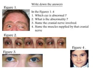
SlideShare uma empresa Scribd logo
1 de 57
Figure 1.
Figure 2.
Figure 3.
Figure 4 .
Write down the answers
In the Figures 1-4
1. Which eye is abnormal ?
2. What is the abnormality ?
3. Name the cranial nerve involved.
4. Name the muscles supplied by that cranial
nerve
EXTRAOCULAR MUSCLES
Extraocular muscles
4 Recti and 2 Obliques
Superior rectus Superior oblique
Inferior rectus Inferior oblique
Medial rectus
Lateral rectus
Levator palpebrae superioris
LEVATOR PALPEBRAE SUPERIORIS
Origin:
Undersurface of lesser wing of
sphenoid above optic canal
Insertion:
 Skin of upper eyelids
Anterior surface of superior tarsus
Muller`s muscle/Superior tarsal
muscle
Superior conjunctival fornix
LEVATOR PALPEBRAE SUPERIORIS
Nerve supply and Actions
Paralysis - PTOSIS
Oculomotor nerve, Sympathetics
Elevates upper lid
Equator
Optical axis/Axis of Gaze – direction of
sight .Primary position of eye
Axis of movements
Axis of muscles
Movements
Abduction
Elevation
Depression
Adduction Intorsion
Extorsion
Elevation & Depression – Around the transverse axis
Adduction & Abduction – Around the vertical axis
Intortion & Extortion – Around the anteroposterior axis
And the RULE is…..(for recti and oblique)
Any muscle inserting
medial to vertical axis – Adduction
lateral to vertical axis - Abduction
superior to AP axis – Intorsion
inferior to AP axis – Extorsion
For muscle inserting in front of equator i.e RECTI
above transverse axis – Elevation
below transverse axis - Depression
ORIGIN OF THE 4 RECTI MUSCLE
Common tendinous ring
(Annulus of Zinn)
•Lateral rectus by 2
heads
–Extra head from
adjoining greater
wing of sphenoid
LEFT EYE
COURSE OF THE 4 RECTI
Muscular cone
Corresponding
wall of orbit
Rectus muscle length – 40mm
Innervated from intraconal
side of the muscle belly at the
junction of anterior 2/3 and
posterior 1/3 of the muscle
INSERTION OF THE 4 RECTI
The line connecting the insertion of the
recti in series is spiral & is known as spiral
line of Tillaux
Pierce
Tenon’scapsule
Sclera in front of the
equator
Medial rectus is susceptible to injury during anterior segment
procedures
AXES OF THE RECTI MUSCLE
Medial and lateral recti in same
horizontal plane
Superior and inferior recti in same
oblique plane, 25⁰lateral to optical
axis
In the abducted eye the axes
coincide
Action of the RECTI
• Medial & lateral recti lie in the same horizontal plane
Around a vertical axis
Medial rectus - adduction Lateral rectus -
abduction
• Superior rectus
 Around the transverse axis – rotates the
eyeball upwards – Elevation (PRIMARY
ACTION)
 Around the vertical axis - Adduction
 Around the anteroposterior axis -
Intortion
• Inferior rectus
 Around the transverse axis – rotates the
eyeball downwards – Depression (PRIMARY
ACTION)
 Around the vertical axis – Adduction
 Around the anteroposterior axis - Extortion
Only in the Abducted position of the eyeball the visual axis coincides with
the axis of superior and inferior recti
In abducted eye
Superior rectus – Elevation only
Inferior rectus - Depression only
Superior Oblique muscle
Body of sphenoid above and medial
to optic canal
Winds around trochlea at
superomedial part of orbit
(functional origin)
Insertion behind the equator
Postero‐superior quadrant
Only eye muscle innervated on the outer
surface of muscle belly.
Retrobulbar anaesthetic block
Origin from orbital surface of
maxilla
Passes backward and laterally
below inferior rectus
Insertion behind equator
parallel to superior oblique
Postero‐superior quadrant
Inferior Oblique Muscle
The oblique muscles always course below the corresponding vertical
rectus muscle
Axis of the Oblique Muscles
The obliques lie in
the same oblique
plane 51⁰medial to
optical axis
In the adducted eye
axes coincide with
the optical axis
• Superior oblique
 Around the anteroposterior axis –
Intorsion(primary action)
 Around the vertical axis Abduction
 Around the transverse eaxis –
Depression
• Inferior oblique
 Extortion(primary action)
 Abduction
 Elevation
Only in the Adducted position of the eyeball the visual axis coincides with the axis of
superior and inferior oblique
In Adducted eye
Superior oblique – Depression only
Inferior oblique – Elevation only
Superior division of oculomotor:- levator palpebrae superioris, superior rectus
Inferior division of oculomotor:- medial rectus, inferior oblique, inferior rectus
Trochlear nerve - superior oblique
Abducent nerve - lateral rectus
Nerve Supply of Extraocular Muscles
Blood supply
Ophthalmic artery
Extraocular Muscles
 Allow accurate positioning of visual axis
 Determine the spatial relationship
between the two eyes
 Responsible for binocular vision
 Have the smallest motor unit among
skeletal muscles – ratio of nerve fibre to
muscle fibre is 1:2(whereas 1:25 in
other skeletal muscles)
 -Yoke Muscles: a muscle of one eye is
paired with another muscle of the fellow
eye to produce a cardinal gaze
-Example: Right LR & Left MR
while looking towards right side
 They develop from ?
Preotic/preoccipital somitomeres
Fascial expansions of Extraocular muscles
RECTI -Adduct
OBLIQUES – Abduct
SUPERIORS – Intort
INFERIORS -Extort
Clinical Testing
Ptosis
Eyeball turned down and out
Ocular movements restricted
Pupil fixed and dilated
Loss of accomodation
OCCULOMOTOR NERVE PALSY
ABDUCENS PALSY – Internal squint
The right eye unable to abduct
External squint- Medial rectus paralysis
The right eye unable to adduct
OPTHALMOPLEGIA / EXTRAOCULAR MUSCLE PALSY
Injury to III, IV, VI cranial nerve Muscle paralysis
Unilateral paralysis produces Strabismus /Squint, Diplopia
TROCHLEAR NERVE
PALSY
Eyeball turned upwards
and inwards
TROCHLEAR NERVE PALSY
 Affected eye rotated up and in.
 Attempts to compensate lead to the patient tilting their head to the contralateral side.
ABDUCENS PALSY
Third nerve palsy results in an inability to move
the eye normally in all directions. Injury to the
third nerve can occur anywhere along its path,
from where it originates within the brain to
where it innervates the muscles that move the
eyeball. Third nerve palsy prevents the proper
functioning of the medial, superior, and
inferior recti, and inferior oblique muscles. As
a result, the eye cannot move up, down, or in.
When at rest, the eye tends to look down and
to the side, due to an inequality of muscle
functioning. The muscle responsible for
keeping the upper eyelid open (levator
palpebrae superioris) is also affected, resulting
in a drooping upper eyelid (ptosis
Movements
Elevation
Depression
Adduction
Abduction
Intortion extortion
phthalmoplegia, also called extraocular muscle palsy, paralysis of the
extraocular muscles that control the movements of the eye. Ophthalmoplegia usually involves the third (oculomotor), fourth
(trochlear), or sixth (abducens)cranial nerves. Double vision is the characteristic symptom in all three cases
The optical axis of the eye (the line from the
center of the cornea to the fovea) points
straight ahead during straight-ahead gaze, but
the axis of the orbit points about 23 degrees
laterally. The superior and inferior recti
originate from the back of the orbit, and so
their direction of pulling is not parallel to the
optical axis. As a result, although the superior
rectus primarily elevates the eye, it also has
smaller adducting and intorting effects.
(Similarly, although not indicated in the Þgure,
the inferior rectus primarily depresses but also
adducts and extorts a little.)
The pulling direction of the obliques is not
aligned with either the optical axis or the
orbital axis, and their actions change with the
direction of gaze. The superior oblique inserts
in the posterior half of the eye and pulls
diagonally forward. A, As a result, during
straight-ahead gaze, although it primarily
intorts the eye, it also pulls the back of the eye
a little bit medially and upward (i.e., abducts
and depresses a little). B, During adduction,
the direction of pull is more nearly in line with
the optical axis, and the same muscle
depresses more and intorts less. C, During
abduction, the direction of pull can wind up
perpendicular to the optical axis, and the
action becomes purely intorsion. (Similarly,
although not indicated in the Þgure, the
inferior oblique primarily extorts when the eye
is abducted, but it also elevates and abducts in
other directions of gaze.)
Extra ocular muscles ppt

Mais conteúdo relacionado

Mais procurados

Mais procurados (20)

Visual pathway ppt
Visual pathway pptVisual pathway ppt
Visual pathway ppt
 
Light reflex
Light reflexLight reflex
Light reflex
 
Extraocular muscles
Extraocular muscles Extraocular muscles
Extraocular muscles
 
Abducens Nerve - Course and relation.
Abducens Nerve - Course and relation.Abducens Nerve - Course and relation.
Abducens Nerve - Course and relation.
 
EXTRA OCULAR MUSCLES
EXTRA OCULAR MUSCLES EXTRA OCULAR MUSCLES
EXTRA OCULAR MUSCLES
 
Anatomy of extraocular muscles and ocular motility
Anatomy of extraocular muscles and ocular motilityAnatomy of extraocular muscles and ocular motility
Anatomy of extraocular muscles and ocular motility
 
Anatomy of lacrimal apparatus
Anatomy of lacrimal apparatusAnatomy of lacrimal apparatus
Anatomy of lacrimal apparatus
 
Extraocular muscles
Extraocular muscles Extraocular muscles
Extraocular muscles
 
Orbital anatomy
Orbital anatomyOrbital anatomy
Orbital anatomy
 
Trochlear nerve
Trochlear nerveTrochlear nerve
Trochlear nerve
 
Lacrimal system ppt.
Lacrimal system ppt.Lacrimal system ppt.
Lacrimal system ppt.
 
Embryology of eye
Embryology of eyeEmbryology of eye
Embryology of eye
 
Pupillary reflexes
Pupillary reflexesPupillary reflexes
Pupillary reflexes
 
Visual Pathway - Ophthalmology - Eye
Visual Pathway - Ophthalmology - EyeVisual Pathway - Ophthalmology - Eye
Visual Pathway - Ophthalmology - Eye
 
EXTRA OCULAR MUSCLES PHYSIOLOGY
EXTRA OCULAR MUSCLES PHYSIOLOGYEXTRA OCULAR MUSCLES PHYSIOLOGY
EXTRA OCULAR MUSCLES PHYSIOLOGY
 
PUPIL - ANATOMY, PHYSIOLOGY AND REFLEXES
PUPIL - ANATOMY, PHYSIOLOGY AND REFLEXESPUPIL - ANATOMY, PHYSIOLOGY AND REFLEXES
PUPIL - ANATOMY, PHYSIOLOGY AND REFLEXES
 
The pupillary pathway and its clinical aspects
The pupillary pathway and its clinical aspectsThe pupillary pathway and its clinical aspects
The pupillary pathway and its clinical aspects
 
Blood Supply Of Eye and Optic Nerve
Blood Supply Of Eye and Optic NerveBlood Supply Of Eye and Optic Nerve
Blood Supply Of Eye and Optic Nerve
 
Visual pathway
Visual pathway Visual pathway
Visual pathway
 
Oculomotor nerve
Oculomotor nerveOculomotor nerve
Oculomotor nerve
 

Semelhante a Extra ocular muscles ppt

EXTRA OCULAR MUSCLES
EXTRA OCULAR MUSCLESEXTRA OCULAR MUSCLES
EXTRA OCULAR MUSCLES
arya das
 
ophthalmology.squint.(dr.khald)
ophthalmology.squint.(dr.khald)ophthalmology.squint.(dr.khald)
ophthalmology.squint.(dr.khald)
student
 
Diseases of ocular motility
Diseases of ocular motilityDiseases of ocular motility
Diseases of ocular motility
meducationdotnet
 

Semelhante a Extra ocular muscles ppt (20)

Orbit and extra ocular muscles
Orbit and extra ocular musclesOrbit and extra ocular muscles
Orbit and extra ocular muscles
 
Extraocular muscles
Extraocular musclesExtraocular muscles
Extraocular muscles
 
EXTRA OCULAR MUSCLES
EXTRA OCULAR MUSCLESEXTRA OCULAR MUSCLES
EXTRA OCULAR MUSCLES
 
Paralytic strabismus
Paralytic strabismusParalytic strabismus
Paralytic strabismus
 
Eye movements
Eye movementsEye movements
Eye movements
 
STRABISMUS new-WPS Office.pptx
STRABISMUS new-WPS Office.pptxSTRABISMUS new-WPS Office.pptx
STRABISMUS new-WPS Office.pptx
 
Extraocular muscles
Extraocular musclesExtraocular muscles
Extraocular muscles
 
The extraocular muscles and ocular movements
The extraocular muscles and ocular movementsThe extraocular muscles and ocular movements
The extraocular muscles and ocular movements
 
ANATOMY AND PHYSIOLOGY OF EXTRAOCULAR MUSCLES.pptx
ANATOMY AND PHYSIOLOGY OF EXTRAOCULAR MUSCLES.pptxANATOMY AND PHYSIOLOGY OF EXTRAOCULAR MUSCLES.pptx
ANATOMY AND PHYSIOLOGY OF EXTRAOCULAR MUSCLES.pptx
 
Extra ocular muscles
Extra ocular musclesExtra ocular muscles
Extra ocular muscles
 
Orbit and Extra-Ocular Muscles
Orbit and Extra-Ocular MusclesOrbit and Extra-Ocular Muscles
Orbit and Extra-Ocular Muscles
 
squint-150215112527-conversion-gate01.pptx
squint-150215112527-conversion-gate01.pptxsquint-150215112527-conversion-gate01.pptx
squint-150215112527-conversion-gate01.pptx
 
Squint
SquintSquint
Squint
 
ophthalmology.squint.(dr.khald)
ophthalmology.squint.(dr.khald)ophthalmology.squint.(dr.khald)
ophthalmology.squint.(dr.khald)
 
Ophthalmology 5th year, 5th lecture (Dr. Tara)
Ophthalmology 5th year, 5th lecture (Dr. Tara)Ophthalmology 5th year, 5th lecture (Dr. Tara)
Ophthalmology 5th year, 5th lecture (Dr. Tara)
 
Motor adaptation in paretic and nonparetic strabismus
Motor adaptation in paretic and nonparetic strabismusMotor adaptation in paretic and nonparetic strabismus
Motor adaptation in paretic and nonparetic strabismus
 
Orbit
OrbitOrbit
Orbit
 
Diseases of ocular motility
Diseases of ocular motilityDiseases of ocular motility
Diseases of ocular motility
 
Strabismus
StrabismusStrabismus
Strabismus
 
Cranial nerve iv and vi
Cranial nerve iv and viCranial nerve iv and vi
Cranial nerve iv and vi
 

Último

1029-Danh muc Sach Giao Khoa khoi 6.pdf
1029-Danh muc Sach Giao Khoa khoi  6.pdf1029-Danh muc Sach Giao Khoa khoi  6.pdf
1029-Danh muc Sach Giao Khoa khoi 6.pdf
QucHHunhnh
 
The basics of sentences session 3pptx.pptx
The basics of sentences session 3pptx.pptxThe basics of sentences session 3pptx.pptx
The basics of sentences session 3pptx.pptx
heathfieldcps1
 

Último (20)

1029-Danh muc Sach Giao Khoa khoi 6.pdf
1029-Danh muc Sach Giao Khoa khoi  6.pdf1029-Danh muc Sach Giao Khoa khoi  6.pdf
1029-Danh muc Sach Giao Khoa khoi 6.pdf
 
Fostering Friendships - Enhancing Social Bonds in the Classroom
Fostering Friendships - Enhancing Social Bonds  in the ClassroomFostering Friendships - Enhancing Social Bonds  in the Classroom
Fostering Friendships - Enhancing Social Bonds in the Classroom
 
The basics of sentences session 3pptx.pptx
The basics of sentences session 3pptx.pptxThe basics of sentences session 3pptx.pptx
The basics of sentences session 3pptx.pptx
 
Accessible Digital Futures project (20/03/2024)
Accessible Digital Futures project (20/03/2024)Accessible Digital Futures project (20/03/2024)
Accessible Digital Futures project (20/03/2024)
 
SKILL OF INTRODUCING THE LESSON MICRO SKILLS.pptx
SKILL OF INTRODUCING THE LESSON MICRO SKILLS.pptxSKILL OF INTRODUCING THE LESSON MICRO SKILLS.pptx
SKILL OF INTRODUCING THE LESSON MICRO SKILLS.pptx
 
Understanding Accommodations and Modifications
Understanding  Accommodations and ModificationsUnderstanding  Accommodations and Modifications
Understanding Accommodations and Modifications
 
TỔNG ÔN TẬP THI VÀO LỚP 10 MÔN TIẾNG ANH NĂM HỌC 2023 - 2024 CÓ ĐÁP ÁN (NGỮ Â...
TỔNG ÔN TẬP THI VÀO LỚP 10 MÔN TIẾNG ANH NĂM HỌC 2023 - 2024 CÓ ĐÁP ÁN (NGỮ Â...TỔNG ÔN TẬP THI VÀO LỚP 10 MÔN TIẾNG ANH NĂM HỌC 2023 - 2024 CÓ ĐÁP ÁN (NGỮ Â...
TỔNG ÔN TẬP THI VÀO LỚP 10 MÔN TIẾNG ANH NĂM HỌC 2023 - 2024 CÓ ĐÁP ÁN (NGỮ Â...
 
ICT Role in 21st Century Education & its Challenges.pptx
ICT Role in 21st Century Education & its Challenges.pptxICT Role in 21st Century Education & its Challenges.pptx
ICT Role in 21st Century Education & its Challenges.pptx
 
Spatium Project Simulation student brief
Spatium Project Simulation student briefSpatium Project Simulation student brief
Spatium Project Simulation student brief
 
Graduate Outcomes Presentation Slides - English
Graduate Outcomes Presentation Slides - EnglishGraduate Outcomes Presentation Slides - English
Graduate Outcomes Presentation Slides - English
 
Basic Civil Engineering first year Notes- Chapter 4 Building.pptx
Basic Civil Engineering first year Notes- Chapter 4 Building.pptxBasic Civil Engineering first year Notes- Chapter 4 Building.pptx
Basic Civil Engineering first year Notes- Chapter 4 Building.pptx
 
General Principles of Intellectual Property: Concepts of Intellectual Proper...
General Principles of Intellectual Property: Concepts of Intellectual  Proper...General Principles of Intellectual Property: Concepts of Intellectual  Proper...
General Principles of Intellectual Property: Concepts of Intellectual Proper...
 
Unit-V; Pricing (Pharma Marketing Management).pptx
Unit-V; Pricing (Pharma Marketing Management).pptxUnit-V; Pricing (Pharma Marketing Management).pptx
Unit-V; Pricing (Pharma Marketing Management).pptx
 
Unit-IV; Professional Sales Representative (PSR).pptx
Unit-IV; Professional Sales Representative (PSR).pptxUnit-IV; Professional Sales Representative (PSR).pptx
Unit-IV; Professional Sales Representative (PSR).pptx
 
Python Notes for mca i year students osmania university.docx
Python Notes for mca i year students osmania university.docxPython Notes for mca i year students osmania university.docx
Python Notes for mca i year students osmania university.docx
 
SOC 101 Demonstration of Learning Presentation
SOC 101 Demonstration of Learning PresentationSOC 101 Demonstration of Learning Presentation
SOC 101 Demonstration of Learning Presentation
 
ICT role in 21st century education and it's challenges.
ICT role in 21st century education and it's challenges.ICT role in 21st century education and it's challenges.
ICT role in 21st century education and it's challenges.
 
Mehran University Newsletter Vol-X, Issue-I, 2024
Mehran University Newsletter Vol-X, Issue-I, 2024Mehran University Newsletter Vol-X, Issue-I, 2024
Mehran University Newsletter Vol-X, Issue-I, 2024
 
Making communications land - Are they received and understood as intended? we...
Making communications land - Are they received and understood as intended? we...Making communications land - Are they received and understood as intended? we...
Making communications land - Are they received and understood as intended? we...
 
Key note speaker Neum_Admir Softic_ENG.pdf
Key note speaker Neum_Admir Softic_ENG.pdfKey note speaker Neum_Admir Softic_ENG.pdf
Key note speaker Neum_Admir Softic_ENG.pdf
 

Extra ocular muscles ppt

  • 1. Figure 1. Figure 2. Figure 3. Figure 4 . Write down the answers In the Figures 1-4 1. Which eye is abnormal ? 2. What is the abnormality ? 3. Name the cranial nerve involved. 4. Name the muscles supplied by that cranial nerve
  • 3. Extraocular muscles 4 Recti and 2 Obliques Superior rectus Superior oblique Inferior rectus Inferior oblique Medial rectus Lateral rectus Levator palpebrae superioris
  • 4. LEVATOR PALPEBRAE SUPERIORIS Origin: Undersurface of lesser wing of sphenoid above optic canal Insertion:  Skin of upper eyelids Anterior surface of superior tarsus Muller`s muscle/Superior tarsal muscle Superior conjunctival fornix
  • 5.
  • 6.
  • 7. LEVATOR PALPEBRAE SUPERIORIS Nerve supply and Actions Paralysis - PTOSIS Oculomotor nerve, Sympathetics Elevates upper lid
  • 8. Equator Optical axis/Axis of Gaze – direction of sight .Primary position of eye Axis of movements Axis of muscles
  • 9. Movements Abduction Elevation Depression Adduction Intorsion Extorsion Elevation & Depression – Around the transverse axis Adduction & Abduction – Around the vertical axis Intortion & Extortion – Around the anteroposterior axis
  • 10. And the RULE is…..(for recti and oblique) Any muscle inserting medial to vertical axis – Adduction lateral to vertical axis - Abduction superior to AP axis – Intorsion inferior to AP axis – Extorsion For muscle inserting in front of equator i.e RECTI above transverse axis – Elevation below transverse axis - Depression
  • 11. ORIGIN OF THE 4 RECTI MUSCLE Common tendinous ring (Annulus of Zinn) •Lateral rectus by 2 heads –Extra head from adjoining greater wing of sphenoid LEFT EYE
  • 12. COURSE OF THE 4 RECTI Muscular cone Corresponding wall of orbit Rectus muscle length – 40mm Innervated from intraconal side of the muscle belly at the junction of anterior 2/3 and posterior 1/3 of the muscle
  • 13. INSERTION OF THE 4 RECTI The line connecting the insertion of the recti in series is spiral & is known as spiral line of Tillaux Pierce Tenon’scapsule Sclera in front of the equator Medial rectus is susceptible to injury during anterior segment procedures
  • 14. AXES OF THE RECTI MUSCLE Medial and lateral recti in same horizontal plane Superior and inferior recti in same oblique plane, 25⁰lateral to optical axis In the abducted eye the axes coincide
  • 15. Action of the RECTI • Medial & lateral recti lie in the same horizontal plane Around a vertical axis Medial rectus - adduction Lateral rectus - abduction
  • 16. • Superior rectus  Around the transverse axis – rotates the eyeball upwards – Elevation (PRIMARY ACTION)  Around the vertical axis - Adduction  Around the anteroposterior axis - Intortion • Inferior rectus  Around the transverse axis – rotates the eyeball downwards – Depression (PRIMARY ACTION)  Around the vertical axis – Adduction  Around the anteroposterior axis - Extortion
  • 17. Only in the Abducted position of the eyeball the visual axis coincides with the axis of superior and inferior recti In abducted eye Superior rectus – Elevation only Inferior rectus - Depression only
  • 18. Superior Oblique muscle Body of sphenoid above and medial to optic canal Winds around trochlea at superomedial part of orbit (functional origin) Insertion behind the equator Postero‐superior quadrant Only eye muscle innervated on the outer surface of muscle belly. Retrobulbar anaesthetic block
  • 19. Origin from orbital surface of maxilla Passes backward and laterally below inferior rectus Insertion behind equator parallel to superior oblique Postero‐superior quadrant Inferior Oblique Muscle The oblique muscles always course below the corresponding vertical rectus muscle
  • 20. Axis of the Oblique Muscles The obliques lie in the same oblique plane 51⁰medial to optical axis In the adducted eye axes coincide with the optical axis
  • 21. • Superior oblique  Around the anteroposterior axis – Intorsion(primary action)  Around the vertical axis Abduction  Around the transverse eaxis – Depression • Inferior oblique  Extortion(primary action)  Abduction  Elevation
  • 22. Only in the Adducted position of the eyeball the visual axis coincides with the axis of superior and inferior oblique In Adducted eye Superior oblique – Depression only Inferior oblique – Elevation only
  • 23. Superior division of oculomotor:- levator palpebrae superioris, superior rectus Inferior division of oculomotor:- medial rectus, inferior oblique, inferior rectus Trochlear nerve - superior oblique Abducent nerve - lateral rectus Nerve Supply of Extraocular Muscles
  • 25. Extraocular Muscles  Allow accurate positioning of visual axis  Determine the spatial relationship between the two eyes  Responsible for binocular vision  Have the smallest motor unit among skeletal muscles – ratio of nerve fibre to muscle fibre is 1:2(whereas 1:25 in other skeletal muscles)  -Yoke Muscles: a muscle of one eye is paired with another muscle of the fellow eye to produce a cardinal gaze -Example: Right LR & Left MR while looking towards right side  They develop from ? Preotic/preoccipital somitomeres
  • 26. Fascial expansions of Extraocular muscles
  • 27. RECTI -Adduct OBLIQUES – Abduct SUPERIORS – Intort INFERIORS -Extort
  • 29. Ptosis Eyeball turned down and out Ocular movements restricted Pupil fixed and dilated Loss of accomodation OCCULOMOTOR NERVE PALSY
  • 30. ABDUCENS PALSY – Internal squint The right eye unable to abduct External squint- Medial rectus paralysis The right eye unable to adduct OPTHALMOPLEGIA / EXTRAOCULAR MUSCLE PALSY Injury to III, IV, VI cranial nerve Muscle paralysis Unilateral paralysis produces Strabismus /Squint, Diplopia TROCHLEAR NERVE PALSY Eyeball turned upwards and inwards
  • 31.
  • 32.
  • 33.
  • 34. TROCHLEAR NERVE PALSY  Affected eye rotated up and in.  Attempts to compensate lead to the patient tilting their head to the contralateral side.
  • 36.
  • 37.
  • 38.
  • 39.
  • 40.
  • 41.
  • 42.
  • 43.
  • 44.
  • 45. Third nerve palsy results in an inability to move the eye normally in all directions. Injury to the third nerve can occur anywhere along its path, from where it originates within the brain to where it innervates the muscles that move the eyeball. Third nerve palsy prevents the proper functioning of the medial, superior, and inferior recti, and inferior oblique muscles. As a result, the eye cannot move up, down, or in. When at rest, the eye tends to look down and to the side, due to an inequality of muscle functioning. The muscle responsible for keeping the upper eyelid open (levator palpebrae superioris) is also affected, resulting in a drooping upper eyelid (ptosis
  • 46.
  • 48. phthalmoplegia, also called extraocular muscle palsy, paralysis of the extraocular muscles that control the movements of the eye. Ophthalmoplegia usually involves the third (oculomotor), fourth (trochlear), or sixth (abducens)cranial nerves. Double vision is the characteristic symptom in all three cases
  • 49.
  • 50.
  • 51.
  • 52.
  • 53.
  • 54. The optical axis of the eye (the line from the center of the cornea to the fovea) points straight ahead during straight-ahead gaze, but the axis of the orbit points about 23 degrees laterally. The superior and inferior recti originate from the back of the orbit, and so their direction of pulling is not parallel to the optical axis. As a result, although the superior rectus primarily elevates the eye, it also has smaller adducting and intorting effects. (Similarly, although not indicated in the Þgure, the inferior rectus primarily depresses but also adducts and extorts a little.)
  • 55. The pulling direction of the obliques is not aligned with either the optical axis or the orbital axis, and their actions change with the direction of gaze. The superior oblique inserts in the posterior half of the eye and pulls diagonally forward. A, As a result, during straight-ahead gaze, although it primarily intorts the eye, it also pulls the back of the eye a little bit medially and upward (i.e., abducts and depresses a little). B, During adduction, the direction of pull is more nearly in line with the optical axis, and the same muscle depresses more and intorts less. C, During abduction, the direction of pull can wind up perpendicular to the optical axis, and the action becomes purely intorsion. (Similarly, although not indicated in the Þgure, the inferior oblique primarily extorts when the eye is abducted, but it also elevates and abducts in other directions of gaze.)

Notas do Editor

  1. A layer of invol smooth muscle fibres arise from the aponeurosis of LPS andis attached to superior tarsal plate, innervated by sympathetics, denervation- ptosis.
  2. Ocular rotations are for the most part under vol. control, whereas torsional movements cant be vol. initiated
  3. When the visual axis in its primary position, directed to the horizon, Medial rectus rotates the eye medially – adduction Lateral rectus rotates the eye laterally – abduction around a vertical axis. Medial & lateral recti lie in the same horizontal plane
  4. The eye's major blood supply comes from the ophthalmic artery. The lateral muscular branch of the ophthalmic artery supplies the lateral rectus, superior rectus, and superior oblique muscles. The medial muscular branch supplies the inferior rectus, medial rectus, and inferior oblique muscles. Medial and lateral muscular branches of the artery give rise to 7 anterior ciliary vessels, which travel with the 4 rectus muscles to provide circulation for the anterior segment of the eye. Each rectus muscle has 2 anterior ciliary vessels, except for the lateral rectus muscle, which has 1 vessel. These vessels pass anteriorly to the episclera and supply the anterior segment of the eye, including the sclera, limbus, and conjunctiva.
  5. The role of eye movts is to bring the image of objects of visual interest onto the fovea of the retina and to hold the image steady in order to achieve the highest level of visual acuity..several types of eye movts are required to ensure that these conditions are met. Moreover the movements of both eyes must be near perfectly matched to achieve the venefits of binocularity
  6. In the setting of an eye movement problem, isolating which muscle or CN is the culprit can be tricky. When trying to isolate a problem, it can help to check movement in the direction in which that muscle is the primary mover. This can be assessed as follows: Superior oblique: Depresses the eye when looking medially Inferior oblique: Elevates the eye when looking medially Superior rectus: Elevates the eye when looking laterally Inferior rectus: Depresses the eye when looking laterally Medial rectus: Adduction when pupil moving along horizontal plane Lateral rectus: Abduction when pupil moving along horizontal plane