
Recomendados
Mais conteúdo relacionado
Mais procurados
Mais procurados (20)
Semelhante a Oral lichen planus in children
Semelhante a Oral lichen planus in children (20)
Último
Último (20)
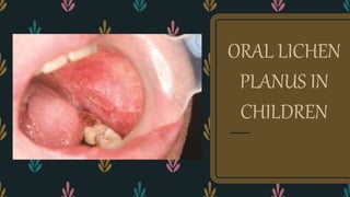
Oral lichen planus in children
- 2. – ORAL LICHEN PLANUS is a mucocutaneous disease described by ERASMUS WILSON In 1869 – The condition can affect either skin or mucosa or both – It can cause bilateral white striations, papules or plaques on the buccal mucosa, tongue, and gingiva – Erythema, erosions and blisters may or may not be present INTRODUCTION
- 3. PREVALENCE – It is predominantly a disease of the middle aged or older, and has a prevalence of 0.5% to 2% in adults. – It is more common in females than in males ratio of approximately 2:1 – It is also seen in children, although it is rare, and they may be present with atypical findings. Familial LP occurs in 1-2% of all childhood cases. – The prevalence of oral lichen planus in children is about 0.03%
- 4. ETIOLOGY – OLP has been associated with multiple disease processes and agents such as Viral And Bacterial Infections, Autoimmune Diseases (such as ulcerative colitis, myasthenia gravis, lupus erythematosus) Medications, Vaccinations and Allergy to Dental Restorative Materials (such as gold and amalgam), Local trauma (Koebner phenomenon) and Systemic diseases (such as diabetes mellitus and hypertension)
- 5. – Childhood lichen planus has been documented as a complication of Hepatitis B Vaccinations (HBV) where the recombinant proteins of the HBV vaccine may trigger a cell mediated autoimmune response targeted at keratinocytes giving rise to a lichenoid reactions. – It is also found in association with predisposing conditions, such as graft vs host disease and chronic Hepatitis C
- 6. PATHOGENESIS CD8+ T CELLS TRIGGER APOPTOSIS OF BASAL CELLS T CELLS MIGRATE INTO EPITHELIUM TOWARDS THE BASAL KERATINOCYTES T CELLS ARE ACTIVATED BY ANTIGEN ON KERATINOCYTE OR THROUGH ACTIVATED CD4+ LYMPHOCYTES THE ACTIVATED T CELLS KILL THE BASAL KERATINOCYTES THROUGH TNF-α ACTIVATED APOPTOSIS
- 8. • The histopathology include the existence of a band of lymphocytic inflammatory infiltrate in the subepithelial connective tissue, hydropic degeneration of the basal layer and the absence of epithelial dysplasia. • If the above three criteria are met, the lesion is considered a typical lichen planus from a histological perspective; and as for those that do not meet one of the histological criteria, they are considered to be lesions that are histologically compatible with lichen planus. HISTOPATHOLOGY
- 10. CLINICAL FEATURES – OLP in childhood was first described in 1920. – The family history of lichen planus is more commonly positive in patients with lichen planus in childhood than in adulthood.
- 11. – No malignant transformation of Oral Lichen Planus has been reported – Bilateral and symmetrical white striations are seen commonly called the “WICKHAMS STRIAE”.
- 12. – Oral lichen planus is chronic and usually persists for several years with periods of exacerbation and quiescence. – During exacerbation areas of erythema or erosion increases with slight increase in pain and sensitivity and vice versa in period of quiescence. – Usually exacerbations are associated with psychological stress, anxiety and mechanical trauma (Koebner’s Phenomenon)
- 13. – Andreason described six types : Reticular Papular Plaque Atrophic Erosive Bullous TYPES
- 14. – Reticular lesions are often asymptomatic, and may appear as symmetrical white lace like pattern on buccal mucosa, affects tongue or gum. – Atrophic OLP may appear as mixture of clinical subtypes (like white and grey streaks in linear or reticular pattern on erythematous background) red lesions often with a whitish border. May cause erosions (superficial ulceration). Most often affects the gums (gingiva) and lips. Can be very painful
- 15. RETICULAR ORAL LICHEN PLANUS PLAQUE TYPE ORAL LICHEN PLANUS
- 16. – Plaque type OLP appears as homogenous white patch resembling leukoplakia. Usually seen in Smokers – Erosive OLP can be present as irregular erosion covered with pseudomembrane. Symptoms range from episodic pain to severe discomfort that interferes with normal masticatory function – Bullous type OLP which is the least common usually present with bullae (diameters about few mm to cm) tends to rupture leaving ulcerated and painful surfaces
- 17. EROSIVE ORAL LICHEN PLANUS ULCERATIVE ORAL LICHEN PLANUS
- 18. – It is an uncommon medication-induced chronic change inside the mouth. It appears the same as idiopathic oral lichen planus clinically and under the microscope, but an oral lichenoid drug eruption resolves if the triggering drug is ceased – An oral lichenoid drug eruption is predominantly a problem seen in adults, probably because adults are the most frequent users of the majority of medications associated with this reaction. However it has also been reported rarely in children. ORAL LICHENOID DRUG REACTION
- 19. LICHENOID REACTION DUE TO DENTAL RESTORATIONS LICHENOID REACTION DUE TO DRUGS
- 20. – Medications associated with this reaction include: – • Penicillamine – • Antimalarials such as hydroxychloroquine – • Antihypertensives including beta-blockers, angiotensin converting enzyme (ACE) inhibitors and diuretics – • Non steroidal anti-inflammatory drugs (NSAID) – • Oral hypoglycaemic agents for type 2 diabetes – • Antiretroviral medications to treat HIV infection
- 21. Grinspans syndrome – Characterised by the presence of the triad DIABETES MELLITUS ,HYPERTENSION and ORAL LICHEN PLANUS – ORAL LICHEN PLANUS is thought to be a result of the drugs used for treatment of hypertension and diabetes mellitus, but this is not confirmed. – Rarely seen in children than adults
- 22. DIAGNOSIS – Complete history – Physical examination by a multidisciplinary group of health care providers – Biopsy to differentiate between OLP and other white chronic lesions – Direct immunofluorescence to distinguish erosive,ulcerative or bullous forms of OLP
- 23. Differential diagnosis – Lupus erythematosus – Psoriasis – Candidiasis – Morsicatiobuccarum (linear alba) – Leukoplakia – Erythema multiforme – Recurrent apthous stomatitis – Several viral infections
- 24. management CORTICOSTEROIDS : TOPICAL CORTICOSTEROIDS such as 0.05% Clobetasol proprionate gel, 0.1 or 0.05% Betamethasone valerate gel, 0.05% Fluocinonide gel, 0.1% Triamcinolone acetonide ointment. Patients are instructed to apply a thin layer up to 3times a day after meals and at bedtime Side effects are fewer. Adverse effects include Candidiasis, Thinning of Oral Mucosa and Discomfort on application Potent corticosteroids can cause adrenal suppression if used in large amounts for longer period of time
- 25. SYSTEMIC CORTICOSTEROIDS is indicated in patients who are unresponsive to topical steroids or mucocutaneous disease in a dosage of 0.5-1mg/kg as a tapering dose over 3-6weeks Systemic PREDNISONE is used to control ulcers and erythema in OLP
- 27. – IMMUNOSUPPRESANTS Tacrolimus Ointment (given two or three times daily for three months with chlorhexidine mouthwash) had better penetration into mucosal surface and tends to be 100 times more potent than cyclosporine – Topical Tacrolimus 0.1% ointment induced better therapeutic response than Triamcinolone Acetonide 0.1% in patients with symptomatic OLP
- 28. RETINOIDS are metabolites of Vitamin A. they have anti- keratinising and immunomodulating effects Retinaldehyde 0.1%, Isotretinoin gel 0.1% shows good clinical efficacy Fenretinide and Tazarotene gel 0.1% has been used successfully
- 30. – ULTRAVIOLET IRRADIATION Photochemotherapy with 8-methoxypsolarine and Long wave UV light (PUVA) is used To avoid side effects photosensitisation with topical 0.01% Trioxsalen is used Side effects nausea, headache, eye symptoms, dizziness, paraesthesia It is also shown to have oncogenic potential
- 31. LASER THERAPY 308nm excimer laser, 980nm diode laser, CO2 laser evaporation are used in treating OLP CO2 laser evaporation showed no problem with wound healing and there was complete epithelisation within 3 weeks It is a good treatment option for OLP when there is no further improvement with steroids
- 32. conclusion OLP is rare in children and the clinical presentations resembles those of adult OLP. However, the prognosis of OLP in childhood seems to be more favourable