The intervertebral disc consists of three main components: the cartilage end plates, the nucleus pulposus, and the annulus fibrosus. Degeneration of the intervertebral disc is a natural aging process but can be exacerbated by factors like poor nutrition, trauma from sports or injuries, and biomechanical factors related to posture and repetitive microtrauma. The degenerative process typically begins with softening of the nucleus pulposus and weakening of the annulus fibrosus, which can lead to protrusion or extrusion of the nucleus through tears in the annulus. Eventually fibrosis and calcification may occur as the body attempts to repair the degenerated disc.
3. THE IV DISC CONSISTS OF THREE DISTINCT
COMPONENTS :
1.THE CARTILAGE END PLATES
2.NUCLEUS PULPOSUS
3.ANNULUS FIBROSUS.
THE CARTILAGE RING ARE THIN LAYERS OF
HYALINE CARTILAGE BETWEEN TWO
ADJACENT VETEBRAL BODIES AND DISC
PROPER.
12. CONGENTIAL
DEVELOPMENT
AL
•POOR NUTRITION
•POOR HEALTH HABITS
•ACCUMATED TRAUMA : SPORTS,AUTOMOBILE INJURY
LEAD TO WEAKING OF I.V.DISC
•BIOMECHANICAL FACTORS
•POSTURE
•REPITITIVE MICROTRAUMA , FLEXION (COMPRESSION
INJURIES) , TENSIONAL INJURIES
13.
14.
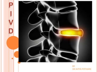
15. IT IS SEQUENCE OF CHANGES OCURRING IN THE DISC WHICH LEAD TO
PROTRUSION OR EXTRUSION OF NP THROUGH A RENT IN THE AF.
THESE CHANGES CONSISTS OF THE FOLLOWING :
A) NUCLEUSDEGENERATION : degenerative changes occur in the disc
before displacement of the nuclear material. these changes are:
(i) Softening of the nucleus and its fragmentation
(ii) Weakening and disintegration of the posterior part of the annulus
16. B) NUCLEUS DISPLACEMENT : the nucleus is under positive pressure
at all the times. When annulus become weak ,either because a small areaof
its entire thickness has been disintegrated spontaneously or because of the
injury , the nucleus tends to bilge through the defect. This is known as disc
protrusion.
This tendency is greatly increased if the nucleus is degenerated and
fragmented. Finally the nucleus comes out of the annulus , and the underthe
post” longitudinal ligament though it has not lost contact between with
parent disc. This is called as disc extrusion.
Once the disc is extruded it cannot go back and PLL is not strong enoughto
prevent the nucleus protruding further. So, the extruded disc, may loose its
contact with parent disc. The sequestrated disc may come to lie behind the
PLL or may become free fragments in the canal.
17.
18. C) STAGE OF FIBROSIS : this the stage of repair.
the begins alongside of degeneration . The residual
nucleus pulposus becomes flattened , fibrosed , and
finally undergoes calcification. At the same time, new
bone formation occurs at the point where the PLL has
been stripped from the vertebral body and
spurformation occurs.
19.
20.
21.
22. ASSESSMENT TREATMENT
ASSESSSMENT :
•POSTURE (rigid posture , loss of curve spine( i.e flattened back
in lumbar spine , posterior pelvic tilt , sciatic scoliosis may present
.)
•MOVEMENT : trunk flexion (may be painful , flexion may produce
pain .)
•TENDERNESS : along midline or lateral to spinous processes.
23.
24. IN CASE OF Cx PIVD :
a) Foraminal compression / spurling test
b) Distraction test
c) ULTT(upper limb tension test)
d) Shoulder abduction test
e) Tinel’s sign
(a)
26. IN CASE OF Lx PIVD:
a) SLR
b) Lasegue’s test
c) Bowstring test
d) Prone knee bending (PKB) test
e) Sensory impairment (over dermatomes motor
weakness over myotomal distraction)