approach to infant with Hydrops fetalis
•Transferir como PPTX, PDF•
2 gostaram•976 visualizações

Immune and non immune Hydrops fetalis

Recomendados
Recomendados
Mais conteúdo relacionado
Mais procurados
Mais procurados (20)
Semelhante a approach to infant with Hydrops fetalis
Semelhante a approach to infant with Hydrops fetalis (20)
Mais de Dr Praman Kushwah
Mais de Dr Praman Kushwah (20)
Último
Último (20)
approach to infant with Hydrops fetalis
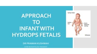
- 1. APPROACH TO INFANTWITH HYDROPS FETALIS DR PRAMAN KUSHWAH DrNB NEONATOLOGY RESIDENT
- 2. Introduction Hydrops fetalis is a Greek term that describes pathological fluid (“ὕdur,” Greek for water) accumulation in fetal soft tissues and serous cavities Defn -The presence of 2 abnormal fluid collections in the fetus detected by ultrasound.
- 3. The overall incidence of fetal hydrops reported in the literature is between 1 in 1500 and 1 in 3000 pregnancies
- 4. Features detected by ultrasound, and are defined as the presence of 2 abnormal fluid collections in the fetus. These include ascites, pleural effusions, pericardial effusion, and Generalized skin edema (defined as skin thickness >5 mm) Other frequent sonographic findings include polyhydramnios placental thickening placental thickness 4 cm in the second trimester 6 cm in the third trimester
- 5. Sub-types Hydrops fetalis is broadly classified based on etiology into Immune estimated 10%-24% Non-immune accounting for 76%-90% of cases (becoming increasingly the main cause of Hydrops)
- 6. Immune Hydrops Fetalis Maternal–fetal blood group incompatibilities and production of Red cell antibodies Maternal– fetal Rhesus (Rh) blood group incompatibility a/k/a Rh Iso-immunization ABO blood group incompatibility (rare) Non-Rh(D) and non-ABO incompatibility (atypical antibodies) Phenomenon of Rh Iso-immunisation was reported by Levine in 1941.
- 7. Causes The five antigens that make up the Rh system are D, C, c, E, and e. Rhesus D, c, and E are the commonest, Incidence of Rh D antibodies is reducing use of anti-D Red cell antibodies that are now commonly associated with immune hydrops- kell antibodies Duffy Lewis p antibodies Last two rarely cause hemolytic disease of the newborn Kell iso-immunization - mechanism for anemia is most likely erythroid suppression rather than hemolysis (Vaughan et al.,1994).
- 8. Development of Rh-DAntibodies Out ofTotal Rh Positives 45% are homozygous and 55 % are heterozygous First-timer Rh Iso-immune , carries 16% risk of stimulating the maternal immune system to produce anti-D antibody First immune response Anti-D IgM antibody, cannot cross placenta, no fetal hemolysis. Subsequent pregnancy, if the fetus is again Rh positive More rapid immune response occurs High titers of anti-D IgG antibody, Antibodies rapidly crosses the placenta Fetal hemolysis and profound fetal anemia occurs.
- 10. Mechanism of Hydrops Fetalis 1. Maternal anti-D IgG antibody attaches itself to the Rh antigen present on fetal red cells. 2. Chemotaxis of phagocytes into the fetal spleen 3. Destruction and hemolysis of fetal red cells 4. Increased Fetal Erythropoietin 5. Fetal bone marrow stimulated to increase red cell production 6. Marrow capacity is exhausted and extra-medullary erythropoiesis begins 7. Red cell production occurs in the fetal liver, spleen, kidney, adrenal glands, and intestine 8. Fetal rbcs at these sites are often immature, are nucleated, and appear in the circulation as erythroblasts. 9. Hence, the synonym erythroblastosis fetalis for immune hydrops
- 12. Pathophysiology Majority of Rh isoimmunization lead to mild or moderate fetal or neonatal hemolytic disease. Only 20% to 25% of cases result in severe hemolytic disease, with immune hydrops Pathophysiology is unclear and several hypothesis being suggested 1. Congestive heart failure secondary to fetal anemia leading to hydrops 2. Severe fetal anemia leading to extensive extramedullary erythropoiesis, with associated hepatosplenomegaly and distortion of intrahepatic architecture, resulting in portal and umbilical venous distortion, portal hypertension, placental edema, and placental hypoperfusion (Bowman, 1999). Deteriorating hepatic synthesis, progressive hypo albuminemia occurs, adding to the generalized edema, anasarca, and pleural and pericardial effusions.
- 13. Non- Immune Hydrops 1. Accounts for 80 - 90% of cases 2. Any other cause besides immune. 3. No identifiable circulating antibody to any red cell antigen. 4. No common mechanism is responsible for the signs of hydrops. 5. Pathogenesis associated with failure of the interstitial fluid (the liquid between the cells of the body) to return into the venous system As an end result of an array of disorders of the fetus, umbilical cord and placenta that leads to deranged fluid homeostasis. A wide range of fetal organs are involved
- 14. Causes ConditionsAssociated with NIH (This list is not exhaustive) Cardiac Cardiomyopathy, Ebstein's anomaly, pulmonary atresia, coarctation of the aorta, hypoplastic left heart, completeAV canal, left sided obstructive lesions, premature closure of the foramen ovale Intracardiac tumors (tuberous sclerosis) Cardiac arrhythmia SVT, flutter, heart block,WPW syndrome Chromosomal /Genetic Syndromes T13,T18,T21, XO (Turners syndrome) , Noonan syndrome , multiple pterygium syndrome, Pena-Shokeir, arthrogryposis Fetal Anemia Alpha (α) thalassemia, parvovirus, fetal hemorrhage, G-6-PD deficiency Infection Parvovirus, CMV, syphilis, coxsackie virus, rubella, toxoplasmosis, herpes,varicella, adenovirus, enterovirus, influenza, listeria
- 15. Thoracic Abnormalities Congenital cystic adenomatoid malformation (CCAM) , chylothorax, diaphragmatic hernia, mediastinal tumor, skeletal dysplasias Twinnning Twin to twin transfusion Severe anemia in the donor twin or high- output failure in the recipient Tumors Fetal sacrococcygeal teratoma, hemangiomas (Hepatic, Klippel- Trenaunay syndrome), fetal adrenal neuroblastoma, placental tumors (chorioangioma) Miscellaneous Cystic hygromas, inheritable disorders of metabolism (lysosomal storage diseases) ,maternal thyroid disease, congenital nephrotic syndrome
- 16. Difficult to find a plausible explanation for the development of hydrops in every case - which may represent chance associations. However, as a general rule, HF presenting before 24 weeks is usually due to chromosomal aberrations Hydrops presenting after this is usually due to structural anomalies (such as cardiac and pulmonary)
- 17. More than 100 causes or associations of NIHF have been found. 10 to 20% of cases are idiopathic Seven major categories: 1. Cardiovascular Pathologies (35 %) 2. Chromosomal Anomalies (20 %) 3. Anemia (15 %) 4. Malformation Syndromes (15 %) 5. Infections (10 %) 6. Liver Diseases (5 %) 7. Miscellaneous Causes (5 %)
- 18. Pathogenesis
- 19. Cardiac causes A. STRUCTURAL DEFECTS 1. KARYOTYPIC ABNORMALITIES Coarctation of aorta withTurner syndrome Arterioventricular canal and septal defects with Down syndrome B. FUNCTIONAL DEFECT 1. Fetal supraventricular tachyarrhythmias (SVT) 1. Important cause of hydrops 2. Detected by fetal echocardiography 3. Amenable to therapy 2. Congenital heart block (CHB) 1. > 60% of pregnancies complicated by collagen disease (esp. lupus). 2. Maternal ANA attack fetal collagen in conduction bundle 3. Not all fetuses with CHB are affected
- 20. CHROMOSOMAL GENETIC SYNDROMES (1/3rd of cases of NIHF) 1. Most common -Turner syndrome – typified by ultrasound finding of cystic hygromas 2. Prognosis is poor in most of cases due to associated structural, functional, and metabolic defects 3. complete family history is important to rule out known inherited disorder 4. Hemoglobinopathies 5. Inborn errors of metabolism (rare)
- 21. SEVERE FETALANEMIA A. ALPHA -THALASSEMIA (Hb Barts) Fetal anemia leading to hypoxia and high output cardiac failure South East Asia, 86 % (25/29) of hydrops were due to Hb Bart’s. B. FETAL HEMORRHAGE 1. Intracranial bleeding from AV malformation or sacrococcygeal teratoma 2. Fetal-maternal hemorrhage (abruptio or placental tumours such as chorioangioma) 3. Hemolysis (Spherocytosis,G6PD or PKD) 4. Failed erythropoiesis (marrow dysfunction Parvovirus B19, leukemia, pure red cell aplasia).
- 22. Malformation syndromes Can involve any of the major system 1. PULMONARY - CCAM M/C thoracic lesion associated with hydrops. Abnormal capillary-alveolar development Functionless cystic lung masses space occupying lesion Compression and underdevelopment of normal lung tissue Hypoplastic lung, shifting of mediastinum and heart, obstruction of cardiac venous return, central venous hypertension leading to hydrops in severe cases. Other pulmonary causes of hydrops probably share the same pathogenesis
- 23. 2. GASTROINTESTINAL - CONGENITAL DIAPHRAGMATIC HERNIA (CDH) Herniated bowel or liver causing intrathoracic mass effect similar to CCAM. Other causes with same pathogenesis- Esophageal Atresia, MidgutVolvulus, Meconium Peritonitis, Duodenal Diverticulum, Intestinal Duplication, Malrotation, And Imperforate Anus. 3. RENALAND UROLOGIC Finnish nephrosis leading to severe hypoproteinemia, hypoplastic kidney(s), polycystic kidneys, renal vein thrombosis, bladder outlet obstruction, and dysplastic kidneys. 4. NEUROLOGIC MALFORMATIONS are a rare cause of hydrops which includes encephalocele, porencephaly with absent corpus callosum, fetal intracranial hemorrhage, and vein of Galen aneurysm. 5. SKELETAL DYSPLASIAS such as achondroplasia, achondrogenesis, osteogenesis imperfecta, osteochondrodystrophy, thanatophoric dwarfism, asphyxiating thoracic dysplasia, chrondrodystrophy and chrondrodysplasia may all be associated with thoracic compression, impairment of venous return resulting in hydrops.
- 24. INTRAUTERINE INFECTIONS CAUSES IMPLICATED Toxoplasmosis, Rubella, Cytomegalovirus, Herpes simplex, Syphilis (collectively known as “TORCHS”), Coxsackie virus and Parvovirus B19 POSSIBLE MECHANISMS fetal anemia from hemolysis, suppressed erythropoesis and myelopoiesis, fetal myocarditis and fetal hepatitis. Parvovirus B19 one third of all cases of NIHF red cell aplastic crisis leading to severe anemia risk of fetal death of 15% at 13-20 weeks and 6 % after 20 weeks PROGNOSIS SYPHILIS - very poor prognosis Parvovirus B19 and other - self limited and may resolve spontaneously and 85% of fetuses treated by in-utero transfusion survive.
- 25. Twin –Twin Transfusion Imbalance in blood flow due to anastomoses in placentas of monochorionic twin pregnancies In severe cases, one or both twins may develop Hydrops Recipient twin hypervolemia and rise central venous pressure Laser therapy - best available therapeutic approach Selective termination via umbilical cord coagulation Radiofrequency ablation of the acardiac twin for severe cases
- 26. APPROACH
- 27. Diagnostic challenge To establish the etiology To determine appropriate therapy and timing of delivery. (cause of hydrops can be determined in ONLY about 60-85% of cases including postnatal evaluation)
- 28. Presentation FETAL PRESENTATION DURING ROUTINE ULTRASOUND EVALUATION Polyhydramnios Size greater than dates Fetal tachycardia Decreased fetal movement Abnormal serum screening Antenatal hemorrhage May be diagnosed on routine sonographic screening or may be diagnosed after fetal death
- 29. Maternal complications MATERNAL PRESENTATION– Edema , hypertension, proteinuria Condition can quickly deteriorate into fulminant pre- eclampsia and eclampsia. Mirror syndrome – An uncommon complication in which the mother develops edema that “mirrors” that of her hydropic fetus – necessitates urgent delivery. Early onset severe pre-eclampsia or polyhydramnios beginning at 28 weeks, hydrops should be excluded by sonography In an attempt to compensate for the fetal hypoxia, placenta increases in size and sometimes also penetrate deeper into the myometrium causing morbid adherence of placenta causing problems for third stage of labor necessitating the manual removal of Placenta.
- 30. OBSTETRIC COMPLICATIONS Polyhydramnios If a/w maternal respiratory distress Preterm birth Tocolytic agents <24 weeks Prostaglandin InhibitorOR Serial Amnioreduction Inutero Ductus closure, PPROM,Abruption NEC,
- 31. Investigations Initial investigations include an indirect Coombs test to exclude immune causes, followed by- determination of routine blood counts and indices to exclude thalassemias; maternal blood chemistry testing for G-6-PD deficiency; Betke-Kleihauer testing for fetal-maternal transfusion; screening for toxoplasmosis, rubella, CMV, herpes simplex (TORCH) and other infections infection during pregnancy.
- 33. Sonography Fetal hydrops is not seen sonographically until the fetal hematocrit has fallen to below one-third of its normal range Axial image of a fetus demonstrating scalp edema due to immune hydrops Axial image demonstrating abdominal ascities and edema of the abdominal wall in a fetus with immune hydrops.
- 34. Sagittal image demonstrating edema of the scalp and face in a fetus with immune hydrops Hydrocele can be early manifestation in Hydrops
- 35. Axial image demonstrating abdominal ascites in a fetus with immune hydrops
- 36. Fetal Echo Axial image demonstrating a pericardial effusion in a fetus with immune hydrops • Consider fetal heart rate monitoring for 12 to 24 hours if fetal arrhythmia is suspected
- 38. Doppler Blood FlowStudies The use of Doppler sonography to predict fetal anemia has been more successful (Mari et al., 1995). Elevated peak systolic velocity (PSV) measured in the middle cerebral artery (MCA) is associated with an increased likelihood of fetal anemia
- 39. Initiated at 16–18 weeks of gestation Reliability decreases after 35 weeks of gestation MCA-PSV >1.5 MoM = severely anemic fetus. Sensitivity =100% (95% CI) for moderate or severe anemia False-positive rate of 12%(Mari et al)
- 41. Diagnostic Invasivemethod Previously invasive methods were used to diagnose alloimmunization. Critical titer levels indicative of high risk of fetal anemia, as ≥ 1:16 for anti-RhD ≥ 1:8 for anti-Kell10, Serial AMNIOCENTESIS for bilirubin levels to estimate the severity of hemolysis. Spectrophotometry was used to quantify bilirubin level, which was expressed as the change in optical density (OD) at a wavelength of 450 nm (OD450). These values were plotted on Liley’s curve or Queenan’s curve (< 27 weeks) to predict fetal anemia
- 43. Amniocentesis andCVS AMNIOCENTESIS Fetal karyotyping, amniotic fluid culturing, PCR (polymerase chain reaction) for infections, testing for thalassemia, and determination of the lecithin-sphingomyelin (L/S) ratio. Fetal liver function and metabolic testing if indicated. RECURRENT NONIMMUNE HYDROPS - test for : Storage disorders such as Gaucher’s, gangliosidosis, sialidosis, beta- glucuronidase deficiency, and mucopolysaccharidosis Enzyme analysis and carrier testing in parents analysis of fetal or neonatal blood or urine. Histological examination of fetal tissues. KARYOTYPING can be performed with chorionic villous sampling (CVS) or with fluid obtained from one of the fetal cavities.
- 46. PERCUTANEOUS UMBILICALBLOOD SAMPLING (PUBS) PUBS OR CORDOCENTESIS most accurate method for diagnosis of Fetal Anemia Include hemoglobin chain analysis for thalassemia and fetal serum albumin level done if MCA velocity >= 1.5 MoM INTRAUTERINETRANSFUSION is done as a part of the same procedure after obtaining the Hb/Hct values To summarize SCREENING: MCA DOPPLER DIAGNOSTIC &THERAPEUTIC: PUBS
- 47. Management Immediately referred to a tertiary care center, with trained perinatologists to perform intrauterine transfusions and neonatologists care. Arrangements to perform PUBS and possible intrauterine fetal transfusion should be expedited as rapidly as possible Antenatal corticosteroid if GA between 24 and 34 weeks, to reduce the complications of prematurity . option of elective termination should be discussed with the parents if the gestational age is less than 24 week.
- 51. Fetal Intervention INTRAUTERINETRANSFUSION INDICATION: - Main indication for IUT is fetal anemia due to red cell allo-immunization MCA-PSV >1.5 MoM and/or If signs of hydrops are present The intrauterine transfusions can be done via 3 access points Intraperitoneal Via Umbilical cord Via Intrahepatic portion of UmbilicalVein
- 52. Transfusion volume calculation A commonly used formula for the volume of blood to be transfused is whereVT is volume of blood transfused, 150 is a placental correction factor, and EFW is estimated fetal weight in kilograms (Kaufman and Paidas, 1994). Fetoplacental volume (V) : 0.1 mL volume/g of estimated fetal weight Fetal erythrocyte treatment has also been reported to be successful in nonimmune etiology
- 54. Fetal monitoring POST - IUT Sonographic surveillance should be instituted, depending on the gestational age. Daily fetal testing with nonstress tests or biophysical profiles is reasonable for all potentially viable fetuses. With sonographic evidence of resolution of hydrops, testing can be decreased to twice weekly. Sonographic surveillance for appropriate fetal growth should continue every 2 weeks
- 55. Delivery Pregnancies not to be allowed beyond 37 to 38 weeks premature delivery indicated if hydrops does not improve if fetal testing non-reassuring despite correction of fetal anemia Delivery by cesarean Because of the risk of soft-tissue dystocia associated with hydrops Minimize chances of maternal and fetal trauma. A trial of labor may be reasonable in cases in which the fetal anemia has been adequately corrected, and in which the hydropic features improve. In carefully selected and monitored cases, up to 80% of fetuses with immune hydrops may successfully deliver vaginally (Bowman, 1999).
- 56. Neonatal Management HYPOPLASTIC LUNGS, HYALINE MEMBRANE DISEASE, PLEURAL EFFUSIONS, PULMONARY EDEMA, SEPSIS, PERINATAL DEPRESSION, HYPOXIA, OR ACIDOSIS Immediate neonatal endotracheal intubation and supportive care in almost all infants Cautious use of inotropic agents, diuretics, blood products, albumin, and fluids to maintain an adequate cardiac function without fluid overload or soft-tissue edema, High-frequency ventilator and high airway pressure settings will often be needed to achieve adequate oxygenation Umbilical lines help with administration of various agents, monitoring of blood gases and arterial and venous pressures.
- 57. Once the neonate is stabilized Full physical examination Relevant echocardiographic, and radiologic investigations to exclude structural malformation Hematologic tests to rule out sepsis, biochemical and karyotypic anomalies; Placenta should be sent for histology and culture, Specific treatment is based on the underlying etiology Appropriate referral to the relevant pediatric subspecialist including clinical geneticist and pediatric surgeon. No Surgical management described.
- 58. Fetal intervention OTHER FORMSOF FETAL INTERVENTION (esp. NIHF) 1. Medical therapy for the mother to correct fetal arrhythmias . Digoxin administration to pregnant women has been successful in the treatment of fetal arrhythmias, with resolution of hydrops in some cases (Knilans, 1995). 2. Fetal thoracentesis may be performed under sonographic guidance with resolution of pleural effusions (Jones, 1995). 3. Thoracoamniotic shunt if repeat thoracentesis are needed 4. Other - Open fetal surgical resection of thoracic masses or sacrococcygeal teratoma (Bullard andHarrison, 1995). …..However, very few data is available confirming whether such invasive approaches have a significant impact on fetal or neonatal outcome…..
- 59. Recurrence risk A postmortem examination indicated in all cases of NIHF This will maximize the number of cases in which a definite underlying cause is identified and will facilitate appropriate genetic counseling and prediction of recurrence risk (Steiner, 1995). Recurrence of idiopathic NIHF is rare, Patients should therefore be made aware that, while idiopathic NIHF is extremely rare, recurrences can and do occur.
- 60. Long term outcome Long-term neurodevelopmental outcome after IUT for hemolytic disease : the LOTUS study Neurodevelopmental outcome till least 2 years of age was assessed. Primary outcome was the incidence of neurodevelopmental impairment defined as at least one of the following: cerebral palsy, severe developmental delay, bilateral deafness, and/or blindness.
- 61. Take home message Identify the cause of Hydrops MCA Doppler in monitoring and treatment of fetal Anemia Improvement in prognosis and long term outcome on use of IUTs. Optimal timing of delivery Capable NICU team and a tertiary centre Anticipation of Preterm delivery and RDS
- 62. Hydrops to Hope – Journey of Jacoby – one of a twin diagnosed with non immune hydrops – Altogether spent 16 months in the NICU and Cleveland Clinic Children's Hospital for Rehabilitation before finally going home…..
- 63. ThankYou