SlideShare uma empresa Scribd logo
1 de 51
Baixar para ler offline
Quantum Dots
NANOPARTICLES
• Nanoparticles are particles that have dimension of 100
nm or less in size.
• The properties of many conventional materials change
when formed from nanoparticles.
• This is typically because nanoparticles have a greater
surface area per weight than larger particles; this causes
them to be more reactive to certain other molecules.
• Iron nanoparticles
are used to clean up carbon tetrachloride
pollution in ground water.
• Designed by Oregon Health & Science
University's OGI School of Science &
Engineering, in collaboration with Pacific
Northwest National Laboratory (PNNL)
and the University of Minnesota.
• Carbon tetrachloride is a manufactured toxic
chemical used mainly in cleaning fluids and
degreasing agents.
• Spillage infiltrates the soil and creates very large
areas of contaminated groundwater and soil in
few areas causing cancer in animals.
• A commercially available product of iron oxide
with a magnetite shell high in sulfur, quickly and
effectively degraded carbon tetrachloride to a
mixture of relatively harmless products.
• Researchers at MIT and Harvard Medical School
have built targeted nanoparticles that can cling to artery
walls and slowly release medicine, an advance that
potentially provides an alternative to drug-releasing
stents in some patients with cardiovascular disease.
• The particles, dubbed “nanoburrs” ,are coated with
tiny protein fragments that allow them to stick to target
proteins.
• They are designed to release their drug payload over
several days and are one of the first such targeted
particles that can precisely home in on damaged vascular
tissue.
QUANTUM DOTS
INTRODUCTION
• Quantum dots are tiny particles,or
“nanoparticles”, of a semiconductor material,
traditionally chalcogenides (selenides or
sulfides) of metals like cadmium or zinc (CdSe or
ZnS), which range from 2 to 10 nanometers in
diameter (about the width of 50 atoms).
• They are the smallest objects that can be synthesized on
the nanoscale.
• Like the name suggests, its structure is much like a small
dot. Common shapes include pyramids, cylinders, lens
shapes, and spheres.
• Different synthesis routes create different kinds of
quantum dots.
• Quantum dots are so important because they confine
electrons in three dimensions.
• The reason 'quantum' prefixes the name is because the
dots exhibit quantum confinement properties in all three
dimensions i.e. electrons within a dot can't freely move
around in any direction.
• The ability to precisely control the size of a quantum
dot enables the manufacturer to determine the
wavelength of the emission, which in turn determines
the colour of light the human eye perceives.
• Quantum dots exhibit quantized energy levels like an
atom. For a given input energy, for instance, a quantum
dot will only emit specific spectra of light.
• With decreasing diameters of quantum dots, there is a
corresponding increase in energy of emitted light.
• Quantum dots can therefore be “tuned” during
production to emit any colour of light desired. The ability
to control, or “tune” the emission from the quantum dot
by changing its core size is called the “size quantisation
effect”.
• Due to excellent confinement properties not seen in
nanowires or quantum wells (in all modern lasers),
quantum dots are extremely efficient at emitting light.
HISTORY
• In the 1970s the first low dimensional structures QW (Quantum
Wells) were developed.
• 1D ( Quantum Wires) and 0D (Quantum Dots) were subsequently
developed.
PROPERTIES OF QUANTUM DOTS
• Being smaller than the wavelength of the visible light,
they cannot be seen under normal conditions.
• Quantum Dots luminesce under ultraviolet light, with
the size of the dots controlling its colour.
• A quantum dot can have anything from a single electron
to a collection of several thousands electrons.
• Quantum Dots fluoresce or stay lit much longer then
dyes conventionally used for tagging cells.
• They can be tagged to proteins and their glow enables
the identification of specific proteins or DNA making it
possible to diagnose various diseases.
The electrons in quantum dots are confined in a small space (quantum box),
and when the radii of the semiconductor nanocrystal is smaller than the
exciton Bohr radius (exciton Bohr radius is the average distance between the electron in the
conduction band and the hole it leaves behind in the valence band), there is quantization of
the energy levels according to Pauli’s exclusion principle.
The discrete, quantized energy levels of quantum dots relate them more
closely to atoms than bulk materials and have resulted in quantum dots
being nicknamed 'artificial atoms'.
As the size of the crystal decreases, the difference in energy between the
highest valence band and the lowest conduction band increases. More energy
is then needed to excite the dot, and more energy is released when the crystal
returns to its ground state, resulting in a color shift from red to blue in the
emitted light. As a result of this phenomenon, quantum dots can emit any
color of light from the same material simply by changing the dot size.
Additionally, because of the high level of control possible over the size of the
nanocrystals produced, quantum dots can be tuned during manufacturing to
emit any color of light.
Quantum dots have a unique electronic structure
Quantum dots can be classified into different types based on their composition
and structure.
Core-Type Quantum Dots
Quantum dots can be single component materials with uniform internal
compositions, such as chalcogenides (selenides or sulfides) of metals like
cadmium or zinc, example, CdSe. The photo- and electroluminescence
properties of core-type nanocrystals can be fine-tuned by simply changing the
crystallite size.
Core-Shell Quantum Dots
The luminescent properties of quantum dots arise from recombination of
electron-hole pairs (exciton decay) through radiative pathways. However, the
exciton decay can also occur through nonradiative methods, reducing the
fluorescence quantum yield. To improve efficiency and brightness of
semiconductor nanocrystals a new method is growing shells of another higher
band gap semiconducting material around them. These quantum dots with
small regions of one material embedded in another with a wider band gap are
known as core-shell quantum dots (CSQDs) or core-shell semiconducting
nanocrystals (CSSNCs).
For example, quantum dots with CdSe in the core and ZnS in the shell exhibit
greater than 80% quantum yield. Coating quantum dots with shells improves
quantum yield by passivizing nonradiative recombination sites and also
makes them more robust to processing conditions for various applications.
This method has been widely explored as a way to adjust the photophysical
properties of quantum dots.
Alloyed Quantum Dots
Tuning the properties by changing crystallite size could cause problems in
many applications with size restrictions. Multicomponent quantum dots is an
alternative method to tune properties without changing crystallite size.
Alloyed semiconductor quantum dots with both homogeneous and gradient
internal structures allow tuning of optical and electronic properties by merely
changing the composition and internal structure without changing the
crystallite size. For example, alloyed quantum dots of the compositions
CdSxSe1-x/ZnS of 6nm diameter emits light of different wavelengths by just
changing the composition. Alloyed semiconductor quantum dots formed by
alloying together two semiconductors with different band gap energies
exhibited interesting properties distinct not only from the properties of their
bulk counterparts but also from those of their parent semiconductors. Thus,
alloyed nanocrystals possess novel and additional composition-tunable
properties aside from properties due to quantum confinement effects.
Visible spectrum
• 5 nm dots: red
• 1.5 nm dots: violet
Quantum dots change color with size because additional
energy is required to “confine” the semiconductor excitation
To a smaller volume
Ordinary light excites all color quantum dots.
(Any light source “bluer” than the dot of interest works.)
COLLOIDAL SYNTHESIS
• Colloidal semiconductor nanocrystals -
synthesized from precursor compounds
dissolved in solutions, much like traditional
chemical processes.
• The synthesis of colloidal quantum dots is based
on a three-component system composed of:
precursors, organic surfactants, and solvents.
• The precursors transform into monomers on
heating to a suitable temperature.
• Once the monomers reach a high enough
supersaturation level, the nanocrystal growth
starts with a nucleation process.
• The temperature during the growth process
must be high enough to allow for rearrangement
and annealing of atoms during the synthesis
process while being low enough to promote
crystal growth.
FABRICATION
• Self-assembled quantum dots are typically between 5 and 50 nm in
size, defined by lithographically patterned gate electrodes, or by
etching on two-dimensional electron gases in semiconductor
heterostructures.
• Some quantum dots are small regions of one material buried in
another with a larger band gap. These can be core-shell structures,
e.g., with CdSe in the core and ZnS in the shell or from special forms
of silica called ormosil.
• Quantum dots sometimes occur spontaneously in quantum well
structures due to monolayer fluctuations in the well's thickness.
• This fabrication method has potential for applications in quantum
computation.
• The main limitations of this method are the cost of fabrication and
the lack of control over positioning of individual dots.
VIRAL ASSEMBLY
• Lee et al. (2002) reported using genetically engineered M13
bacteriophage viruses to create quantum dot biocomposite
structures.
• Genetically engineered viruses can recognize specific
semiconductor surfaces.
• It is also known that liquid crystalline structures of wild-
type viruses (Fd, M13, and TMV) are adjustable by
controlling the solution concentrations, ionic strength, and
the external magnetic field applied to the solutions. Thus,
the specific recognition properties of the virus can be used
to organize inorganic nanocrystals, forming ordered arrays
over the length scale defined by liquid crystal formation.
• Using this information, Lee et al. (2000) were
able to create self-assembled, highly oriented,
self-supporting films from a phage and ZnS
precursor solution. This system allowed them to
vary both the length of bacteriophage and the
type of inorganic material through genetic
modification and selection.
ELECTROCHEMICAL ASSEMBLY
• Highly ordered arrays of quantum dots may also be self-
assembled by electrochemical techniques.
• A template is created by causing an ionic reaction at an
electrolyte-metal interface which results in the spontaneous
assembly of nanostructures, including quantum dots, onto the
metal which is then used as a mask for mesa-etching these
nanostructures on a chosen substrate.
BULK MANUFACTURING
• Conventional, small-scale quantum dot manufacturing relies on a
process called “high temperature dual injection” which is
impractical for most commercial applications that require large
quantities of quantum dots.
• A reproducible method for creating larger quantities of consistent,
high-quality quantum dots involves producing nanoparticles
from chemical precursors in the presence of a molecular
cluster compound under conditions whereby the integrity of the
molecular cluster is maintained and acts as a prefabricated seed
template.
• Individual molecules of a cluster compound act as a seed or
nucleation point upon which nanoparticle growth can be initiated.
In this way, a high temperature nucleation step is not necessary to
initiate nanoparticle growth because suitable nucleation sites are
already provided in the system by the molecular clusters.
• A significant advantage of this method is that it is highly scalable.
CADMIUM FREE QUANTUM DOTS
• For commercial viability, a range of restricted, heavy metal-free quantum
dots has been developed showing bright emissions in the visible and near
infra-red region of the spectrum and have similar optical properties to those
of CdSe quantum dots.
• A new type of CFQD can be made from rare earth (RE) doped oxide
colloidal phosphor nanoparticles. Unlike semiconductor nanoparticles,
excitation was due to UV absorption of host material, which is same for
different RE doped materials using same host.
• Multiplexing applications can be thus realized. The emission depends on
the type of RE, which enables very large stokes shift and is narrower than
CdSe QDs. The synthesis is aqueous based, which eliminated issues of water
solubility for biological applications.
31
Applications
• Photovoltaic devices: solar cells
• QD Solar paint
• Biology: biosensors, imaging
• Light emitting diodes: LEDs
• Quantum computation
• Photodetectors
• Lasers
31
32
Solar Cells
• Photovoltaic effect:
▫ p-n junction.
▫ Sunlight excites electrons and
creates electron-hole pairs.
▫ Electrons concentrate on one
side of the cell and holes on the
other side.
▫ Connecting the 2 sides creates
electricity.
32
33
Different Generations of Solar Cells
• First generation:
▫ Single crystal silicon wafer.
▫ Advantages: high carrier mobility.
▫ Disadvantages: most of photon energy is
wasted as heat, expensive.
• Second generation:
▫ Thin-film technology.
▫ Advantages: less expensive.
▫ Disadvantages: efficiency lower
compared with silicon solar cells.
• Third generation:
▫ Nanocrystal solar cells.
▫ Enhance electrical performances of the
second generation while maintaining low
production costs.
34
• The quantum dot band gap is tunable and can be used to
create intermediate bandgaps. The maximum theoretical
efficiency of the solar cell is as high as 63.2% with this
method.
How Can Quantum Dots Improve the
Efficiency?
Cheap quantum dot solar paint
• “Sun Believable solar paint,” consists of a yellow or brown paste made
of quantum dots.
• The scientists experimented with three types of quantum dots: CdS,
CdSe, and TiO2, all of which are powder-like, with water and tert
butanol as the solvent.
• Instead of adding dye to give the paint a desired color, they added
colored semiconductor nanocrystals to the solar paint to achieve the
desired optical and electronic properties.
Quantum Dot LEDs
• Used to produce inexpensive,
industrial quality white light.
• Marked improvement over
traditional LED-phosphor
integration by dot’s ability to
absorb and emit at any desired
wavelength.
• Produce white light by
intermixing red, green and blue
emitting dots homogenously
within the phosphor difficult to
accomplish with the traditional
LED-phosphor set up.
Biolabelling
• Multicolor labeling of cells is a powerful technique for visualizing many
structures simultaneously, such as cytoskeletal proteins or organelles, and
to elucidate intracellular processes.
• QDs have been used to label cellular structures both within and external to
the cell membrane. They are delivered inside cells via receptor-mediated
pathways where specific ligands were attached to QDs to induce cellular
uptake, as well as nonspecific endocytosis (ie, pinocytosis) where cells were
incubated with a concentrated QD solution.
Bioimaging
• Non invasive, real-time in vivo fluorescence imaging requires exciting
fluorophores and detecting their emission through tissue which is
invariably hindered by scattering and absorption of both the excitation and
emission wavelengths.
• Using a filtered halogen source and an IR camera, the collection of QDs
within tissue was monitored in real-time to identify a region for surgical
resection.
PHOTODETECTORS
• Photodetectors based on single quantum dots are expected to find uses in
opto-electrical interfaces in future quantum computers, where single
photons will carry information over long distances and single electrons will
be used for computation.
Futuristic applications
• Anti counterfeiting applications: inject dots into liquid mixtures,
fabrics, polymer matrices, etc. Ability to specifically control absorption and
emission spectra to produce unique validation signatures. Almost
impossible to mimic with traditional semi-conductors.
• Counter-espionage/Defense applications: Integrate quantum dots
into dust that tracks enemies. Protection against friendly-fire events.
• New research provides evidence for significant differences between new and
old red blood cells used for transfusions and could provide a cheap, rapid
and effective way to monitor the quality of blood .
• Scientists have discover nanoparticles that can disrupt intracellular
transport pathways.
Imaging is an important clinical modality used in determining appropriate
cancer therapy.
x-ray, computed tomography, ultrasound, radionuclide imaging and MRI, :
used widely for cancer screening and staging, determining the efficacy of
cancer therapy and monitoring recurrence.
2 major limitations
1. do not have sufficient sensitivity to detect small numbers of malignant
cells in the primary or metastatic sites.
2. The imaging techniques have not been developed to detect specific cancer
cell-surface markers.
In many instances, these cell-surface markers might be targets for cancer
therapy and might assist in the diagnosis and staging of cancer.
Quantum dot (QD) imaging probes, although still in the early development
stage, provide the potential to fulfill these requirements for in vivo cancer
imaging.
QDs In Cancer Therapy
QDs vs Organic Fluorescent dyes
QDs offer great advantages over traditional organic fluorescent dyes and
present a number of beneficial characteristics for spectroscopy, such as
1. high fluorescence intensity (brightness)
2. long lifetime
3. good resistance to photobleaching.
4. have broad excitation and narrow and symmetric emission spectra, which
make it feasible to perform 'multiplexing' (simultaneous detection of multiple
signals) imaging using a single excitation source
5. high sensitivity for simultaneous cancer molecular imaging and targeted
therapy.
the sensitivity of QD-based molecular imaging can be two to three orders
larger than that of routine fluorescent dyes.
Furthermore, the fluorescence in near infrared of NIR-QDs can be detected in
deep tissues, making them suitable for in vivo imaging with high signal-to-
background ratio
Biocompatibility of QDs
QDs are highly hydrophobic and, therefore, only soluble in organic solvents.
Thus often encapsulated by amphiphilic molecules.
The hydrophobic segment of these molecules interacts with the hydrophobic
molecules on the QD surface, whereas the hydrophilic segment interacts with the
aqueous medium, solublizing the QDs.
Several types of amphiphilic polymers, including polyethylene glycol (PEG)-derived
phospholipids, triblock copolymers, octylamine-modified polyacrylic acid,
oligomeric phosphine and copolymers of alkyl monomers and anhydrides, can
serve this encapsulation function.
After encapsulation, the hydrodynamic radius of QDs increases to 10-20 nm.
To functionalize QDs with biomolecules, amphiphilic polymers are engineered to
carry reactive groups, such as amines and carboxylic acids. Biomolecules, such as
peptides, antibodies, DNA or siRNA, can react with these functional groups to form
covalent linkages mediated by various coupling reagents.
In addition, biomolecules can be conjugated with QDs through noncovalent affinity
binding, such as the interactions of biotin/avidin, or nickel nitrilotriacetic acid (Ni-
NTA)/histidine-tagged peptides.
Controlling ratio of reactive groups and reaction time, the average number of
biomolecules conjugated to each QD can be controlled average of two or a few
biomolecules are typically conjugated to one QD.
Encapsulation and bioconjugation do not usually alter the optical property of QDs
significantly.
Recently, QDs based on silicon have drawn much attention because of their potential
lower toxicity than heavy metal QDs, such as Cd/Se dots. Silicon QDs are typically 2-8
nm in size. The surface of QDs can be passivated with organic ligands, such as
octadecene and dodecene, resulting in QDs that are more stable during storage with
dramatically increased quantum yield and solubility in organic solvents.
Some silicon QDs emit fluorescence at red and infrared range, which is ideal for in
vivo imaging. However, their excitation wavelength normally lies in the ultraviolet
region, which penetrates poorly into tissues and might be damaging to cells.
Detection of primary tumor in vitro
Biocompatible QDs were introduced for imaging of cancer cells in vitro in 1998.
Researchers have synthesized QD-based probes conjugated with cancer specific
ligands, antibodies, or peptides for cancer imaging and diagnosis in vitro.
Compared with traditional immunohistochemistry, QD-IHC is more accurate and
precise at low protein expression levels and can achieve quantitative detection which
will provide much more information for personalized treatment.
Prostate cancer
Gao et al. labeled human prostate cancer cells based on the conjugate of QDs with an
antibody for prostate specific membrane antigen (PSMA). QD-based immunolabelling
has more stable photo-intensity compared with conventional fluorescent
immunolabelling.
Superior quality of QD-IHC compared with conventional IHC has been demonstrated
along with simultaneous detection of androgen receptor and PSA in prostate cancer
cells based on multiplexing QDs. The detection sensitivity of QD-based prostate cancer
biomarkers can be enhanced by surface plasmon-coupled emission which has been
introduced as a novel biosensing technology for detecting biosensors and biochips.
Breast Cancer
Human epidermal growth factor receptor 2 (HER2) is overexpressed in approximately
25% to 30% BC patients and has important function in cancer progression. Recent
studies have validated the value of HER2 detection for BC treatment and prognosis.
Compared with the golden standard method of fluorescence in situ hybridization (FISH),
the advantages of QD-based IHC have been well documented.
This method is easier, cheaper and less time-consuming. Various studies have reported
the successful detection for BC by QD-HER2 conjugates. This approach has been
extended to selectively label MCF-7 and BT-474 BC cells for HER2, epidermal growth
factor receptor (EGFR), estrogen receptor (ER), progesterone receptor (PR), and
mammalian target of rapamycin (m-TOR) by visible and NIR QDs which indicated that
QD-based nanotechnology is an efficient approach to offer multiplexed cancer
biomarker imaging in situ on intact tumor tissue specimens for tumor pathology study at
the histological and molecular levels simultaneously. BC was successfully detected with
QD-based probes which demonstrated that lower expression of HER2 could be clearly
detected by QD-IHC compared with conventional IHC.
Thus, QD-based multiplexed imaging will provide more information for the individual
events of tumor, personalized diagnosis, prognosis, and treatment
Ovarian cancer
QDs can also be used to detect the ovarian carcinoma marker CA125 in different
types of specimens, such as fixed cells, tissue sections, and xenograft piece.
Additionally, the photostability of QD signals is more specific and brighter than that
of conventional organic dye. Liu et al. synthesized pH-sensitive photoluminescent
CdSe/ZnSe/ZnS QDs in SKOV-3 human ovarian cancer cells that are pH-dependent,
suggesting applications for intracellular pH sensors.
Gastrointestinal cancer
Bostick et al. detected five biomarkers on the same tissue slide by QD-based
multiplexed imaging.
both efficient and convenient, takes only 7 h to analyze five biomarkers, which
was advantageous for clinical application.
Pancreatic cancer
QD-based imaging probes can target pancreatic cancer at a very early stage with
the help of proteins/peptides directed against over-expressed surface receptors on
cancer cells/tissues, such as transferring receptor, antigen claudin-4, and
urokinase plasminogen activator receptor.
Both CdSe/CdS/ZnS QDs and non-cadmium-based QDs with improved
photoluminescence efficiency and stability as optical agents have been used for the
imaging of pancreatic cancer cells using transferring and anti-Claudin-4.
In vivo Tumor imaging
Directly demonstrate the evolution mechanism of tumor progression.
More convincing evidence could be obtained from in vivo tumor imaging compared
with in vitro molecular imaging.
Sensitive and specific imaging agents are required for high-quality in vivo tumor
imaging and less biological impacts on the animal model.
QD-based imaging agents can meet this demand by “enhanced permeability and
retention” (EPR) or targeted molecular imaging. The principle of EPR-based tumor
imaging is the leakiness of tumor blood vessels. Compared with normal tissues, tumor
vasculature is quantitatively higher, but irregular, leaky, dilated, and vascular
endothelial cells are poorly aligned.
The morphology results in increased leakage of macromolecules and nanocarriers out
of the circulatory system into the tumor tissue. These finally accumulate in the tumor
microenvironment because of the lack of lymphatic drainage. Many studies have
reported that non-targeted QDs can be used for cell trafficking, vasculature imaging,
sentinel lymph node (SLN) mapping, and neural imaging.
SLN diagnosis contributes to operation strategy in cancer surgery. During lymph node
metastasis, cancer cells first reach the SLN via lymph flow. The cancer cells can be
detected with high sensitivity in the SLN connected to the tumor site. The superiority of
NIR QDs has been demonstrated in SLN mapping, a common procedure in BC surgery,
whereby the lymph node closest to the targeted organ is monitored for the presence of
locally disseminated cancer cells.
Recently highly sensitive, real-time intra-operative SLN mapping of the gastrointestinal
tract by NIR QDs allowing image guidance throughout the entire procedure, virtually
free of any background have been reported.
These findings contribute to our understanding of metastasis, which remains a
fundamental barrier to the development of effective cancer therapy. Given the high
sensitivity and penetration of NIR QD fluorescence, the application of QD-based SLN
mapping allows the surgeon to define the tumor border accurately and minimize the
size of the dissection.
QDs need to be effectively, specifically, and reliably directed to a specific organ or
disease site without alteration to make them more beneficial for biomedical applications.
Specific targeting can be achieved by attaching targeting molecules to the QD surface.
Standard protocols for in vivo imaging of liver cancer xenograft animal models have
been developed. Animal imaging by injecting human hepatocellular carcinoma cell
lines (HCCLM6) that overexpress alpha-fetoprotein (AFP) with antiAFP monoclonal
antibody and QD-IgG probes has been established. HCCLM6 has increased potential
for lung metastasis, thus it allows construction of a platform for the early monitoring
of cancer metastasis

Mais conteúdo relacionado

Mais procurados

Thin films in nano particles
Thin films in nano particlesThin films in nano particles
Thin films in nano particlesN.MANI KANDAN
 
Introduction to nanoscience and nanotechnology
Introduction to nanoscience and nanotechnologyIntroduction to nanoscience and nanotechnology
Introduction to nanoscience and nanotechnologyMazhar Laliwala
 
Arc discharge method
Arc discharge methodArc discharge method
Arc discharge methodSudama04
 
nano science and nano technology
nano science and nano technologynano science and nano technology
nano science and nano technologyAnmol Bagga
 
Semiconductor Nanomaterials
Semiconductor NanomaterialsSemiconductor Nanomaterials
Semiconductor NanomaterialsSantanu Paria
 
13_Properties of Nanomaterials.pptx
13_Properties of Nanomaterials.pptx13_Properties of Nanomaterials.pptx
13_Properties of Nanomaterials.pptxRABEYABASORI
 
ppt of Phy.(Nanophysics)
ppt of Phy.(Nanophysics)ppt of Phy.(Nanophysics)
ppt of Phy.(Nanophysics)Nirali Akabari
 
Nanomaterials dr.surendran prambadath
Nanomaterials dr.surendran prambadathNanomaterials dr.surendran prambadath
Nanomaterials dr.surendran prambadathSurendran Parambadath
 
Introduction to nanoparticles and bionanomaterials
Introduction to nanoparticles and bionanomaterialsIntroduction to nanoparticles and bionanomaterials
Introduction to nanoparticles and bionanomaterialsShreyaBhatt23
 

Mais procurados (20)

Quantum dot
Quantum dotQuantum dot
Quantum dot
 
Thin films in nano particles
Thin films in nano particlesThin films in nano particles
Thin films in nano particles
 
Quantum dots ppt
Quantum dots pptQuantum dots ppt
Quantum dots ppt
 
Nanomaterials
NanomaterialsNanomaterials
Nanomaterials
 
Introduction to nanoscience and nanotechnology
Introduction to nanoscience and nanotechnologyIntroduction to nanoscience and nanotechnology
Introduction to nanoscience and nanotechnology
 
Synthesis of nanomaterials
Synthesis of nanomaterialsSynthesis of nanomaterials
Synthesis of nanomaterials
 
Arc discharge method
Arc discharge methodArc discharge method
Arc discharge method
 
Quantum dots
Quantum dots Quantum dots
Quantum dots
 
nano science and nano technology
nano science and nano technologynano science and nano technology
nano science and nano technology
 
Semiconductor Nanomaterials
Semiconductor NanomaterialsSemiconductor Nanomaterials
Semiconductor Nanomaterials
 
quantum dots
quantum dotsquantum dots
quantum dots
 
13_Properties of Nanomaterials.pptx
13_Properties of Nanomaterials.pptx13_Properties of Nanomaterials.pptx
13_Properties of Nanomaterials.pptx
 
ppt of Phy.(Nanophysics)
ppt of Phy.(Nanophysics)ppt of Phy.(Nanophysics)
ppt of Phy.(Nanophysics)
 
Nanomaterials dr.surendran prambadath
Nanomaterials dr.surendran prambadathNanomaterials dr.surendran prambadath
Nanomaterials dr.surendran prambadath
 
Nano lithography techniques
Nano lithography techniquesNano lithography techniques
Nano lithography techniques
 
Carbon nanotube
Carbon nanotubeCarbon nanotube
Carbon nanotube
 
Carbon Nanotubes
Carbon NanotubesCarbon Nanotubes
Carbon Nanotubes
 
Quantum Dots
Quantum DotsQuantum Dots
Quantum Dots
 
Nanomaterials
NanomaterialsNanomaterials
Nanomaterials
 
Introduction to nanoparticles and bionanomaterials
Introduction to nanoparticles and bionanomaterialsIntroduction to nanoparticles and bionanomaterials
Introduction to nanoparticles and bionanomaterials
 

Semelhante a Quantum Dots.pdf

20120406183244644
2012040618324464420120406183244644
20120406183244644Mantosh Gpt
 
quantum dots and forms of nanomaterials.ppt
quantum dots and forms of nanomaterials.pptquantum dots and forms of nanomaterials.ppt
quantum dots and forms of nanomaterials.pptManju923187
 
Nanotechnology In Bio Medical Applications
Nanotechnology In Bio Medical ApplicationsNanotechnology In Bio Medical Applications
Nanotechnology In Bio Medical Applicationslusik
 
CARBON NANO TUBE -- PREPARATION – METHODS
CARBON NANO TUBE -- PREPARATION – METHODSCARBON NANO TUBE -- PREPARATION – METHODS
CARBON NANO TUBE -- PREPARATION – METHODSArjun K Gopi
 
NanoBiotechnology Lecture-1-3-2023.pdf
NanoBiotechnology Lecture-1-3-2023.pdfNanoBiotechnology Lecture-1-3-2023.pdf
NanoBiotechnology Lecture-1-3-2023.pdfSudeshnaChakraborty38
 
Nanotechnology in Engineering
Nanotechnology in EngineeringNanotechnology in Engineering
Nanotechnology in EngineeringDr. Bircan Dinç
 
Ankit quantum-dots-semina1r
Ankit quantum-dots-semina1rAnkit quantum-dots-semina1r
Ankit quantum-dots-semina1rANKIT NAUTIYAL
 
Case study presentation
Case study presentationCase study presentation
Case study presentationLucyhague
 
growth of carbon nanotubes
growth of carbon nanotubesgrowth of carbon nanotubes
growth of carbon nanotubessumit__kumar
 
Structure, Synthesis and Functionalization of CNTs & fullerene
  Structure, Synthesis and Functionalization of CNTs & fullerene   Structure, Synthesis and Functionalization of CNTs & fullerene
Structure, Synthesis and Functionalization of CNTs & fullerene raosandy11
 
NT0209 UNIT IV_SupportingPPT-2.pdf
NT0209 UNIT IV_SupportingPPT-2.pdfNT0209 UNIT IV_SupportingPPT-2.pdf
NT0209 UNIT IV_SupportingPPT-2.pdfJa Veed
 
ETE444-lec4-Carbon Nanotubes.pdf
ETE444-lec4-Carbon Nanotubes.pdfETE444-lec4-Carbon Nanotubes.pdf
ETE444-lec4-Carbon Nanotubes.pdfmashiur
 

Semelhante a Quantum Dots.pdf (20)

Nano technology.pptx
Nano technology.pptxNano technology.pptx
Nano technology.pptx
 
Ashok quantum dots
Ashok quantum dotsAshok quantum dots
Ashok quantum dots
 
20120406183244644
2012040618324464420120406183244644
20120406183244644
 
Anusha
AnushaAnusha
Anusha
 
zagros
zagroszagros
zagros
 
quantum dots and forms of nanomaterials.ppt
quantum dots and forms of nanomaterials.pptquantum dots and forms of nanomaterials.ppt
quantum dots and forms of nanomaterials.ppt
 
Nanotechnology In Bio Medical Applications
Nanotechnology In Bio Medical ApplicationsNanotechnology In Bio Medical Applications
Nanotechnology In Bio Medical Applications
 
CARBON NANO TUBE -- PREPARATION – METHODS
CARBON NANO TUBE -- PREPARATION – METHODSCARBON NANO TUBE -- PREPARATION – METHODS
CARBON NANO TUBE -- PREPARATION – METHODS
 
Dr.CK_Unit- 1_Adv.Nano.pdf
Dr.CK_Unit- 1_Adv.Nano.pdfDr.CK_Unit- 1_Adv.Nano.pdf
Dr.CK_Unit- 1_Adv.Nano.pdf
 
Nano technology
Nano technologyNano technology
Nano technology
 
NanoBiotechnology Lecture-1-3-2023.pdf
NanoBiotechnology Lecture-1-3-2023.pdfNanoBiotechnology Lecture-1-3-2023.pdf
NanoBiotechnology Lecture-1-3-2023.pdf
 
Nanometirals
NanometiralsNanometirals
Nanometirals
 
Nanotechnology in Engineering
Nanotechnology in EngineeringNanotechnology in Engineering
Nanotechnology in Engineering
 
Ankit quantum-dots-semina1r
Ankit quantum-dots-semina1rAnkit quantum-dots-semina1r
Ankit quantum-dots-semina1r
 
Case study presentation
Case study presentationCase study presentation
Case study presentation
 
QUANTUM DOTS
QUANTUM DOTSQUANTUM DOTS
QUANTUM DOTS
 
growth of carbon nanotubes
growth of carbon nanotubesgrowth of carbon nanotubes
growth of carbon nanotubes
 
Structure, Synthesis and Functionalization of CNTs & fullerene
  Structure, Synthesis and Functionalization of CNTs & fullerene   Structure, Synthesis and Functionalization of CNTs & fullerene
Structure, Synthesis and Functionalization of CNTs & fullerene
 
NT0209 UNIT IV_SupportingPPT-2.pdf
NT0209 UNIT IV_SupportingPPT-2.pdfNT0209 UNIT IV_SupportingPPT-2.pdf
NT0209 UNIT IV_SupportingPPT-2.pdf
 
ETE444-lec4-Carbon Nanotubes.pdf
ETE444-lec4-Carbon Nanotubes.pdfETE444-lec4-Carbon Nanotubes.pdf
ETE444-lec4-Carbon Nanotubes.pdf
 

Último

Online food ordering system project report.pdf
Online food ordering system project report.pdfOnline food ordering system project report.pdf
Online food ordering system project report.pdfKamal Acharya
 
kiln thermal load.pptx kiln tgermal load
kiln thermal load.pptx kiln tgermal loadkiln thermal load.pptx kiln tgermal load
kiln thermal load.pptx kiln tgermal loadhamedmustafa094
 
Introduction to Serverless with AWS Lambda
Introduction to Serverless with AWS LambdaIntroduction to Serverless with AWS Lambda
Introduction to Serverless with AWS LambdaOmar Fathy
 
DeepFakes presentation : brief idea of DeepFakes
DeepFakes presentation : brief idea of DeepFakesDeepFakes presentation : brief idea of DeepFakes
DeepFakes presentation : brief idea of DeepFakesMayuraD1
 
Verification of thevenin's theorem for BEEE Lab (1).pptx
Verification of thevenin's theorem for BEEE Lab (1).pptxVerification of thevenin's theorem for BEEE Lab (1).pptx
Verification of thevenin's theorem for BEEE Lab (1).pptxchumtiyababu
 
COST-EFFETIVE and Energy Efficient BUILDINGS ptx
COST-EFFETIVE  and Energy Efficient BUILDINGS ptxCOST-EFFETIVE  and Energy Efficient BUILDINGS ptx
COST-EFFETIVE and Energy Efficient BUILDINGS ptxJIT KUMAR GUPTA
 
Moment Distribution Method For Btech Civil
Moment Distribution Method For Btech CivilMoment Distribution Method For Btech Civil
Moment Distribution Method For Btech CivilVinayVitekari
 
Hostel management system project report..pdf
Hostel management system project report..pdfHostel management system project report..pdf
Hostel management system project report..pdfKamal Acharya
 
Employee leave management system project.
Employee leave management system project.Employee leave management system project.
Employee leave management system project.Kamal Acharya
 
"Lesotho Leaps Forward: A Chronicle of Transformative Developments"
"Lesotho Leaps Forward: A Chronicle of Transformative Developments""Lesotho Leaps Forward: A Chronicle of Transformative Developments"
"Lesotho Leaps Forward: A Chronicle of Transformative Developments"mphochane1998
 
A Study of Urban Area Plan for Pabna Municipality
A Study of Urban Area Plan for Pabna MunicipalityA Study of Urban Area Plan for Pabna Municipality
A Study of Urban Area Plan for Pabna MunicipalityMorshed Ahmed Rahath
 
Work-Permit-Receiver-in-Saudi-Aramco.pptx
Work-Permit-Receiver-in-Saudi-Aramco.pptxWork-Permit-Receiver-in-Saudi-Aramco.pptx
Work-Permit-Receiver-in-Saudi-Aramco.pptxJuliansyahHarahap1
 
Computer Networks Basics of Network Devices
Computer Networks  Basics of Network DevicesComputer Networks  Basics of Network Devices
Computer Networks Basics of Network DevicesChandrakantDivate1
 
Online electricity billing project report..pdf
Online electricity billing project report..pdfOnline electricity billing project report..pdf
Online electricity billing project report..pdfKamal Acharya
 
Generative AI or GenAI technology based PPT
Generative AI or GenAI technology based PPTGenerative AI or GenAI technology based PPT
Generative AI or GenAI technology based PPTbhaskargani46
 
Hospital management system project report.pdf
Hospital management system project report.pdfHospital management system project report.pdf
Hospital management system project report.pdfKamal Acharya
 
HAND TOOLS USED AT ELECTRONICS WORK PRESENTED BY KOUSTAV SARKAR
HAND TOOLS USED AT ELECTRONICS WORK PRESENTED BY KOUSTAV SARKARHAND TOOLS USED AT ELECTRONICS WORK PRESENTED BY KOUSTAV SARKAR
HAND TOOLS USED AT ELECTRONICS WORK PRESENTED BY KOUSTAV SARKARKOUSTAV SARKAR
 

Último (20)

Online food ordering system project report.pdf
Online food ordering system project report.pdfOnline food ordering system project report.pdf
Online food ordering system project report.pdf
 
kiln thermal load.pptx kiln tgermal load
kiln thermal load.pptx kiln tgermal loadkiln thermal load.pptx kiln tgermal load
kiln thermal load.pptx kiln tgermal load
 
FEA Based Level 3 Assessment of Deformed Tanks with Fluid Induced Loads
FEA Based Level 3 Assessment of Deformed Tanks with Fluid Induced LoadsFEA Based Level 3 Assessment of Deformed Tanks with Fluid Induced Loads
FEA Based Level 3 Assessment of Deformed Tanks with Fluid Induced Loads
 
Introduction to Serverless with AWS Lambda
Introduction to Serverless with AWS LambdaIntroduction to Serverless with AWS Lambda
Introduction to Serverless with AWS Lambda
 
DeepFakes presentation : brief idea of DeepFakes
DeepFakes presentation : brief idea of DeepFakesDeepFakes presentation : brief idea of DeepFakes
DeepFakes presentation : brief idea of DeepFakes
 
Verification of thevenin's theorem for BEEE Lab (1).pptx
Verification of thevenin's theorem for BEEE Lab (1).pptxVerification of thevenin's theorem for BEEE Lab (1).pptx
Verification of thevenin's theorem for BEEE Lab (1).pptx
 
COST-EFFETIVE and Energy Efficient BUILDINGS ptx
COST-EFFETIVE  and Energy Efficient BUILDINGS ptxCOST-EFFETIVE  and Energy Efficient BUILDINGS ptx
COST-EFFETIVE and Energy Efficient BUILDINGS ptx
 
Moment Distribution Method For Btech Civil
Moment Distribution Method For Btech CivilMoment Distribution Method For Btech Civil
Moment Distribution Method For Btech Civil
 
Hostel management system project report..pdf
Hostel management system project report..pdfHostel management system project report..pdf
Hostel management system project report..pdf
 
Employee leave management system project.
Employee leave management system project.Employee leave management system project.
Employee leave management system project.
 
"Lesotho Leaps Forward: A Chronicle of Transformative Developments"
"Lesotho Leaps Forward: A Chronicle of Transformative Developments""Lesotho Leaps Forward: A Chronicle of Transformative Developments"
"Lesotho Leaps Forward: A Chronicle of Transformative Developments"
 
Cara Menggugurkan Sperma Yang Masuk Rahim Biyar Tidak Hamil
Cara Menggugurkan Sperma Yang Masuk Rahim Biyar Tidak HamilCara Menggugurkan Sperma Yang Masuk Rahim Biyar Tidak Hamil
Cara Menggugurkan Sperma Yang Masuk Rahim Biyar Tidak Hamil
 
A Study of Urban Area Plan for Pabna Municipality
A Study of Urban Area Plan for Pabna MunicipalityA Study of Urban Area Plan for Pabna Municipality
A Study of Urban Area Plan for Pabna Municipality
 
Work-Permit-Receiver-in-Saudi-Aramco.pptx
Work-Permit-Receiver-in-Saudi-Aramco.pptxWork-Permit-Receiver-in-Saudi-Aramco.pptx
Work-Permit-Receiver-in-Saudi-Aramco.pptx
 
Computer Networks Basics of Network Devices
Computer Networks  Basics of Network DevicesComputer Networks  Basics of Network Devices
Computer Networks Basics of Network Devices
 
Online electricity billing project report..pdf
Online electricity billing project report..pdfOnline electricity billing project report..pdf
Online electricity billing project report..pdf
 
Generative AI or GenAI technology based PPT
Generative AI or GenAI technology based PPTGenerative AI or GenAI technology based PPT
Generative AI or GenAI technology based PPT
 
Hospital management system project report.pdf
Hospital management system project report.pdfHospital management system project report.pdf
Hospital management system project report.pdf
 
Integrated Test Rig For HTFE-25 - Neometrix
Integrated Test Rig For HTFE-25 - NeometrixIntegrated Test Rig For HTFE-25 - Neometrix
Integrated Test Rig For HTFE-25 - Neometrix
 
HAND TOOLS USED AT ELECTRONICS WORK PRESENTED BY KOUSTAV SARKAR
HAND TOOLS USED AT ELECTRONICS WORK PRESENTED BY KOUSTAV SARKARHAND TOOLS USED AT ELECTRONICS WORK PRESENTED BY KOUSTAV SARKAR
HAND TOOLS USED AT ELECTRONICS WORK PRESENTED BY KOUSTAV SARKAR
 

Quantum Dots.pdf

  • 3. • Nanoparticles are particles that have dimension of 100 nm or less in size. • The properties of many conventional materials change when formed from nanoparticles. • This is typically because nanoparticles have a greater surface area per weight than larger particles; this causes them to be more reactive to certain other molecules.
  • 4. • Iron nanoparticles are used to clean up carbon tetrachloride pollution in ground water. • Designed by Oregon Health & Science University's OGI School of Science & Engineering, in collaboration with Pacific Northwest National Laboratory (PNNL) and the University of Minnesota.
  • 5. • Carbon tetrachloride is a manufactured toxic chemical used mainly in cleaning fluids and degreasing agents. • Spillage infiltrates the soil and creates very large areas of contaminated groundwater and soil in few areas causing cancer in animals. • A commercially available product of iron oxide with a magnetite shell high in sulfur, quickly and effectively degraded carbon tetrachloride to a mixture of relatively harmless products.
  • 6. • Researchers at MIT and Harvard Medical School have built targeted nanoparticles that can cling to artery walls and slowly release medicine, an advance that potentially provides an alternative to drug-releasing stents in some patients with cardiovascular disease.
  • 7. • The particles, dubbed “nanoburrs” ,are coated with tiny protein fragments that allow them to stick to target proteins. • They are designed to release their drug payload over several days and are one of the first such targeted particles that can precisely home in on damaged vascular tissue.
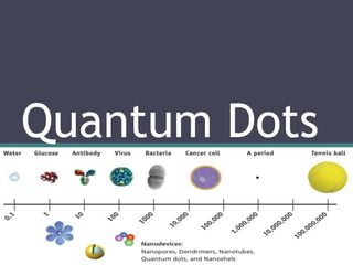
  • 9. INTRODUCTION • Quantum dots are tiny particles,or “nanoparticles”, of a semiconductor material, traditionally chalcogenides (selenides or sulfides) of metals like cadmium or zinc (CdSe or ZnS), which range from 2 to 10 nanometers in diameter (about the width of 50 atoms).
  • 10. • They are the smallest objects that can be synthesized on the nanoscale. • Like the name suggests, its structure is much like a small dot. Common shapes include pyramids, cylinders, lens shapes, and spheres. • Different synthesis routes create different kinds of quantum dots.
  • 11. • Quantum dots are so important because they confine electrons in three dimensions. • The reason 'quantum' prefixes the name is because the dots exhibit quantum confinement properties in all three dimensions i.e. electrons within a dot can't freely move around in any direction.
  • 12. • The ability to precisely control the size of a quantum dot enables the manufacturer to determine the wavelength of the emission, which in turn determines the colour of light the human eye perceives. • Quantum dots exhibit quantized energy levels like an atom. For a given input energy, for instance, a quantum dot will only emit specific spectra of light. • With decreasing diameters of quantum dots, there is a corresponding increase in energy of emitted light.
  • 13. • Quantum dots can therefore be “tuned” during production to emit any colour of light desired. The ability to control, or “tune” the emission from the quantum dot by changing its core size is called the “size quantisation effect”. • Due to excellent confinement properties not seen in nanowires or quantum wells (in all modern lasers), quantum dots are extremely efficient at emitting light.
  • 14. HISTORY • In the 1970s the first low dimensional structures QW (Quantum Wells) were developed. • 1D ( Quantum Wires) and 0D (Quantum Dots) were subsequently developed.
  • 15. PROPERTIES OF QUANTUM DOTS • Being smaller than the wavelength of the visible light, they cannot be seen under normal conditions. • Quantum Dots luminesce under ultraviolet light, with the size of the dots controlling its colour. • A quantum dot can have anything from a single electron to a collection of several thousands electrons. • Quantum Dots fluoresce or stay lit much longer then dyes conventionally used for tagging cells. • They can be tagged to proteins and their glow enables the identification of specific proteins or DNA making it possible to diagnose various diseases.
  • 16. The electrons in quantum dots are confined in a small space (quantum box), and when the radii of the semiconductor nanocrystal is smaller than the exciton Bohr radius (exciton Bohr radius is the average distance between the electron in the conduction band and the hole it leaves behind in the valence band), there is quantization of the energy levels according to Pauli’s exclusion principle. The discrete, quantized energy levels of quantum dots relate them more closely to atoms than bulk materials and have resulted in quantum dots being nicknamed 'artificial atoms'. As the size of the crystal decreases, the difference in energy between the highest valence band and the lowest conduction band increases. More energy is then needed to excite the dot, and more energy is released when the crystal returns to its ground state, resulting in a color shift from red to blue in the emitted light. As a result of this phenomenon, quantum dots can emit any color of light from the same material simply by changing the dot size. Additionally, because of the high level of control possible over the size of the nanocrystals produced, quantum dots can be tuned during manufacturing to emit any color of light. Quantum dots have a unique electronic structure
  • 17.
  • 18. Quantum dots can be classified into different types based on their composition and structure. Core-Type Quantum Dots Quantum dots can be single component materials with uniform internal compositions, such as chalcogenides (selenides or sulfides) of metals like cadmium or zinc, example, CdSe. The photo- and electroluminescence properties of core-type nanocrystals can be fine-tuned by simply changing the crystallite size. Core-Shell Quantum Dots The luminescent properties of quantum dots arise from recombination of electron-hole pairs (exciton decay) through radiative pathways. However, the exciton decay can also occur through nonradiative methods, reducing the fluorescence quantum yield. To improve efficiency and brightness of semiconductor nanocrystals a new method is growing shells of another higher band gap semiconducting material around them. These quantum dots with small regions of one material embedded in another with a wider band gap are known as core-shell quantum dots (CSQDs) or core-shell semiconducting nanocrystals (CSSNCs).
  • 19. For example, quantum dots with CdSe in the core and ZnS in the shell exhibit greater than 80% quantum yield. Coating quantum dots with shells improves quantum yield by passivizing nonradiative recombination sites and also makes them more robust to processing conditions for various applications. This method has been widely explored as a way to adjust the photophysical properties of quantum dots. Alloyed Quantum Dots Tuning the properties by changing crystallite size could cause problems in many applications with size restrictions. Multicomponent quantum dots is an alternative method to tune properties without changing crystallite size. Alloyed semiconductor quantum dots with both homogeneous and gradient internal structures allow tuning of optical and electronic properties by merely changing the composition and internal structure without changing the crystallite size. For example, alloyed quantum dots of the compositions CdSxSe1-x/ZnS of 6nm diameter emits light of different wavelengths by just changing the composition. Alloyed semiconductor quantum dots formed by alloying together two semiconductors with different band gap energies exhibited interesting properties distinct not only from the properties of their bulk counterparts but also from those of their parent semiconductors. Thus, alloyed nanocrystals possess novel and additional composition-tunable properties aside from properties due to quantum confinement effects.
  • 20. Visible spectrum • 5 nm dots: red • 1.5 nm dots: violet
  • 21. Quantum dots change color with size because additional energy is required to “confine” the semiconductor excitation To a smaller volume Ordinary light excites all color quantum dots. (Any light source “bluer” than the dot of interest works.)
  • 22. COLLOIDAL SYNTHESIS • Colloidal semiconductor nanocrystals - synthesized from precursor compounds dissolved in solutions, much like traditional chemical processes. • The synthesis of colloidal quantum dots is based on a three-component system composed of: precursors, organic surfactants, and solvents.
  • 23. • The precursors transform into monomers on heating to a suitable temperature. • Once the monomers reach a high enough supersaturation level, the nanocrystal growth starts with a nucleation process. • The temperature during the growth process must be high enough to allow for rearrangement and annealing of atoms during the synthesis process while being low enough to promote crystal growth.
  • 24. FABRICATION • Self-assembled quantum dots are typically between 5 and 50 nm in size, defined by lithographically patterned gate electrodes, or by etching on two-dimensional electron gases in semiconductor heterostructures. • Some quantum dots are small regions of one material buried in another with a larger band gap. These can be core-shell structures, e.g., with CdSe in the core and ZnS in the shell or from special forms of silica called ormosil. • Quantum dots sometimes occur spontaneously in quantum well structures due to monolayer fluctuations in the well's thickness. • This fabrication method has potential for applications in quantum computation. • The main limitations of this method are the cost of fabrication and the lack of control over positioning of individual dots.
  • 25. VIRAL ASSEMBLY • Lee et al. (2002) reported using genetically engineered M13 bacteriophage viruses to create quantum dot biocomposite structures. • Genetically engineered viruses can recognize specific semiconductor surfaces. • It is also known that liquid crystalline structures of wild- type viruses (Fd, M13, and TMV) are adjustable by controlling the solution concentrations, ionic strength, and the external magnetic field applied to the solutions. Thus, the specific recognition properties of the virus can be used to organize inorganic nanocrystals, forming ordered arrays over the length scale defined by liquid crystal formation.
  • 26. • Using this information, Lee et al. (2000) were able to create self-assembled, highly oriented, self-supporting films from a phage and ZnS precursor solution. This system allowed them to vary both the length of bacteriophage and the type of inorganic material through genetic modification and selection.
  • 27. ELECTROCHEMICAL ASSEMBLY • Highly ordered arrays of quantum dots may also be self- assembled by electrochemical techniques. • A template is created by causing an ionic reaction at an electrolyte-metal interface which results in the spontaneous assembly of nanostructures, including quantum dots, onto the metal which is then used as a mask for mesa-etching these nanostructures on a chosen substrate.
  • 28. BULK MANUFACTURING • Conventional, small-scale quantum dot manufacturing relies on a process called “high temperature dual injection” which is impractical for most commercial applications that require large quantities of quantum dots. • A reproducible method for creating larger quantities of consistent, high-quality quantum dots involves producing nanoparticles from chemical precursors in the presence of a molecular cluster compound under conditions whereby the integrity of the molecular cluster is maintained and acts as a prefabricated seed template. • Individual molecules of a cluster compound act as a seed or nucleation point upon which nanoparticle growth can be initiated. In this way, a high temperature nucleation step is not necessary to initiate nanoparticle growth because suitable nucleation sites are already provided in the system by the molecular clusters. • A significant advantage of this method is that it is highly scalable.
  • 29. CADMIUM FREE QUANTUM DOTS • For commercial viability, a range of restricted, heavy metal-free quantum dots has been developed showing bright emissions in the visible and near infra-red region of the spectrum and have similar optical properties to those of CdSe quantum dots. • A new type of CFQD can be made from rare earth (RE) doped oxide colloidal phosphor nanoparticles. Unlike semiconductor nanoparticles, excitation was due to UV absorption of host material, which is same for different RE doped materials using same host. • Multiplexing applications can be thus realized. The emission depends on the type of RE, which enables very large stokes shift and is narrower than CdSe QDs. The synthesis is aqueous based, which eliminated issues of water solubility for biological applications.
  • 30.
  • 31. 31 Applications • Photovoltaic devices: solar cells • QD Solar paint • Biology: biosensors, imaging • Light emitting diodes: LEDs • Quantum computation • Photodetectors • Lasers 31
  • 32. 32 Solar Cells • Photovoltaic effect: ▫ p-n junction. ▫ Sunlight excites electrons and creates electron-hole pairs. ▫ Electrons concentrate on one side of the cell and holes on the other side. ▫ Connecting the 2 sides creates electricity. 32
  • 33. 33 Different Generations of Solar Cells • First generation: ▫ Single crystal silicon wafer. ▫ Advantages: high carrier mobility. ▫ Disadvantages: most of photon energy is wasted as heat, expensive. • Second generation: ▫ Thin-film technology. ▫ Advantages: less expensive. ▫ Disadvantages: efficiency lower compared with silicon solar cells. • Third generation: ▫ Nanocrystal solar cells. ▫ Enhance electrical performances of the second generation while maintaining low production costs.
  • 34. 34 • The quantum dot band gap is tunable and can be used to create intermediate bandgaps. The maximum theoretical efficiency of the solar cell is as high as 63.2% with this method. How Can Quantum Dots Improve the Efficiency?
  • 35. Cheap quantum dot solar paint • “Sun Believable solar paint,” consists of a yellow or brown paste made of quantum dots. • The scientists experimented with three types of quantum dots: CdS, CdSe, and TiO2, all of which are powder-like, with water and tert butanol as the solvent. • Instead of adding dye to give the paint a desired color, they added colored semiconductor nanocrystals to the solar paint to achieve the desired optical and electronic properties.
  • 36. Quantum Dot LEDs • Used to produce inexpensive, industrial quality white light. • Marked improvement over traditional LED-phosphor integration by dot’s ability to absorb and emit at any desired wavelength. • Produce white light by intermixing red, green and blue emitting dots homogenously within the phosphor difficult to accomplish with the traditional LED-phosphor set up.
  • 37. Biolabelling • Multicolor labeling of cells is a powerful technique for visualizing many structures simultaneously, such as cytoskeletal proteins or organelles, and to elucidate intracellular processes. • QDs have been used to label cellular structures both within and external to the cell membrane. They are delivered inside cells via receptor-mediated pathways where specific ligands were attached to QDs to induce cellular uptake, as well as nonspecific endocytosis (ie, pinocytosis) where cells were incubated with a concentrated QD solution.
  • 38. Bioimaging • Non invasive, real-time in vivo fluorescence imaging requires exciting fluorophores and detecting their emission through tissue which is invariably hindered by scattering and absorption of both the excitation and emission wavelengths. • Using a filtered halogen source and an IR camera, the collection of QDs within tissue was monitored in real-time to identify a region for surgical resection.
  • 39. PHOTODETECTORS • Photodetectors based on single quantum dots are expected to find uses in opto-electrical interfaces in future quantum computers, where single photons will carry information over long distances and single electrons will be used for computation.
  • 40. Futuristic applications • Anti counterfeiting applications: inject dots into liquid mixtures, fabrics, polymer matrices, etc. Ability to specifically control absorption and emission spectra to produce unique validation signatures. Almost impossible to mimic with traditional semi-conductors. • Counter-espionage/Defense applications: Integrate quantum dots into dust that tracks enemies. Protection against friendly-fire events. • New research provides evidence for significant differences between new and old red blood cells used for transfusions and could provide a cheap, rapid and effective way to monitor the quality of blood . • Scientists have discover nanoparticles that can disrupt intracellular transport pathways.
  • 41. Imaging is an important clinical modality used in determining appropriate cancer therapy. x-ray, computed tomography, ultrasound, radionuclide imaging and MRI, : used widely for cancer screening and staging, determining the efficacy of cancer therapy and monitoring recurrence. 2 major limitations 1. do not have sufficient sensitivity to detect small numbers of malignant cells in the primary or metastatic sites. 2. The imaging techniques have not been developed to detect specific cancer cell-surface markers. In many instances, these cell-surface markers might be targets for cancer therapy and might assist in the diagnosis and staging of cancer. Quantum dot (QD) imaging probes, although still in the early development stage, provide the potential to fulfill these requirements for in vivo cancer imaging. QDs In Cancer Therapy
  • 42. QDs vs Organic Fluorescent dyes QDs offer great advantages over traditional organic fluorescent dyes and present a number of beneficial characteristics for spectroscopy, such as 1. high fluorescence intensity (brightness) 2. long lifetime 3. good resistance to photobleaching. 4. have broad excitation and narrow and symmetric emission spectra, which make it feasible to perform 'multiplexing' (simultaneous detection of multiple signals) imaging using a single excitation source 5. high sensitivity for simultaneous cancer molecular imaging and targeted therapy. the sensitivity of QD-based molecular imaging can be two to three orders larger than that of routine fluorescent dyes. Furthermore, the fluorescence in near infrared of NIR-QDs can be detected in deep tissues, making them suitable for in vivo imaging with high signal-to- background ratio
  • 43. Biocompatibility of QDs QDs are highly hydrophobic and, therefore, only soluble in organic solvents. Thus often encapsulated by amphiphilic molecules. The hydrophobic segment of these molecules interacts with the hydrophobic molecules on the QD surface, whereas the hydrophilic segment interacts with the aqueous medium, solublizing the QDs. Several types of amphiphilic polymers, including polyethylene glycol (PEG)-derived phospholipids, triblock copolymers, octylamine-modified polyacrylic acid, oligomeric phosphine and copolymers of alkyl monomers and anhydrides, can serve this encapsulation function. After encapsulation, the hydrodynamic radius of QDs increases to 10-20 nm. To functionalize QDs with biomolecules, amphiphilic polymers are engineered to carry reactive groups, such as amines and carboxylic acids. Biomolecules, such as peptides, antibodies, DNA or siRNA, can react with these functional groups to form covalent linkages mediated by various coupling reagents.
  • 44. In addition, biomolecules can be conjugated with QDs through noncovalent affinity binding, such as the interactions of biotin/avidin, or nickel nitrilotriacetic acid (Ni- NTA)/histidine-tagged peptides. Controlling ratio of reactive groups and reaction time, the average number of biomolecules conjugated to each QD can be controlled average of two or a few biomolecules are typically conjugated to one QD. Encapsulation and bioconjugation do not usually alter the optical property of QDs significantly. Recently, QDs based on silicon have drawn much attention because of their potential lower toxicity than heavy metal QDs, such as Cd/Se dots. Silicon QDs are typically 2-8 nm in size. The surface of QDs can be passivated with organic ligands, such as octadecene and dodecene, resulting in QDs that are more stable during storage with dramatically increased quantum yield and solubility in organic solvents. Some silicon QDs emit fluorescence at red and infrared range, which is ideal for in vivo imaging. However, their excitation wavelength normally lies in the ultraviolet region, which penetrates poorly into tissues and might be damaging to cells.
  • 45. Detection of primary tumor in vitro Biocompatible QDs were introduced for imaging of cancer cells in vitro in 1998. Researchers have synthesized QD-based probes conjugated with cancer specific ligands, antibodies, or peptides for cancer imaging and diagnosis in vitro. Compared with traditional immunohistochemistry, QD-IHC is more accurate and precise at low protein expression levels and can achieve quantitative detection which will provide much more information for personalized treatment. Prostate cancer Gao et al. labeled human prostate cancer cells based on the conjugate of QDs with an antibody for prostate specific membrane antigen (PSMA). QD-based immunolabelling has more stable photo-intensity compared with conventional fluorescent immunolabelling. Superior quality of QD-IHC compared with conventional IHC has been demonstrated along with simultaneous detection of androgen receptor and PSA in prostate cancer cells based on multiplexing QDs. The detection sensitivity of QD-based prostate cancer biomarkers can be enhanced by surface plasmon-coupled emission which has been introduced as a novel biosensing technology for detecting biosensors and biochips.
  • 46. Breast Cancer Human epidermal growth factor receptor 2 (HER2) is overexpressed in approximately 25% to 30% BC patients and has important function in cancer progression. Recent studies have validated the value of HER2 detection for BC treatment and prognosis. Compared with the golden standard method of fluorescence in situ hybridization (FISH), the advantages of QD-based IHC have been well documented. This method is easier, cheaper and less time-consuming. Various studies have reported the successful detection for BC by QD-HER2 conjugates. This approach has been extended to selectively label MCF-7 and BT-474 BC cells for HER2, epidermal growth factor receptor (EGFR), estrogen receptor (ER), progesterone receptor (PR), and mammalian target of rapamycin (m-TOR) by visible and NIR QDs which indicated that QD-based nanotechnology is an efficient approach to offer multiplexed cancer biomarker imaging in situ on intact tumor tissue specimens for tumor pathology study at the histological and molecular levels simultaneously. BC was successfully detected with QD-based probes which demonstrated that lower expression of HER2 could be clearly detected by QD-IHC compared with conventional IHC. Thus, QD-based multiplexed imaging will provide more information for the individual events of tumor, personalized diagnosis, prognosis, and treatment
  • 47. Ovarian cancer QDs can also be used to detect the ovarian carcinoma marker CA125 in different types of specimens, such as fixed cells, tissue sections, and xenograft piece. Additionally, the photostability of QD signals is more specific and brighter than that of conventional organic dye. Liu et al. synthesized pH-sensitive photoluminescent CdSe/ZnSe/ZnS QDs in SKOV-3 human ovarian cancer cells that are pH-dependent, suggesting applications for intracellular pH sensors.
  • 48. Gastrointestinal cancer Bostick et al. detected five biomarkers on the same tissue slide by QD-based multiplexed imaging. both efficient and convenient, takes only 7 h to analyze five biomarkers, which was advantageous for clinical application. Pancreatic cancer QD-based imaging probes can target pancreatic cancer at a very early stage with the help of proteins/peptides directed against over-expressed surface receptors on cancer cells/tissues, such as transferring receptor, antigen claudin-4, and urokinase plasminogen activator receptor. Both CdSe/CdS/ZnS QDs and non-cadmium-based QDs with improved photoluminescence efficiency and stability as optical agents have been used for the imaging of pancreatic cancer cells using transferring and anti-Claudin-4.
  • 49. In vivo Tumor imaging Directly demonstrate the evolution mechanism of tumor progression. More convincing evidence could be obtained from in vivo tumor imaging compared with in vitro molecular imaging. Sensitive and specific imaging agents are required for high-quality in vivo tumor imaging and less biological impacts on the animal model. QD-based imaging agents can meet this demand by “enhanced permeability and retention” (EPR) or targeted molecular imaging. The principle of EPR-based tumor imaging is the leakiness of tumor blood vessels. Compared with normal tissues, tumor vasculature is quantitatively higher, but irregular, leaky, dilated, and vascular endothelial cells are poorly aligned. The morphology results in increased leakage of macromolecules and nanocarriers out of the circulatory system into the tumor tissue. These finally accumulate in the tumor microenvironment because of the lack of lymphatic drainage. Many studies have reported that non-targeted QDs can be used for cell trafficking, vasculature imaging, sentinel lymph node (SLN) mapping, and neural imaging.
  • 50. SLN diagnosis contributes to operation strategy in cancer surgery. During lymph node metastasis, cancer cells first reach the SLN via lymph flow. The cancer cells can be detected with high sensitivity in the SLN connected to the tumor site. The superiority of NIR QDs has been demonstrated in SLN mapping, a common procedure in BC surgery, whereby the lymph node closest to the targeted organ is monitored for the presence of locally disseminated cancer cells. Recently highly sensitive, real-time intra-operative SLN mapping of the gastrointestinal tract by NIR QDs allowing image guidance throughout the entire procedure, virtually free of any background have been reported. These findings contribute to our understanding of metastasis, which remains a fundamental barrier to the development of effective cancer therapy. Given the high sensitivity and penetration of NIR QD fluorescence, the application of QD-based SLN mapping allows the surgeon to define the tumor border accurately and minimize the size of the dissection. QDs need to be effectively, specifically, and reliably directed to a specific organ or disease site without alteration to make them more beneficial for biomedical applications. Specific targeting can be achieved by attaching targeting molecules to the QD surface.
  • 51. Standard protocols for in vivo imaging of liver cancer xenograft animal models have been developed. Animal imaging by injecting human hepatocellular carcinoma cell lines (HCCLM6) that overexpress alpha-fetoprotein (AFP) with antiAFP monoclonal antibody and QD-IgG probes has been established. HCCLM6 has increased potential for lung metastasis, thus it allows construction of a platform for the early monitoring of cancer metastasis