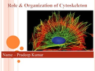
SlideShare uma empresa Scribd logo
1 de 37
Name – Pradeep Kumar
 The cytoskeleton is a network of fibers extending throughout the
cytoplasm
 It organizes the cell’s structures and activities, anchoring many
organelles
 It is composed of three types of molecular structures:
 Microfilaments
 Microtubules
 Intermediate filaments
Microfilaments:
 Actin
7nm
Microtubules:
 Tubulins (a, b)
25 nm
Intermediate filaments:
 Lamin
 cell specific prot. (e.g.
Vimentin)
8-12 nm
Microfilament
 The cytoskeleton helps to support the cell and maintain
its shape
 It interacts with motor proteins to produce motility
 Inside the cell, vesicles can travel along “monorails”
provided by the cytoskeleton
 Recent evidence suggests that the cytoskeleton may
help regulate biochemical activities
Properties Microfilament Microtubules Intermediate
filaments
Subunits incorporated
into polymer
ATP - actin
monomers
GTP - αβ - tubulin
heterodimer
Various globular
proteins
Preferential site of
incorporation
+ End barbed + End β - tubulin Internal
Polarity Yes Yes No
Enzymatic activity ATPase GTPase None
Motor proteins Myosin's Kinesins, Dyneins None
Major group of
associated proteins
Actin – binding
proteins
Maps Plakins
Structure
Flexible, helical
filament
Stiff, hollow tube Tough, ropelike fibers
Dimensions 8 nm diam. 25 nm outer diam. 10 nm diam.
Distribution All eukaryotes All eukaryotes Animals
Primary functions Motility, contractility
Support, intracellular
transport, cell
organization
Structural support
ATP-actin monomers
10 µm
Column of tubulin dimers
Tubulin dimer 
25 nm
Fibrous subunit (keratins
coiled together)
8–12 nm
5 µm
Keratin proteins
Tissue level
Muscle
movement
Determines shape of
the cell
Motility of the cells
Cell adhesion
 Mitosis, meiosis
Cellular level
Subcellular level
Anchors organelles
Organization of organelles
Provides tensile strength
 Movement of
chromosomes
Organizing cell polarity
Intracellular movement of
vesicles
 Endocytosis – clathrin-
mediated endocytosis and
phagocytosis
Dynamic
Adaptable
Stable
Strong
o Cytoskeletal filaments are all constructed
from smaller protein subunits
o All form as helical assemblies
of subunits
o Noncovalent interactions: rapid assembly
and disassembly
Intermediate filaments- resistant to stretching forces
 Microfilaments are solid rods about 7 nm in diameter,
built as a twisted double chain of actin subunits
 The structural role of microfilaments is to bear tension,
resisting pulling forces within the cell
 They form a 3-D network called the cortex just inside
the plasma membrane to help support the cell’s shape
 Bundles of microfilaments make up the core of
microvilli of intestinal cells.
Microvillus
Plasma membrane
Microfilaments (actin
filaments)
Intermediate filaments
0.25 µm
F-actin
G-actin
Two parallel protofilaments
that twist around each other in
a right-handed helix
 Microfilaments that function in cellular motility contain the
protein myosin in addition to actin
 In muscle cells, thousands of actin filaments are arranged parallel
to one another
 Thicker filaments composed of myosin interdigitate with the
thinner actin fibers
 Myosin molecules walk along the actin filament, pulling stacks of
actin fibers together and shortening
the cell.
o Localized contraction brought about by actin and myosin also
drives amoeboid movement
o Pseudopodia (cellular extensions) extend and contract through
the reversible assembly and contraction of actin subunits into
microfilaments
 Cytoplasmic streaming is a circular flow of cytoplasm within
cells
 This streaming speeds distribution of materials within the cell
 In plant cells, actin-myosin interactions and sol-gel
transformations drive cytoplasmic streaming
Cortex (outer cytoplasm):
gel with actin network
Inner cytoplasm: sol
with actin subunits
Extending
pseudopodium
(b) Amoeboid movement
Nonmoving cortical
cytoplasm (gel)
Chloroplast
Cell wall
Streaming
cytoplasm
(sol)
Parallel actin
filaments
(c) Cytoplasmic streaming in plant cells
Vacuole
 Microtubules are hollow
rods about 25 nm in
diameter and about 200 nm
to 25 microns long
 Functions of microtubules:
 Shaping the cell
 Guiding movement of
organelles
 Separating chromosomes
during cell division
Interphase cell
Dividing cell
Neuron
centrosome
Basal body
Cilla
spindle
centrosome
axon
-Centrosome
-Cilia / flagellum
-Mitotic system
- Vesicular transport
o In many cells, microtubules grow out from a centrosome near the
nucleus
o The centrosome is a “microtubule-organizing center”
o In animal cells, the centrosome has a pair of centrioles, each with
nine triplets of microtubules arranged in a ring
5 µm
Direction of swimming
(a) Motion of flagella
Direction of organism’s
movement
Power stroke Recovery stroke
(b) Motion of cilia
15 µm
 Cilia and flagella share a
common ultrastructure:
 A core of microtubules sheathed
by the plasma membrane
 A basal body that anchors the
cilium or flagellum
 A motor protein called dynein,
which drives the bending
movements of a cilium or
flagellum
Role of the dynein arms in beating cilia
Telescopic effect Beating
 Intermediate filaments range in diameter from 8–12 nanometers, larger
than microfilaments but smaller than microtubules
 They support cell shape and fix organelles in place
 Intermediate filaments are more permanent cytoskeleton fixtures than the other
two classes
 The extracellular matrix (ECM) of animal cells
 Intercellular junctions
 Animal cells lack cell walls but are covered by an elaborate
extracellular matrix (ECM)
 The ECM is made up of glycoprotein's such as collagen,
proteoglycans, and fibronectin
 ECM proteins bind to receptor proteins in the plasma membrane
called integrins
 Functions of the ECM:
 Support
 Adhesion
 Movement
 Regulation
EXTRACELLULAR MATRIX (ECM)
 Neighboring cells in tissues, organs, or organ systems often
adhere, interact, and communicate through direct physical
contact
 Intercellular junctions facilitate this contact
 There are several types of intercellular junctions
 Plasmodesmata
 Tight junctions
 Desmosomes
 Gap junctions
 Plasmodesmata are channels
that perforate plant cell walls
 Through plasmodesmata, water
and small solutes (and
sometimes proteins and RNA)
can pass from cell to cell
 At tight junctions, membranes of neighboring cells are pressed
together, preventing leakage of extracellular fluid
 Desmosomes (anchoring junctions) fasten cells together into
strong sheets
 Gap junctions (communicating junctions) provide cytoplasmic
channels between adjacent cells
Thank you

Mais conteúdo relacionado

Mais procurados

Mais procurados (20)

Cell cell interaction
Cell cell interactionCell cell interaction
Cell cell interaction
 
Microtubules
MicrotubulesMicrotubules
Microtubules
 
Cytoskeleton presentation (introduction structure & function)
Cytoskeleton presentation (introduction structure & function)Cytoskeleton presentation (introduction structure & function)
Cytoskeleton presentation (introduction structure & function)
 
THE CYTOSKELETON
THE CYTOSKELETONTHE CYTOSKELETON
THE CYTOSKELETON
 
Golgi complex structure and functions
Golgi complex structure and functionsGolgi complex structure and functions
Golgi complex structure and functions
 
Structure and functon of golgi apparatus
Structure and functon of golgi apparatusStructure and functon of golgi apparatus
Structure and functon of golgi apparatus
 
Cytoskeleton
CytoskeletonCytoskeleton
Cytoskeleton
 
Membrane Dynamics:Properties of biological membrane (plasma membrane)
Membrane Dynamics:Properties of biological membrane (plasma membrane)Membrane Dynamics:Properties of biological membrane (plasma membrane)
Membrane Dynamics:Properties of biological membrane (plasma membrane)
 
Chloroplast
ChloroplastChloroplast
Chloroplast
 
Cell Adhesion Molecules(cams) and its types, Cadherins and Integrins and inte...
Cell Adhesion Molecules(cams) and its types, Cadherins and Integrins and inte...Cell Adhesion Molecules(cams) and its types, Cadherins and Integrins and inte...
Cell Adhesion Molecules(cams) and its types, Cadherins and Integrins and inte...
 
Cytoskeleton , cell shape and cell motility
Cytoskeleton , cell shape and cell motilityCytoskeleton , cell shape and cell motility
Cytoskeleton , cell shape and cell motility
 
Cytoskeletal structures
Cytoskeletal structuresCytoskeletal structures
Cytoskeletal structures
 
Cell adhesion molecules
Cell adhesion moleculesCell adhesion molecules
Cell adhesion molecules
 
Ribosome structure and assembly
Ribosome structure and assemblyRibosome structure and assembly
Ribosome structure and assembly
 
Structure and functions of endoplasmic reticulum
Structure and functions of endoplasmic reticulumStructure and functions of endoplasmic reticulum
Structure and functions of endoplasmic reticulum
 
Cytoskeleton
CytoskeletonCytoskeleton
Cytoskeleton
 
Cell motility cilia and flagella
Cell motility  cilia and flagellaCell motility  cilia and flagella
Cell motility cilia and flagella
 
Micro filaments
Micro filamentsMicro filaments
Micro filaments
 
Membrane carbohydrate and their significance in cellular recognition
Membrane carbohydrate and their significance in cellular recognitionMembrane carbohydrate and their significance in cellular recognition
Membrane carbohydrate and their significance in cellular recognition
 
Cilia and flagella ppt by ramana babu
Cilia and flagella ppt by ramana babuCilia and flagella ppt by ramana babu
Cilia and flagella ppt by ramana babu
 

Semelhante a Orginagition and role of cytoskeleton

5- Cell II
5- Cell II5- Cell II
5- Cell II
tchubb
 

Semelhante a Orginagition and role of cytoskeleton (20)

Cytoskelton.ppt
Cytoskelton.pptCytoskelton.ppt
Cytoskelton.ppt
 
Cytoskeleton
CytoskeletonCytoskeleton
Cytoskeleton
 
Cytoskeleton
CytoskeletonCytoskeleton
Cytoskeleton
 
Copy of cytoskeleton
Copy of cytoskeletonCopy of cytoskeleton
Copy of cytoskeleton
 
Cytoskeleton
CytoskeletonCytoskeleton
Cytoskeleton
 
Cytoskeleton
CytoskeletonCytoskeleton
Cytoskeleton
 
Microtubules by jimmy
Microtubules by jimmyMicrotubules by jimmy
Microtubules by jimmy
 
The Cytoskeleton
The CytoskeletonThe Cytoskeleton
The Cytoskeleton
 
Cytoskeleton[1]
Cytoskeleton[1]Cytoskeleton[1]
Cytoskeleton[1]
 
Cytoskeleton
Cytoskeleton Cytoskeleton
Cytoskeleton
 
Cytoskeleton
CytoskeletonCytoskeleton
Cytoskeleton
 
unit 1 cytoskeletal structures ECM docx.pdf sh.pdf
unit 1 cytoskeletal structures ECM docx.pdf sh.pdfunit 1 cytoskeletal structures ECM docx.pdf sh.pdf
unit 1 cytoskeletal structures ECM docx.pdf sh.pdf
 
Final cytoplasm and cytoskeleton
Final cytoplasm and cytoskeletonFinal cytoplasm and cytoskeleton
Final cytoplasm and cytoskeleton
 
CMB_L28_2008s.ppt
CMB_L28_2008s.pptCMB_L28_2008s.ppt
CMB_L28_2008s.ppt
 
Cytoskeleton
CytoskeletonCytoskeleton
Cytoskeleton
 
Cytoskeleton
CytoskeletonCytoskeleton
Cytoskeleton
 
Cell cytoskeleton (filaments and their roles)
Cell cytoskeleton (filaments and their roles)Cell cytoskeleton (filaments and their roles)
Cell cytoskeleton (filaments and their roles)
 
Biology 12 - Cytoskeleton and Cell Membrane
Biology 12 - Cytoskeleton and Cell MembraneBiology 12 - Cytoskeleton and Cell Membrane
Biology 12 - Cytoskeleton and Cell Membrane
 
Eukaryotic Cell Functions
Eukaryotic Cell FunctionsEukaryotic Cell Functions
Eukaryotic Cell Functions
 
5- Cell II
5- Cell II5- Cell II
5- Cell II
 

Mais de Pradeep Kumar

Mais de Pradeep Kumar (7)

Pgs 502 Laboratory precautions
Pgs 502 Laboratory precautionsPgs 502 Laboratory precautions
Pgs 502 Laboratory precautions
 
Genome Assembly copy
Genome Assembly   copyGenome Assembly   copy
Genome Assembly copy
 
Plant tissue culture, Biotechnology
Plant tissue culture, BiotechnologyPlant tissue culture, Biotechnology
Plant tissue culture, Biotechnology
 
Gus staining and reporter gene
Gus staining and reporter geneGus staining and reporter gene
Gus staining and reporter gene
 
Chromosome packaging
Chromosome  packagingChromosome  packaging
Chromosome packaging
 
Tissue engineering and stem cell by regenerative medicine.pptx badal 2014
Tissue engineering and stem cell by regenerative medicine.pptx badal 2014Tissue engineering and stem cell by regenerative medicine.pptx badal 2014
Tissue engineering and stem cell by regenerative medicine.pptx badal 2014
 
Androgenesis of rice
Androgenesis of riceAndrogenesis of rice
Androgenesis of rice
 

Último

Activity 01 - Artificial Culture (1).pdf
Activity 01 - Artificial Culture (1).pdfActivity 01 - Artificial Culture (1).pdf
Activity 01 - Artificial Culture (1).pdf
ciinovamais
 
1029 - Danh muc Sach Giao Khoa 10 . pdf
1029 -  Danh muc Sach Giao Khoa 10 . pdf1029 -  Danh muc Sach Giao Khoa 10 . pdf
1029 - Danh muc Sach Giao Khoa 10 . pdf
QucHHunhnh
 
Jual Obat Aborsi Hongkong ( Asli No.1 ) 085657271886 Obat Penggugur Kandungan...
Jual Obat Aborsi Hongkong ( Asli No.1 ) 085657271886 Obat Penggugur Kandungan...Jual Obat Aborsi Hongkong ( Asli No.1 ) 085657271886 Obat Penggugur Kandungan...
Jual Obat Aborsi Hongkong ( Asli No.1 ) 085657271886 Obat Penggugur Kandungan...
ZurliaSoop
 

Último (20)

Graduate Outcomes Presentation Slides - English
Graduate Outcomes Presentation Slides - EnglishGraduate Outcomes Presentation Slides - English
Graduate Outcomes Presentation Slides - English
 
Basic Civil Engineering first year Notes- Chapter 4 Building.pptx
Basic Civil Engineering first year Notes- Chapter 4 Building.pptxBasic Civil Engineering first year Notes- Chapter 4 Building.pptx
Basic Civil Engineering first year Notes- Chapter 4 Building.pptx
 
Mehran University Newsletter Vol-X, Issue-I, 2024
Mehran University Newsletter Vol-X, Issue-I, 2024Mehran University Newsletter Vol-X, Issue-I, 2024
Mehran University Newsletter Vol-X, Issue-I, 2024
 
Accessible Digital Futures project (20/03/2024)
Accessible Digital Futures project (20/03/2024)Accessible Digital Futures project (20/03/2024)
Accessible Digital Futures project (20/03/2024)
 
Mixin Classes in Odoo 17 How to Extend Models Using Mixin Classes
Mixin Classes in Odoo 17  How to Extend Models Using Mixin ClassesMixin Classes in Odoo 17  How to Extend Models Using Mixin Classes
Mixin Classes in Odoo 17 How to Extend Models Using Mixin Classes
 
UGC NET Paper 1 Mathematical Reasoning & Aptitude.pdf
UGC NET Paper 1 Mathematical Reasoning & Aptitude.pdfUGC NET Paper 1 Mathematical Reasoning & Aptitude.pdf
UGC NET Paper 1 Mathematical Reasoning & Aptitude.pdf
 
Sociology 101 Demonstration of Learning Exhibit
Sociology 101 Demonstration of Learning ExhibitSociology 101 Demonstration of Learning Exhibit
Sociology 101 Demonstration of Learning Exhibit
 
Key note speaker Neum_Admir Softic_ENG.pdf
Key note speaker Neum_Admir Softic_ENG.pdfKey note speaker Neum_Admir Softic_ENG.pdf
Key note speaker Neum_Admir Softic_ENG.pdf
 
How to Manage Global Discount in Odoo 17 POS
How to Manage Global Discount in Odoo 17 POSHow to Manage Global Discount in Odoo 17 POS
How to Manage Global Discount in Odoo 17 POS
 
SOC 101 Demonstration of Learning Presentation
SOC 101 Demonstration of Learning PresentationSOC 101 Demonstration of Learning Presentation
SOC 101 Demonstration of Learning Presentation
 
Explore beautiful and ugly buildings. Mathematics helps us create beautiful d...
Explore beautiful and ugly buildings. Mathematics helps us create beautiful d...Explore beautiful and ugly buildings. Mathematics helps us create beautiful d...
Explore beautiful and ugly buildings. Mathematics helps us create beautiful d...
 
Food safety_Challenges food safety laboratories_.pdf
Food safety_Challenges food safety laboratories_.pdfFood safety_Challenges food safety laboratories_.pdf
Food safety_Challenges food safety laboratories_.pdf
 
Activity 01 - Artificial Culture (1).pdf
Activity 01 - Artificial Culture (1).pdfActivity 01 - Artificial Culture (1).pdf
Activity 01 - Artificial Culture (1).pdf
 
Python Notes for mca i year students osmania university.docx
Python Notes for mca i year students osmania university.docxPython Notes for mca i year students osmania university.docx
Python Notes for mca i year students osmania university.docx
 
1029 - Danh muc Sach Giao Khoa 10 . pdf
1029 -  Danh muc Sach Giao Khoa 10 . pdf1029 -  Danh muc Sach Giao Khoa 10 . pdf
1029 - Danh muc Sach Giao Khoa 10 . pdf
 
Dyslexia AI Workshop for Slideshare.pptx
Dyslexia AI Workshop for Slideshare.pptxDyslexia AI Workshop for Slideshare.pptx
Dyslexia AI Workshop for Slideshare.pptx
 
Micro-Scholarship, What it is, How can it help me.pdf
Micro-Scholarship, What it is, How can it help me.pdfMicro-Scholarship, What it is, How can it help me.pdf
Micro-Scholarship, What it is, How can it help me.pdf
 
Understanding Accommodations and Modifications
Understanding  Accommodations and ModificationsUnderstanding  Accommodations and Modifications
Understanding Accommodations and Modifications
 
Jual Obat Aborsi Hongkong ( Asli No.1 ) 085657271886 Obat Penggugur Kandungan...
Jual Obat Aborsi Hongkong ( Asli No.1 ) 085657271886 Obat Penggugur Kandungan...Jual Obat Aborsi Hongkong ( Asli No.1 ) 085657271886 Obat Penggugur Kandungan...
Jual Obat Aborsi Hongkong ( Asli No.1 ) 085657271886 Obat Penggugur Kandungan...
 
ComPTIA Overview | Comptia Security+ Book SY0-701
ComPTIA Overview | Comptia Security+ Book SY0-701ComPTIA Overview | Comptia Security+ Book SY0-701
ComPTIA Overview | Comptia Security+ Book SY0-701
 

Orginagition and role of cytoskeleton

  • 2.  The cytoskeleton is a network of fibers extending throughout the cytoplasm  It organizes the cell’s structures and activities, anchoring many organelles  It is composed of three types of molecular structures:  Microfilaments  Microtubules  Intermediate filaments
  • 3. Microfilaments:  Actin 7nm Microtubules:  Tubulins (a, b) 25 nm Intermediate filaments:  Lamin  cell specific prot. (e.g. Vimentin) 8-12 nm Microfilament
  • 4.  The cytoskeleton helps to support the cell and maintain its shape  It interacts with motor proteins to produce motility  Inside the cell, vesicles can travel along “monorails” provided by the cytoskeleton  Recent evidence suggests that the cytoskeleton may help regulate biochemical activities
  • 5. Properties Microfilament Microtubules Intermediate filaments Subunits incorporated into polymer ATP - actin monomers GTP - αβ - tubulin heterodimer Various globular proteins Preferential site of incorporation + End barbed + End β - tubulin Internal Polarity Yes Yes No Enzymatic activity ATPase GTPase None Motor proteins Myosin's Kinesins, Dyneins None Major group of associated proteins Actin – binding proteins Maps Plakins Structure Flexible, helical filament Stiff, hollow tube Tough, ropelike fibers Dimensions 8 nm diam. 25 nm outer diam. 10 nm diam. Distribution All eukaryotes All eukaryotes Animals Primary functions Motility, contractility Support, intracellular transport, cell organization Structural support
  • 7. 10 µm Column of tubulin dimers Tubulin dimer  25 nm
  • 8. Fibrous subunit (keratins coiled together) 8–12 nm 5 µm Keratin proteins
  • 9. Tissue level Muscle movement Determines shape of the cell Motility of the cells Cell adhesion  Mitosis, meiosis Cellular level Subcellular level Anchors organelles Organization of organelles Provides tensile strength  Movement of chromosomes Organizing cell polarity Intracellular movement of vesicles  Endocytosis – clathrin- mediated endocytosis and phagocytosis Dynamic Adaptable Stable Strong
  • 10. o Cytoskeletal filaments are all constructed from smaller protein subunits o All form as helical assemblies of subunits o Noncovalent interactions: rapid assembly and disassembly
  • 11.
  • 12. Intermediate filaments- resistant to stretching forces
  • 13.  Microfilaments are solid rods about 7 nm in diameter, built as a twisted double chain of actin subunits  The structural role of microfilaments is to bear tension, resisting pulling forces within the cell  They form a 3-D network called the cortex just inside the plasma membrane to help support the cell’s shape  Bundles of microfilaments make up the core of microvilli of intestinal cells.
  • 15. F-actin G-actin Two parallel protofilaments that twist around each other in a right-handed helix
  • 16.  Microfilaments that function in cellular motility contain the protein myosin in addition to actin  In muscle cells, thousands of actin filaments are arranged parallel to one another  Thicker filaments composed of myosin interdigitate with the thinner actin fibers  Myosin molecules walk along the actin filament, pulling stacks of actin fibers together and shortening the cell.
  • 17.
  • 18. o Localized contraction brought about by actin and myosin also drives amoeboid movement o Pseudopodia (cellular extensions) extend and contract through the reversible assembly and contraction of actin subunits into microfilaments  Cytoplasmic streaming is a circular flow of cytoplasm within cells  This streaming speeds distribution of materials within the cell  In plant cells, actin-myosin interactions and sol-gel transformations drive cytoplasmic streaming
  • 19. Cortex (outer cytoplasm): gel with actin network Inner cytoplasm: sol with actin subunits Extending pseudopodium (b) Amoeboid movement Nonmoving cortical cytoplasm (gel) Chloroplast Cell wall Streaming cytoplasm (sol) Parallel actin filaments (c) Cytoplasmic streaming in plant cells Vacuole
  • 20.  Microtubules are hollow rods about 25 nm in diameter and about 200 nm to 25 microns long  Functions of microtubules:  Shaping the cell  Guiding movement of organelles  Separating chromosomes during cell division
  • 21. Interphase cell Dividing cell Neuron centrosome Basal body Cilla spindle centrosome axon -Centrosome -Cilia / flagellum -Mitotic system - Vesicular transport
  • 22.
  • 23. o In many cells, microtubules grow out from a centrosome near the nucleus o The centrosome is a “microtubule-organizing center” o In animal cells, the centrosome has a pair of centrioles, each with nine triplets of microtubules arranged in a ring
  • 24. 5 µm Direction of swimming (a) Motion of flagella Direction of organism’s movement Power stroke Recovery stroke (b) Motion of cilia 15 µm  Cilia and flagella share a common ultrastructure:  A core of microtubules sheathed by the plasma membrane  A basal body that anchors the cilium or flagellum  A motor protein called dynein, which drives the bending movements of a cilium or flagellum
  • 25.
  • 26. Role of the dynein arms in beating cilia Telescopic effect Beating
  • 27.  Intermediate filaments range in diameter from 8–12 nanometers, larger than microfilaments but smaller than microtubules  They support cell shape and fix organelles in place  Intermediate filaments are more permanent cytoskeleton fixtures than the other two classes  The extracellular matrix (ECM) of animal cells  Intercellular junctions
  • 28.
  • 29.
  • 30.
  • 31.  Animal cells lack cell walls but are covered by an elaborate extracellular matrix (ECM)  The ECM is made up of glycoprotein's such as collagen, proteoglycans, and fibronectin  ECM proteins bind to receptor proteins in the plasma membrane called integrins  Functions of the ECM:  Support  Adhesion  Movement  Regulation
  • 33.  Neighboring cells in tissues, organs, or organ systems often adhere, interact, and communicate through direct physical contact  Intercellular junctions facilitate this contact  There are several types of intercellular junctions  Plasmodesmata  Tight junctions  Desmosomes  Gap junctions
  • 34.  Plasmodesmata are channels that perforate plant cell walls  Through plasmodesmata, water and small solutes (and sometimes proteins and RNA) can pass from cell to cell
  • 35.  At tight junctions, membranes of neighboring cells are pressed together, preventing leakage of extracellular fluid  Desmosomes (anchoring junctions) fasten cells together into strong sheets  Gap junctions (communicating junctions) provide cytoplasmic channels between adjacent cells
  • 36.