The Cytoskeleton- An overview
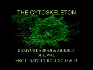
- 1. THE CYTOSKELETON DARIYUS KABRAJI & ABHIJEET SHEJWAL MSC 1 BATCH 2 ROLL NO 18 & 15
- 2. INTRODUCTION • The cytoskeleton is an integral cell component present in all cells (except most bacterial species) • It is the basic unit of the cell maintaining its structural integrity • While the Cytoplasm is the semi-fluid substance that fills the space between the nucleus and the cell membrane that holds all of the organelles in suspension, the Cytoskeleton is what holds the organelles in place, or moves them and aids in cell movement
- 3. BASICS • The cytoskeleton is made up of a network of fibres composed of proteins within a cell's cytoplasm. • The term "cytoskeleton" refers to the filamentous structures themselves, which are composed of a set of proteins, each with their own shape and designation • Although it appears to be fixed, it is actually an ever-changing dynamic configuration, with components regularly destroyed, repaired or newly formed
- 4. BASICS
- 5. FUNCTIONS • It imparts the cell shape • It provides protection by resistance to deformation • It can actively contract, deforming the cell and it's environment, allowing cells to migrate • It is involved in cell signalling pathways • It separates chromosomes during cell division • It provides a blueprint to organize the organelles in cytoplasm and for the construction of a cell wall • It gives rise to structures such as flagella and cilia
- 6. THE EUKARYOTIC CYTOSKELETON • Prokaryotic cytoskeleton is a recent discovery & unwise choice for a presentation • Eukaryotic cytoskeleton is made up of three structures, each with their own design and function: 1. Microfilaments 2. Intermediate filaments 3. Microtubules
- 8. MICROFILAMENTS • Monomers of the protein actin polymerize to form long, thin fibres i.e. G-actin polymerising to form F-actin filaments • These are about 7 nm in diameter and, being the thinnest of the cytoskeletal filaments, are also called microfilaments • They represent the active or motile part of the cytoskeleton
- 9. MICROFILAMENTS FORMATION OF ACTIN POLYMERS
- 10. CHARACTERISTICS • Found in the cortical regions of the cell beneath the plasma membrane • Extend into cell processes involving movement • The concentration of actin in the body’s non-muscle cells add up to 10% of net protein • There are three types of actin filaments- α, β, & γ of which only α is found in muscle cells • In non- muscle cells, actin binds to heavy myosin which results in an arrow shaped pattern, indicating the existence of polarity in actin, crucial to mediating cell movement • Heavy myosin (HMM) is thus used to identify and localize microfilaments
- 11. SENSITIVITY • Microfilaments are sensitive to Cytochalasin- B, an alkaloid • This compound impairs cell activities like • Heartbeat • Cell migration • Cytokinesis • Endocytosis • Exocytosis
- 12. SENSITIVITY
- 13. THE DYNAMIC ASSEMBLY • Unlike other polymers such as DNA, bound together with covalent bonds, the monomers of actin filaments are assembled by weaker bonds. • These bonds give the advantage that the filament ends can easily release or incorporate monomers. • This means that the filaments can be rapidly remodelled and can change cellular structure in response to an environmental stimulus.
- 15. FUNCTIONS • Forms a band just beneath the plasma membrane that provides mechanical strength to the cell • Links transmembrane proteins to cytoplasmic proteins • Separates dividing animal cells during cytokinesis • Generates cytoplasmic streaming in some cells • Generates locomotion in cells such as white blood cells and the amoeba by giving rise to pseudopodia • Interacts with myosin filaments in skeletal muscle fibres to provide the force of muscular contraction • Also involved in movement associated with furrow formation in cell division • Responsible for cytoplasmic streaming in plant cells • Cell migration during embryonic development
- 16. INTERMEDIATE FILAMENTS • Tough protein fibres in higher eukaryotic cells • Mostly between 8-10 nm in diameter • Named ‘intermediate’ for their moderate size between smaller actin filaments and massive microtubules • In animal cells they are stable structures which compensate for the absence of cell walls • They can also be anchored between membranes for extra support
- 18. STRUCTURE • In a cross-section, intermediary filaments have a tubular appearance • Each tube is made up of 4 or 5 parallel protofilaments • They are comprised of polypeptides from 40-130 Kilo- Daltons • All IF proteins have a single common central region of around 310 amino acid residues forming extended helixes with short interruptions. The domains at the terminal ends (amino & carboxyl) are non helical and vary in size and sequence per type
- 19. STRUCTURE
- 20. TYPES OF IFs • There are 4 types of intermediary filaments, each with its own mass and function: • Type 1: Acidic, neutral & basic Keratin (40-70 Kd) in epidermal derivatives and epithelial cells • Type 2: Vimentin (53 Kd), Desmin (52 Kd), Synemin (230 Kd), Glial filaments (45 Kd) in muscle and Glial cells • Type 3: Neurofilament proteins (60-130 Kd) in Neurons • Type 4: Nuclear lamins (65-75 Kd) in Nuclear lamellae
- 21. FUNCTIONS • Main function involves mechanical support to the cell • The nucleus is guarded by an IF mesh network that extends to the cell boundaries • Neurofilaments protect neurons from mechanical damage caused by the animal’s movement • Filaments in epithelia form a tough network to avoid incursion from external factors • Desmin filaments support sacromeres in muscle cells • Vimentin filaments surround and guard fat cell droplets
- 22. MICROTUBULES - ABHIJEET SHEJWAL • First observed in axoplasm of myelinated nerve fibers by Robertis and Franchi 1953. • Microtubules of plant cells observed in 1963 by Ledbetter and Porter. • Found in all eukaryotes, either free in cytoplasm or form part of centriole, cilia and flagella • High densities of microtubule in axon & dendrites of nerve cells.
- 23. STRUCTURE • Rigid hollow rods, approx. 25 nm in diameter. • Single type of globular protein Tubulin M.W.- 55 kD • Tubulin contains 2 dimers, α & β Tubulin. Each contain 450 amino acids. • Tubulin dimers polymerize to form microtubule • 13 linear protofilament around hollow core. • Polar structure of microtubules have 2 ends: a) Fast growing plus end b) Slow growing minus end • Polarity helps determine direction of movement. • Undergo continuous assembly & disassembly in structure. • Initiation of microtubule assembly is done by γ-tubulin in centrosome.
- 25. STRUCTURE • Minus end of cytoplasmic microtubule bound to MTOCs from which polymerization starts. • Plus end is protected by capping protein to avoid disassembly. • MAPs associate protein with surface of microtubule • MAPs of 2 types: a) HMW proteins (M.W. 200 to 300 D) b) tau proteins (M.W. 40 to 60 D) • MTOCs initiation of assembly, serve template for polymerization, exists in basal bodies, in centriole. • ON-OFF of MTOCs regulated by changes in nucleation center, changes in Ca 2+ concentration, involvement of MAPs.
- 26. FORMATION • Tubulin dimers depolymerize as well as polymerizes, α & β tubulin bind to GTP to regulate polymerization. • GTP bound to β tubulin is hydrolyzed to GDP during polymerization. • GTP hydrolysis weakens binding affinity of tubulin which favours depolymerization. • Tubulin bound to GDP are lost from minus end while replaced by addition of tubulin bound to GTP at plus end continuously. • GTP Cap is observed at plus end which leads to continuous growth of microtubule. • Dynamic instability described by Tim Mitchisan & Marc Kirschner in 1984. • Rapid turnover of microtubule is imp for remodeling of cytoskeleton occurs during mitosis.
- 27. FORMATION • Polymerized tubulins are high at interphase, metaphase while low at prophase, anaphase. • Phosphorylation of tubulin monomer by cAMP kinase • Assembly inhibited by drugs like colchicine,vinblastine, vincristine • In vivo control of polymerization involves Ca 2+ and calcium binding protein calmodulin. • King 1986 in vitro assembly of microtubule occur in presence of low Ca 2+ concentration, MAPs , GTP, level of free tubulin monomers.
- 28. FUNCTION • Determine cell shape. • Determine variety of cell movements (cell locomotion, intracellular transport of organelles, separation of chromosomes during mitosis) • Determine intrinsic polarity of cells and motility • Contraction of spindle
- 29. APPLICATION • Drugs affect microtubule assembly are useful in treatment of cancer. e.g. i. Colchicine & Colcemid – binds tubulin, inhibit polymerisation which blocks mitosis. ii. Vin cristine & vin blastine – selectively inhibit rapidly dividing cells. iii. Taxol – stabilizes microtubule, blocks cell division. Used as anticancer agent.
- 30. FUTURE PROSPECTS • Advisable to study the cytoskeleton in prokaryotes since so little is known about them • Probably more undiscovered functions carried out by the cytoskeleton, might be interesting field of specialization • Diseases or disorders of any kind concerning these components • Basic formation and destruction pathways of the components
