SlideShare uma empresa Scribd logo
1 de 85
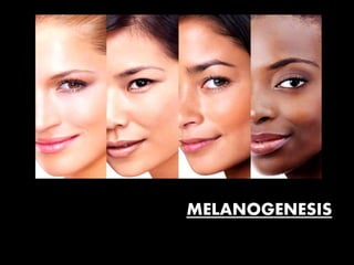
MELANOGENESIS
•The skin has epidermal units - responsible for melanin production and
distribution - melanogenesis
•Melanin is the primary determinant of skin, hair, and eye color.
critical role in photoprotection d/t ability to absorb ultraviolet radiation
(UVR)
• The Fitzpatrick system - mc distinguish different skin pigmentation
phenotypes.
It characterizes six phototypes (I-VI) by grading erythema and acquired
pigmentation after exposure to UVR
•Skin colour results from :
1. Concentration and admixture of the types of melanins in melanocyte
2. Carotenoid pigments
3. Haemoglobin (in both the oxygenated and reduced state).
•Two types of melanin pigmentation occurs -
constitutive skin colour, which is amount of melanin pigmentation
genetically determined in absence of sun exposure and other
influences.
 facultative (inducible) skin colour or ‘tan’, results from sun
exposure.
Increased pigmentation - due to endocrine, paracrine and autocrine
factors
•The major events are:
• the origin and function of the melanocyte
• the formation and function of the melanosome
• Melanin biosynthesis and its regulations
•The melanocyte is a neural crest-derived cell.
•During embryogenesis , Melanoblasts originate in neural crest , migrate
first to dermis & then to basal lamina of epidermis
•Immunocytochemical marker studies with melanocyte‐specific
antibody - melanoblasts appear in epidermis by 7 weeks gestation
•They first appear in the head and neck region at ~10 weeks of gestation.
•By the end of gestation, active dermal melanocytes have
“disappeared”, except in three primary anatomic locations –
- the head and neck,
- the dorsal aspects of the distal extremities,
- and the presacral area.
Distribution of melanocytes
•Number shows little variation between different races or sexes.
•Capacity of melanocytes - synthesize melanin, in basal state and after
stimulation by sunlight varies.
•Melanocytes in dark skin - great capacity to synthesize melanin and to
transfer it to surrounding keratinocytes.
(in contrast with fair skin)
•↓ in number of melanocytes occur - ageing, ↓se in melanocyte density
of about 6–8% per decade.
•Melanocytes - most numerous in epidermis, hair follicles and the eye
• Ear
Melanins found in Striae vascularis of Inner ear.
•Eye
Melanocytes - present in Iris stroma in front of Iris, Iris pigment
epithelium on the back of Iris, Retinal pigment epithelium.
•Adrenal glands
seen in Medula and Zona reticularis.
•Others
Melanin is also found in heart, liver, muscles and intestine
Melanoblast migration and differentiation
Melanoblasts - predominantly found in basal layer of epidermis & hair
follicles , identified by expression of melanocyte-specific markers
•tyrosinase (TYR),
•tyrosinase-related protein 1 (TYRP1),
•tyrosinase-related protein-2 (TYRP2),
•Premelanosome protein 17 (Pmel17/gp1000),
•melan-A or melanoma antigen recognized by T cells 1 (MART-1) and
•microphthalmia-associated transcription factor (MITF)
• Initial segregation of melanocyte lineage - the Wnt/β‐catenin pathway .
•The transcription factor mi is specific for melanocytes differentiation.
•Mi encodes transcription factor MITF, regulates several melanocyte
specific genes
• mutations in mi - Waardenburg syndrome type 2A,AD characterized by
deafness and patchy abnormal pigmentation
•Pax3 - prime cells for differentiation, whereas Wnt signalling allows cells
to proceed along this route
•Variation - density of epidermal melanocytes/mm2 when different
regions of the body are analyzed ,
• e.g. the density of melanocytes is greater in genital region
(~1500/mm2) than with back (~900/mm2)
•There are smaller differences between individuals when same
anatomic site is examined.
•The major determinant of skin color is activity of the
melanocytes, i.e. quantity and quality of pigment production, not
density of melanocytes.
•Several factors play a role in melanocyte activity:
- specific characteristics of individual melanosomes
- baseline (constitutive) and stimulated (facultative) levels
- activity of the enzymes in the melanin biosynthetic pathway.
SURVIVAL
OF
NEURAL CREST DERIVED CELLS
1. Depends upon interactions between cell surface specific receptors
and extracellular ligands.
- Eg, KIT ligand( steel factor or stem cell growth factor) binds to the
transmembrane KIT receptor on melanocytes and melanoblasts
• Melanoblasts require expression of KIT receptor to maintain normal
chemotactic migration directed by production of KIT ligand .
.
•Germline mutations in KIT- that decrease ability of KIT receptor to be
activated by KIT ligand - Piebaldism
•In developing embryo, melanoblasts express endothelin receptor type
B (EDNRB) -stimulated to migrate by endothelin-3 (ET3 [EDN3])
• Mutations in one or both copies of EDN3 or EDNRB can result in
Waardenburg syndrome
2. Transcription factors
• group of proteins - essential role during embryogenesis.
•They bind DNA and influence the activity of other genes, able to
regulate the complex interplay of genes required for embryonic
development.
FORMATION
AND
FUNCTION OF
THE MELANOSOME
•Within cytoplasm of melanocytes - unique organelle - melanosome -
melanin pigments are synthesized, deposited & transported.
•The melanosome - closely related to lysosome.
•Both organelles provide protection for the cell –
lysosomes protect against pro-enzymes (proteases ) &
melanosomes protect against melanin precursors (e.g. phenols,
quinones) that can oxidize lipid membranes.
•The melanosomes transported in melanocytes from cell centre →
periphery.
•Melanosome transport depends upon effective dendrite formation by
melanocytes.
• UV radiation and
melanocyte‐stimulating hormone –
stimulate this process .
Melanocyte
dendrite
formation
requires
actin
polymerizati
on,
controlled by
(GTP)‐bindin
g proteins
Rac &
Rho(regulato
ry associated
proteins)
transfer of
melanosom
e along
dendrites
occurs on
microtubul
es, process
driven by
dynein and
kinesin.
Dynein
binds
microtubul
es & ATP -
produces
forces
which
move
dynein and
melanoso
me
complex
along the
microtubul
e
Both dynein
& kinesin
regulates
direction of
melanosom
e
movmnt
along the
microtubule
s
Once
melanosomes
arrive at
cortical
regions of
melanocyte, 3
individual
proteins work
together in the
final stages of
melanosome
trafficking.
•Myosin Va, ‘capture’ of melanosomes at actin‐rich tip of the dendrite.
• Another protein, Rab27A, associates with membrane of melanocytes
and forms complex with myosin Va and a third protein, melanophylin.
•The ability of melanophylin to bind actin - final part of transport
process prior to melanosome transfer .
•The importance of interactions between these 3 proteins – is
illustrated by the three different forms of Griscelli syndrome
Melanosome transfer to keratinocytes
•Both UV radiation and hormone(MSH) stimulate transfer, while
niacinamide suppress it .
•the exact mechanism unclear.
• One possibility is exocytosis of melanosomes from the tips of
dendrites with subsequent keratinocyte uptake by endocytosis
.
•In vivo high resolution time‐lapse digital images identified
long dynamic filopedia arising from melanocyte dendrite tips packed
with melanosomes
•The filopedia attach and detach from the keratinocyte membrane:
melanocytes observed travelling both directions within the filopedia
•other hypotheses –
- melanosome‐laden protrusions from dendritic tips of melanocyte
breach cell membrane of keratinocyte & protrusions engulfed by the
keratinocyte.
•A final theory suggests -
formation of membrane vesicles containing melanosome
globules released from melanocytes & fusion of these with
keratinocyte cell membrane or phagocytosis.
STAGES OF MELANOSOMES
Stage 1
Melanosomes - spherical vacuoles lacking (TYR) activity and no
internal structural components.
No melanin is present yet.
They contain the melanosomal protein Pmel17 within the organelle.
The presence of Pmel17 gives rise to structurally important
intraluminal fibrils that characterise stage II melanosomes
Stage 2
Melanosomes - ellipsoidal, 0.5 micro-mm diameter.
the presence Pmel17, determine transformation of stage I to
elongated, fibrillar organelles → stage II melanosomes.
They contain tyrosinase, TYRP1.
They exhibit minimal deposition of melanin.
Melanin is deposited within cross-linked longitudinal filaments.
Stage 3
Melanosomes - ellipsoidal.
Melanin deposition increases by enzymatic activity
The pigment is uniformly deposited on the internal fibrils
Stage 4
Melanosomes are ellipsoidal.
Melanosome is fully developed and is filled with electron-opaque
organelles.
Melanin production is through polymerization.
EPIDERMAL- MELANIN UNIT
Approx,
every tenth cell in basal layer is a melanocyte.
Melanosomes are transferred from the dendrites of
the melanocyte into neighboring keratinocytes of the
epidermis, hair matrices and mucous membranes; no
transfer occurs in the pigment epithelium of the
retina.
The epidermal melanin unit refers to the
association of a melanocyte with ~30–40
surrounding keratinocytes to which it transfers
melanosomes.
MELANIN BIOSYNTHESIS
•The “starting material”
for production of melanin, both the brown–black eumelanin and
the yellow–red pheomelanin, is amino acid tyrosine.
•The key regulatory enzyme in the pathway is Tyrosinase, which controls
the initial biochemical reactions in this pathway (RATE LIMITING STEP)
•Melanin from tyrosine through a series of enzymatic and spontaneous
chemical reactions is termed the -
Raper–Mason pathway
MAIN TYPES OF EPIDERMAL MELANIN PIGMENTS.
Eumelanins
• Brown or black nitrogenous pigments, insoluble in all solvents, arise by
oxidative polymerization of 5,6‐dihydroxyindoles from tyrosine
Phaeomelanins
• Alkali‐soluble pigments, yellow to reddish brown; arise by oxidative
polymerization of cysteinyl‐dopa
Trichochromes
• A variety of sulphur‐containing phaeomelanic pigments with a
well‐defined structure, characterized by a bi(1,4‐benzothiazine)
chromophore
ENZYMES OF MELANOGENESIS
•Tyrosinase - glycoprotein - melanosomal membrane, with an internal
transmembrane & a cytoplasmic domain.
•copper dependent enzyme , rate-limiting stage in melanin synthesis
•Mutations inactivating enzyme -severe form of Albinism - OCA type 1 .
cytoplasmic domain → transport of
enzyme from nucleus to
melanosomes.
Internal domain contains catalytic
region ( 90% of the protein) with
histidine residues, where the Cu2+ ions
bind
•Enzyme use superoxide anion as substrate for melanogenesis, protect
melanocytes from ROS
•The phosphorylation of two serine residues from the cytoplasmic
domain by protein kinase C-β (PKC-β) - important for tyrosinase
activation.
•In OCA1A - mutations in both copies of the tyrosinase gene lead to
complete loss of enzyme activity,
no melanin is found in the hair, skin, or eyes
•In OCA1B, there is decreased enzyme activity, pheomelanin is produced,
especially in the hair as the patient ages.
• The activity of tyrosinase - enhanced by DOPA and is stabilized by
tyrosinase-related protein 1 (TYRP1).
• Competitive inhibitors of tyrosinase activity – hydroquinone( melasma)
and L-phenylalanine.
.
•In phenylketonuria (PKU), ↑ L-phenylalanine(deficiency enzyme L-
phenylalanine hydroxylase )
• The characteristic blonde hair of PKU undergo darkening when pt is on a
low-phenylalanine diet.
• Tyrosinase is a copper-requiring enzyme
• In patients with Menkes disease, a transmembrane Cu2+-transporting
ATPase (delivers copper to melanosomes) is dysfunctional, the kinky hair
is hypopigmented
•Two proteins similar to tyrosinase,
tyrosinase-related protein-1 (TRP-1) and tyrosinase-related protein-2
(TRP-2), - membrane of melanosomes.
• TRP-1 - role in activation & stabilization of tyrosinase, melanosome
synthesis, oxidative stress due to its peroxidase effect
•The premature death of melanocytes in Vitiligo -increased sensitivity to
oxidative stress caused by changes in TRP-1.
• Mutations in TRP-1, present in OCA type 3, skin and hair
hypopigmentation
•TRP-2 acts similarly to tyrosinase, requires a metal ion for its activity,
zinc instead of copper
Core Molecular Pathways
Influencing Melanin
Production
Melanocortin 1 receptor (MC1-R)
•Melanocortin receptors belong to the family of G-protein receptors
• includes MC1R to MC5R
•Eumelanin synthesis stimulated via MC1R agonists - ᾳ-MSH & ACTH
while pheomelanin synthesis via ASP
•ᾳ-MSH cleaved from →pro-opiomelanocortin (POMC) produced by
pituitary gland & keratinocytes
• UVR stimulates POMC gene expression, UVR-triggered oxidative stress
leads to POMC peptide production
•this signaling pathway - involved in physiological adaptations of skin to
environmental factors such as UV exposure
•The Agouti signaling protein, is only known antagonist of MC1-R,
competing with α-MSH – stimulating pheomelanogenesis.
• MC1-R activation by POMC peptides stimulates the accumulation
of eumelanin instead of pheomelanin.
•Addison’s Disease with high levels of ACTH, ACTH-producing tumors
(Nelson Syndrome), are associated with hyperpigmentation,
particularly in sun-exposed areas.
•MC1-R genetic polymorphisms -responsible for ethnic differences of
constitutive pigmentation & for different responses to UVR exposure.
•In individuals with red hair and light skin - high incidence of MC1-R
mutations, responsible for ↓sed response to α-MSH, reduced
pigmentation induced by UVR exposure
•The SCF-KIT receptor tyrosine kinase pathway --
involved in melanocyte pigmentation & development via the
activation of the MITF transcription factor
REGULATION OF MELANOGENESIS
I. Regulation of Enzyme Activity in Melanogenesis
•ᾳ-Melanocyte-Stimulating Hormone (ᾳ-MSH)
•Microphthalmia-Associated Transcription Factor (MITF)
•Protein Kinase C
•Sox Family
1.ᾳ-Melanocyte-Stimulating Hormone (ᾳ-MSH)
•The activity of tyrosinase is stimulated by ᾳ-MSH through the cAMP
pathway.
• ᾳ-MSH binds to MC1R (melanocortin-1 receptor) on cell surface
and activates adenylate cyclase - an ↑ intracellular cAMP
•The expression of tyrosinase, TYRP1 and TYRP2 is induced by cAMP
2.MITF
• only member of microphthalmia family of transcription factors -
essential for melanocyte development
• contains multiple promoters. The M promoter is selectively used in
melanocytes and targeted by transcriptional factors -PAX3, SOX9,
SOX10, and MITF itself
•MITF regulates transcription of TYR, TYRP1 and TYRP2 ;
•The regulation of multiple pigmentation and differentiation related-
genes by MITF shows it as a central regulator of melanogenesis.
3.protein kinase C (PKC)-dependent pathway also regulates
melanogenesis .
• Through phosphorylation and activation of tyrosinase .
•The activity of tyrosinase - dependent on phosphorylation of serine
residues in its cytoplasmic domain.
4.SOX family –
• About 20 transcription factors containing domains that mediate
sequence-specific DNA binding.
•9 groups of SOX proteins known in mammals (SOXA, B1, B2 and C–H).
•SOXE includes SOX9 and 10 - essential developmental regulators of
melanogenesis.
•Melanocytes originate from neuroectodermal tissue under SOX
proteins influence
( SOX8, 9 and 10 are expressed in the dorsal neural tube and the neural
crest )
•SOX10 controls transcription of MITF ;critical for melanogenesis
•In the absence of SOX10, MITF cannot induce tyrosinase.
• The most important role of SOX9 in melanoblast development - ability
to induce the expression of SOX10
II. Melanocyte regulation by endocrine factors
•↑ levels oestrogens - ↑sed pigmentation
( face, areola, lower central abdomen and genitalia.)
Melanocytes - oestrogen receptors and ↑ oestradiol stimulate enzymes
involved in melanogenesis
•In Addison disease,diffuse brown hyperpigmentation results from
melanocortins from pituitary.
• In Cushing syndrome, hyperpigmentation is caused by an
overproduction of ACTH from a corticotrophic adenoma or an ectopic
non‐pituitary tumour.
III. Melanocyte regulation by paracrine and autocrine factors
•Human melanocytes synthesize IL‐1α and IL‐1β, an autocrine as well as
paracrine regulatory role .
•Melanocytes respond to PGE2 with ↑sed melanogenesis and dendrite
formation .
Prostaglandins, leukotrienes and thromboxanes are the main inducers of
tyrosinase .
•Basic fibroblast growth factor - first paracrine factor for melanocytes
- identified.
•exerts effect by binding to a tyrosine kinase receptor expressed on
melanocytes .
•Endothelins - important group of peptides that act upon melanocytes in
a paracrine manner.
•ET1 acts synergistically with α‐MSH and basic fibroblast growth factor -
stimulate melanocytes proliferation
•Furthermore, endothelins appear to have a role in protecting
melanocytes:
treatment of melanocytes with ET1 reduced UVR‐induced apoptosis and
prolonged melanocyte survival
IV. Melanocyte response to UV radiation
•UVR - most important extrinsic factor -regulation of melanogenesis.
•The main stimulus for induced or acquired pigmentation, known as
“tanning”
Immediate Pigmentation,
appears 5-10 minutes after exposure
, disappears mins or days later,
largely due to UVA, dependent on
oxidation
of pre-existing melanin &
redistribution of melanosomes to
epidermal upper layers.
Delayed pigmentation, occurs 3-4
days after exposure to UVR,
disappears within weeks, due to UVA
&
mainly UVB radiation, results from an
↑sed epidermal melanin, particularly
eumelanin, providing
photoprotection.
• UVR ↑ses proliferation / recruitment of melanocytes, no. of
dendrites, transfer of melanosomes to keratinocytes for DNA
photoprotection.
•The expression of POMC peptides, MC1-R, and melanogenic enzymes
↑ses in keratinocytes and melanocytes .
•DNA, directly absorbs UVR with formation of thymine dimers &
pyrimidine derivatives, and defects in DNA repair increase the risk of
skin cancer.
•UVR - enhances (ROS) formation in keratinocytes and melanocytes→
consequent DNA damage.
•An elderly individual, depending on constitutive pigmentation &
cumulative UVR dose, may have hyperpigmented lesions (solar
lentigines) indicate photoaging.
•Explained - aged melanocytes possess enhanced functional activity
after years of cumulative UVR exposure. However, with aging, there is
↓se in number of functional melanocytes
•Eumelanin acts as a natural sunscreen against photoaging and
photocarcinogenesis, by reducing ROS and increasing repair of
DNA damage.
Melanin Production in Hair Shaft
•Hair follicle pigmentary unit
interactions - follicular melanocytes,
keratinocytes & dermal papilla
fibroblasts → production of hair shaft
melanin.
•
•Melanin in follicular melanocytes→ transfer of melanin granules →
cortical and medullary keratinocytes → pigmented hair shafts.
•Hair pigmentation active only during anagen stage (growth phase) of
hair cycle. Melanogenesis is switched off in catagen stage and remains
absent through telogen.
•Epidermal and follicular melanins - independent units and the co-
expression of white hair on highly pigmented skin - clear affirmation
•Melanocytes of hair follicle produce larger melanosomes than those
in epidermis.
•Follicular-melanin units are larger, more dendritic, and have more
extensive Golgi and rough endoplasmic reticulum
Defects in
melanocyte lineage
migration
Inheritance Genes Clinical features
Piebaldism AD KIT
SLUG
Well‐demarcated
ventral midline
hypopigmentary
macules, white
forelock
Waardenburg
syndrome
WS1-4
AD PAX3 White forelock,
hypopigmented
patches, iris
heterochromia,
deafness, and mild
facial dysmorphism
(broad
nose root)
Hypopigmentation disorders
Albinism: defects in
melanin synthesis
OCA1A AR TYR Absent skin and hair
pigmentation, no
ability to tan
(0CA1A). Partial
albinism, hair
darkens with age
(OCA1B)
OCA2 AR OCA2 Prevalent
in black people,
blond to red‐brown
hair with age,
ephelides
AR OCA2 Prevalent in
black people, blond
to red‐brown hair
with age
OCA3 AR TYRP1 Rufus albinism in
black people
Defects in lysosomal biogenesis and
transport, including melanosomes
Hermansky–Pudlak syndrome
Chediak–Higashi syndrome
Griscelli–Pruniéras syndrome
Hyperpigmentation disorders
Generalized/diffuse
Familial progressive hyperpigmentation
Linear
Incontinentia pigmenti
Linear and whorled naevoid
Hypermelanosis
Punctate/reticulate
Dyskeratosis congenita
Dowling–Degos disease
Melanogenesis

Mais conteúdo relacionado

Mais procurados

Dermo epidermal junction
Dermo epidermal junctionDermo epidermal junction
Dermo epidermal junctionHimani tandon
 
Keratinization disorders by M.Y.Abdel_Mawla,MD
Keratinization disorders by M.Y.Abdel_Mawla,MDKeratinization disorders by M.Y.Abdel_Mawla,MD
Keratinization disorders by M.Y.Abdel_Mawla,MDM.YOUSRY Abdel-Mawla
 
Skin Keratinization & Its Disorders
Skin Keratinization & Its DisordersSkin Keratinization & Its Disorders
Skin Keratinization & Its DisordersJerriton Brewin
 
Structure of hair
Structure of hairStructure of hair
Structure of hairAnusha Dsza
 
Anatomy of hair and hair cycle
Anatomy of hair and hair cycleAnatomy of hair and hair cycle
Anatomy of hair and hair cycleKriti Maheshwari
 
Basic Pathological Reactions of the Skin - Dr Zainab Almossalli
Basic Pathological Reactions of the Skin - Dr Zainab AlmossalliBasic Pathological Reactions of the Skin - Dr Zainab Almossalli
Basic Pathological Reactions of the Skin - Dr Zainab Almossalliaskadermatologist
 
Anatomy & physiology of sweat glands, sebaceous
Anatomy & physiology of sweat glands, sebaceousAnatomy & physiology of sweat glands, sebaceous
Anatomy & physiology of sweat glands, sebaceousAhmed Amer
 
Inflammatory cells of skin
Inflammatory  cells of skinInflammatory  cells of skin
Inflammatory cells of skinDr Yugandar
 
Epidermal kinetics
Epidermal kineticsEpidermal kinetics
Epidermal kineticsRohit Singh
 
STRUCTURE & FUNCTIONS OF NAIL.pptx
STRUCTURE & FUNCTIONS OF NAIL.pptxSTRUCTURE & FUNCTIONS OF NAIL.pptx
STRUCTURE & FUNCTIONS OF NAIL.pptxchandrapratap21
 
keratin and keratinization
keratin and keratinizationkeratin and keratinization
keratin and keratinizationSuman Mukherjee
 

Mais procurados (20)

Dermo epidermal junction
Dermo epidermal junctionDermo epidermal junction
Dermo epidermal junction
 
Apocrine and eccrine glands
Apocrine and eccrine  glandsApocrine and eccrine  glands
Apocrine and eccrine glands
 
The melanocyte & melanin synthesis.pptx
The melanocyte & melanin synthesis.pptxThe melanocyte & melanin synthesis.pptx
The melanocyte & melanin synthesis.pptx
 
Keratinization disorders by M.Y.Abdel_Mawla,MD
Keratinization disorders by M.Y.Abdel_Mawla,MDKeratinization disorders by M.Y.Abdel_Mawla,MD
Keratinization disorders by M.Y.Abdel_Mawla,MD
 
Hair cycle by aseem
Hair cycle by aseemHair cycle by aseem
Hair cycle by aseem
 
Skin Keratinization & Its Disorders
Skin Keratinization & Its DisordersSkin Keratinization & Its Disorders
Skin Keratinization & Its Disorders
 
Dermoscopy an overview
Dermoscopy  an overviewDermoscopy  an overview
Dermoscopy an overview
 
Hair follicle
Hair follicleHair follicle
Hair follicle
 
Structure of hair
Structure of hairStructure of hair
Structure of hair
 
Elastic fibres
Elastic fibresElastic fibres
Elastic fibres
 
Dermo epidermal junction
Dermo epidermal junctionDermo epidermal junction
Dermo epidermal junction
 
Anatomy of hair and hair cycle
Anatomy of hair and hair cycleAnatomy of hair and hair cycle
Anatomy of hair and hair cycle
 
HAIR FOLLICLE.pptx
HAIR FOLLICLE.pptxHAIR FOLLICLE.pptx
HAIR FOLLICLE.pptx
 
Basic Pathological Reactions of the Skin - Dr Zainab Almossalli
Basic Pathological Reactions of the Skin - Dr Zainab AlmossalliBasic Pathological Reactions of the Skin - Dr Zainab Almossalli
Basic Pathological Reactions of the Skin - Dr Zainab Almossalli
 
K E R A T I N I Z A T I O N
K E R A T I N I Z A T I O NK E R A T I N I Z A T I O N
K E R A T I N I Z A T I O N
 
Anatomy & physiology of sweat glands, sebaceous
Anatomy & physiology of sweat glands, sebaceousAnatomy & physiology of sweat glands, sebaceous
Anatomy & physiology of sweat glands, sebaceous
 
Inflammatory cells of skin
Inflammatory  cells of skinInflammatory  cells of skin
Inflammatory cells of skin
 
Epidermal kinetics
Epidermal kineticsEpidermal kinetics
Epidermal kinetics
 
STRUCTURE & FUNCTIONS OF NAIL.pptx
STRUCTURE & FUNCTIONS OF NAIL.pptxSTRUCTURE & FUNCTIONS OF NAIL.pptx
STRUCTURE & FUNCTIONS OF NAIL.pptx
 
keratin and keratinization
keratin and keratinizationkeratin and keratinization
keratin and keratinization
 

Semelhante a Melanogenesis

Pigmented lesions of the oral cavity
Pigmented lesions of the oral cavityPigmented lesions of the oral cavity
Pigmented lesions of the oral cavityNAVIN NAIR
 
Melanocyte physiology and pathophysiology by Prof. Torello Lotti
Melanocyte physiology and pathophysiology by Prof. Torello LottiMelanocyte physiology and pathophysiology by Prof. Torello Lotti
Melanocyte physiology and pathophysiology by Prof. Torello LottiVR Foundation
 
Presentastion of birendra_kumar_singh_06.04.2013
Presentastion of birendra_kumar_singh_06.04.2013Presentastion of birendra_kumar_singh_06.04.2013
Presentastion of birendra_kumar_singh_06.04.2013Contents Bio Culture
 
STRUCTURE AND FUNCTIONS OF THE SKIN.pptx
STRUCTURE AND FUNCTIONS OF THE SKIN.pptxSTRUCTURE AND FUNCTIONS OF THE SKIN.pptx
STRUCTURE AND FUNCTIONS OF THE SKIN.pptxPrernaYadav80
 
Melanin presentation(the pigment which is responsible for color).pptx
Melanin presentation(the pigment which is responsible for color).pptxMelanin presentation(the pigment which is responsible for color).pptx
Melanin presentation(the pigment which is responsible for color).pptxPrernaYadav80
 
Biology of melanocyte - Professor Torello Lotti, MD - University G.Marconi,...
Biology of melanocyte  - Professor Torello Lotti, MD  - University G.Marconi,...Biology of melanocyte  - Professor Torello Lotti, MD  - University G.Marconi,...
Biology of melanocyte - Professor Torello Lotti, MD - University G.Marconi,...VR Foundation
 
Non keratinocytes and basal lamina
Non keratinocytes and basal laminaNon keratinocytes and basal lamina
Non keratinocytes and basal laminaNarendra Reddy
 
Epidermopoeisis - development of skin
Epidermopoeisis - development of skin Epidermopoeisis - development of skin
Epidermopoeisis - development of skin Kriti Maheshwari
 
Dendritic cells
Dendritic cellsDendritic cells
Dendritic cellsJai Kumar
 
Melanocytic lesions. Pathology
Melanocytic lesions. Pathology Melanocytic lesions. Pathology
Melanocytic lesions. Pathology Dr. Lucky Sinha
 
Biology of keratinocytes
Biology of keratinocytesBiology of keratinocytes
Biology of keratinocytesHima Farag
 
Skin , Appendages Presentation.pdf
Skin , Appendages Presentation.pdfSkin , Appendages Presentation.pdf
Skin , Appendages Presentation.pdfNadiaElAkabawy1
 
Structure of Skin | Layers of Skin
Structure of Skin | Layers of Skin Structure of Skin | Layers of Skin
Structure of Skin | Layers of Skin DrNagendraKumarngdni
 
Melasma – epidemiology, classification - Prof. Torello Lotti, MD
Melasma – epidemiology, classification - Prof. Torello Lotti, MDMelasma – epidemiology, classification - Prof. Torello Lotti, MD
Melasma – epidemiology, classification - Prof. Torello Lotti, MDVR Foundation
 

Semelhante a Melanogenesis (20)

Pigmented lesions of the oral cavity
Pigmented lesions of the oral cavityPigmented lesions of the oral cavity
Pigmented lesions of the oral cavity
 
Melanocyte physiology and pathophysiology by Prof. Torello Lotti
Melanocyte physiology and pathophysiology by Prof. Torello LottiMelanocyte physiology and pathophysiology by Prof. Torello Lotti
Melanocyte physiology and pathophysiology by Prof. Torello Lotti
 
Melanocytes/ dental implant courses
Melanocytes/ dental implant coursesMelanocytes/ dental implant courses
Melanocytes/ dental implant courses
 
Presentastion of birendra_kumar_singh_06.04.2013
Presentastion of birendra_kumar_singh_06.04.2013Presentastion of birendra_kumar_singh_06.04.2013
Presentastion of birendra_kumar_singh_06.04.2013
 
Melanin
MelaninMelanin
Melanin
 
STRUCTURE AND FUNCTIONS OF THE SKIN.pptx
STRUCTURE AND FUNCTIONS OF THE SKIN.pptxSTRUCTURE AND FUNCTIONS OF THE SKIN.pptx
STRUCTURE AND FUNCTIONS OF THE SKIN.pptx
 
Integument
IntegumentIntegument
Integument
 
Melanin presentation(the pigment which is responsible for color).pptx
Melanin presentation(the pigment which is responsible for color).pptxMelanin presentation(the pigment which is responsible for color).pptx
Melanin presentation(the pigment which is responsible for color).pptx
 
Integument
IntegumentIntegument
Integument
 
The Skin ....
The  Skin .... The  Skin ....
The Skin ....
 
Biology of melanocyte - Professor Torello Lotti, MD - University G.Marconi,...
Biology of melanocyte  - Professor Torello Lotti, MD  - University G.Marconi,...Biology of melanocyte  - Professor Torello Lotti, MD  - University G.Marconi,...
Biology of melanocyte - Professor Torello Lotti, MD - University G.Marconi,...
 
Non keratinocytes and basal lamina
Non keratinocytes and basal laminaNon keratinocytes and basal lamina
Non keratinocytes and basal lamina
 
Epidermopoeisis - development of skin
Epidermopoeisis - development of skin Epidermopoeisis - development of skin
Epidermopoeisis - development of skin
 
Dendritic cells
Dendritic cellsDendritic cells
Dendritic cells
 
Ultra structure of epidermis .pptx
Ultra structure of epidermis .pptxUltra structure of epidermis .pptx
Ultra structure of epidermis .pptx
 
Melanocytic lesions. Pathology
Melanocytic lesions. Pathology Melanocytic lesions. Pathology
Melanocytic lesions. Pathology
 
Biology of keratinocytes
Biology of keratinocytesBiology of keratinocytes
Biology of keratinocytes
 
Skin , Appendages Presentation.pdf
Skin , Appendages Presentation.pdfSkin , Appendages Presentation.pdf
Skin , Appendages Presentation.pdf
 
Structure of Skin | Layers of Skin
Structure of Skin | Layers of Skin Structure of Skin | Layers of Skin
Structure of Skin | Layers of Skin
 
Melasma – epidemiology, classification - Prof. Torello Lotti, MD
Melasma – epidemiology, classification - Prof. Torello Lotti, MDMelasma – epidemiology, classification - Prof. Torello Lotti, MD
Melasma – epidemiology, classification - Prof. Torello Lotti, MD
 

Último

Call Girls in Lucknow Just Call 👉👉8630512678 Top Class Call Girl Service Avai...
Call Girls in Lucknow Just Call 👉👉8630512678 Top Class Call Girl Service Avai...Call Girls in Lucknow Just Call 👉👉8630512678 Top Class Call Girl Service Avai...
Call Girls in Lucknow Just Call 👉👉8630512678 Top Class Call Girl Service Avai...soniyagrag336
 
7 steps How to prevent Thalassemia : Dr Sharda Jain & Vandana Gupta
7 steps How to prevent Thalassemia : Dr Sharda Jain & Vandana Gupta7 steps How to prevent Thalassemia : Dr Sharda Jain & Vandana Gupta
7 steps How to prevent Thalassemia : Dr Sharda Jain & Vandana GuptaLifecare Centre
 
ANATOMY AND PHYSIOLOGY OF RESPIRATORY SYSTEM.pptx
ANATOMY AND PHYSIOLOGY OF RESPIRATORY SYSTEM.pptxANATOMY AND PHYSIOLOGY OF RESPIRATORY SYSTEM.pptx
ANATOMY AND PHYSIOLOGY OF RESPIRATORY SYSTEM.pptxSwetaba Besh
 
💚Chandigarh Call Girls Service 💯Piya 📲🔝8868886958🔝Call Girls In Chandigarh No...
💚Chandigarh Call Girls Service 💯Piya 📲🔝8868886958🔝Call Girls In Chandigarh No...💚Chandigarh Call Girls Service 💯Piya 📲🔝8868886958🔝Call Girls In Chandigarh No...
💚Chandigarh Call Girls Service 💯Piya 📲🔝8868886958🔝Call Girls In Chandigarh No...Sheetaleventcompany
 
Cardiac Output, Venous Return, and Their Regulation
Cardiac Output, Venous Return, and Their RegulationCardiac Output, Venous Return, and Their Regulation
Cardiac Output, Venous Return, and Their RegulationMedicoseAcademics
 
Kolkata Call Girls Naktala 💯Call Us 🔝 8005736733 🔝 💃 Top Class Call Girl Se...
Kolkata Call Girls Naktala  💯Call Us 🔝 8005736733 🔝 💃  Top Class Call Girl Se...Kolkata Call Girls Naktala  💯Call Us 🔝 8005736733 🔝 💃  Top Class Call Girl Se...
Kolkata Call Girls Naktala 💯Call Us 🔝 8005736733 🔝 💃 Top Class Call Girl Se...Namrata Singh
 
ANATOMY AND PHYSIOLOGY OF REPRODUCTIVE SYSTEM.pptx
ANATOMY AND PHYSIOLOGY OF REPRODUCTIVE SYSTEM.pptxANATOMY AND PHYSIOLOGY OF REPRODUCTIVE SYSTEM.pptx
ANATOMY AND PHYSIOLOGY OF REPRODUCTIVE SYSTEM.pptxSwetaba Besh
 
Kolkata Call Girls Shobhabazar 💯Call Us 🔝 8005736733 🔝 💃 Top Class Call Gir...
Kolkata Call Girls Shobhabazar  💯Call Us 🔝 8005736733 🔝 💃  Top Class Call Gir...Kolkata Call Girls Shobhabazar  💯Call Us 🔝 8005736733 🔝 💃  Top Class Call Gir...
Kolkata Call Girls Shobhabazar 💯Call Us 🔝 8005736733 🔝 💃 Top Class Call Gir...Namrata Singh
 
tongue disease lecture Dr Assadawy legacy
tongue disease lecture Dr Assadawy legacytongue disease lecture Dr Assadawy legacy
tongue disease lecture Dr Assadawy legacyDrMohamed Assadawy
 
Call Girl In Chandigarh 📞9809698092📞 Just📲 Call Inaaya Chandigarh Call Girls ...
Call Girl In Chandigarh 📞9809698092📞 Just📲 Call Inaaya Chandigarh Call Girls ...Call Girl In Chandigarh 📞9809698092📞 Just📲 Call Inaaya Chandigarh Call Girls ...
Call Girl In Chandigarh 📞9809698092📞 Just📲 Call Inaaya Chandigarh Call Girls ...Sheetaleventcompany
 
Premium Call Girls Dehradun {8854095900} ❤️VVIP ANJU Call Girls in Dehradun U...
Premium Call Girls Dehradun {8854095900} ❤️VVIP ANJU Call Girls in Dehradun U...Premium Call Girls Dehradun {8854095900} ❤️VVIP ANJU Call Girls in Dehradun U...
Premium Call Girls Dehradun {8854095900} ❤️VVIP ANJU Call Girls in Dehradun U...Sheetaleventcompany
 
Call 8250092165 Patna Call Girls ₹4.5k Cash Payment With Room Delivery
Call 8250092165 Patna Call Girls ₹4.5k Cash Payment With Room DeliveryCall 8250092165 Patna Call Girls ₹4.5k Cash Payment With Room Delivery
Call 8250092165 Patna Call Girls ₹4.5k Cash Payment With Room DeliveryJyoti singh
 
💰Call Girl In Bangalore☎️63788-78445💰 Call Girl service in Bangalore☎️Bangalo...
💰Call Girl In Bangalore☎️63788-78445💰 Call Girl service in Bangalore☎️Bangalo...💰Call Girl In Bangalore☎️63788-78445💰 Call Girl service in Bangalore☎️Bangalo...
💰Call Girl In Bangalore☎️63788-78445💰 Call Girl service in Bangalore☎️Bangalo...gragneelam30
 
❤️Amritsar Escorts Service☎️9815674956☎️ Call Girl service in Amritsar☎️ Amri...
❤️Amritsar Escorts Service☎️9815674956☎️ Call Girl service in Amritsar☎️ Amri...❤️Amritsar Escorts Service☎️9815674956☎️ Call Girl service in Amritsar☎️ Amri...
❤️Amritsar Escorts Service☎️9815674956☎️ Call Girl service in Amritsar☎️ Amri...Sheetaleventcompany
 
Cheap Rate Call Girls Bangalore {9179660964} ❤️VVIP BEBO Call Girls in Bangal...
Cheap Rate Call Girls Bangalore {9179660964} ❤️VVIP BEBO Call Girls in Bangal...Cheap Rate Call Girls Bangalore {9179660964} ❤️VVIP BEBO Call Girls in Bangal...
Cheap Rate Call Girls Bangalore {9179660964} ❤️VVIP BEBO Call Girls in Bangal...Sheetaleventcompany
 
Exclusive Call Girls Bangalore {7304373326} ❤️VVIP POOJA Call Girls in Bangal...
Exclusive Call Girls Bangalore {7304373326} ❤️VVIP POOJA Call Girls in Bangal...Exclusive Call Girls Bangalore {7304373326} ❤️VVIP POOJA Call Girls in Bangal...
Exclusive Call Girls Bangalore {7304373326} ❤️VVIP POOJA Call Girls in Bangal...Sheetaleventcompany
 
Call Girls Bangalore - 450+ Call Girl Cash Payment 💯Call Us 🔝 6378878445 🔝 💃 ...
Call Girls Bangalore - 450+ Call Girl Cash Payment 💯Call Us 🔝 6378878445 🔝 💃 ...Call Girls Bangalore - 450+ Call Girl Cash Payment 💯Call Us 🔝 6378878445 🔝 💃 ...
Call Girls Bangalore - 450+ Call Girl Cash Payment 💯Call Us 🔝 6378878445 🔝 💃 ...gragneelam30
 
Call Girl in Chennai | Whatsapp No 📞 7427069034 📞 VIP Escorts Service Availab...
Call Girl in Chennai | Whatsapp No 📞 7427069034 📞 VIP Escorts Service Availab...Call Girl in Chennai | Whatsapp No 📞 7427069034 📞 VIP Escorts Service Availab...
Call Girl in Chennai | Whatsapp No 📞 7427069034 📞 VIP Escorts Service Availab...amritaverma53
 
💰Call Girl In Bangalore☎️7304373326💰 Call Girl service in Bangalore☎️Bangalor...
💰Call Girl In Bangalore☎️7304373326💰 Call Girl service in Bangalore☎️Bangalor...💰Call Girl In Bangalore☎️7304373326💰 Call Girl service in Bangalore☎️Bangalor...
💰Call Girl In Bangalore☎️7304373326💰 Call Girl service in Bangalore☎️Bangalor...Sheetaleventcompany
 
(RIYA)🎄Airhostess Call Girl Jaipur Call Now 8445551418 Premium Collection Of ...
(RIYA)🎄Airhostess Call Girl Jaipur Call Now 8445551418 Premium Collection Of ...(RIYA)🎄Airhostess Call Girl Jaipur Call Now 8445551418 Premium Collection Of ...
(RIYA)🎄Airhostess Call Girl Jaipur Call Now 8445551418 Premium Collection Of ...TanyaAhuja34
 

Último (20)

Call Girls in Lucknow Just Call 👉👉8630512678 Top Class Call Girl Service Avai...
Call Girls in Lucknow Just Call 👉👉8630512678 Top Class Call Girl Service Avai...Call Girls in Lucknow Just Call 👉👉8630512678 Top Class Call Girl Service Avai...
Call Girls in Lucknow Just Call 👉👉8630512678 Top Class Call Girl Service Avai...
 
7 steps How to prevent Thalassemia : Dr Sharda Jain & Vandana Gupta
7 steps How to prevent Thalassemia : Dr Sharda Jain & Vandana Gupta7 steps How to prevent Thalassemia : Dr Sharda Jain & Vandana Gupta
7 steps How to prevent Thalassemia : Dr Sharda Jain & Vandana Gupta
 
ANATOMY AND PHYSIOLOGY OF RESPIRATORY SYSTEM.pptx
ANATOMY AND PHYSIOLOGY OF RESPIRATORY SYSTEM.pptxANATOMY AND PHYSIOLOGY OF RESPIRATORY SYSTEM.pptx
ANATOMY AND PHYSIOLOGY OF RESPIRATORY SYSTEM.pptx
 
💚Chandigarh Call Girls Service 💯Piya 📲🔝8868886958🔝Call Girls In Chandigarh No...
💚Chandigarh Call Girls Service 💯Piya 📲🔝8868886958🔝Call Girls In Chandigarh No...💚Chandigarh Call Girls Service 💯Piya 📲🔝8868886958🔝Call Girls In Chandigarh No...
💚Chandigarh Call Girls Service 💯Piya 📲🔝8868886958🔝Call Girls In Chandigarh No...
 
Cardiac Output, Venous Return, and Their Regulation
Cardiac Output, Venous Return, and Their RegulationCardiac Output, Venous Return, and Their Regulation
Cardiac Output, Venous Return, and Their Regulation
 
Kolkata Call Girls Naktala 💯Call Us 🔝 8005736733 🔝 💃 Top Class Call Girl Se...
Kolkata Call Girls Naktala  💯Call Us 🔝 8005736733 🔝 💃  Top Class Call Girl Se...Kolkata Call Girls Naktala  💯Call Us 🔝 8005736733 🔝 💃  Top Class Call Girl Se...
Kolkata Call Girls Naktala 💯Call Us 🔝 8005736733 🔝 💃 Top Class Call Girl Se...
 
ANATOMY AND PHYSIOLOGY OF REPRODUCTIVE SYSTEM.pptx
ANATOMY AND PHYSIOLOGY OF REPRODUCTIVE SYSTEM.pptxANATOMY AND PHYSIOLOGY OF REPRODUCTIVE SYSTEM.pptx
ANATOMY AND PHYSIOLOGY OF REPRODUCTIVE SYSTEM.pptx
 
Kolkata Call Girls Shobhabazar 💯Call Us 🔝 8005736733 🔝 💃 Top Class Call Gir...
Kolkata Call Girls Shobhabazar  💯Call Us 🔝 8005736733 🔝 💃  Top Class Call Gir...Kolkata Call Girls Shobhabazar  💯Call Us 🔝 8005736733 🔝 💃  Top Class Call Gir...
Kolkata Call Girls Shobhabazar 💯Call Us 🔝 8005736733 🔝 💃 Top Class Call Gir...
 
tongue disease lecture Dr Assadawy legacy
tongue disease lecture Dr Assadawy legacytongue disease lecture Dr Assadawy legacy
tongue disease lecture Dr Assadawy legacy
 
Call Girl In Chandigarh 📞9809698092📞 Just📲 Call Inaaya Chandigarh Call Girls ...
Call Girl In Chandigarh 📞9809698092📞 Just📲 Call Inaaya Chandigarh Call Girls ...Call Girl In Chandigarh 📞9809698092📞 Just📲 Call Inaaya Chandigarh Call Girls ...
Call Girl In Chandigarh 📞9809698092📞 Just📲 Call Inaaya Chandigarh Call Girls ...
 
Premium Call Girls Dehradun {8854095900} ❤️VVIP ANJU Call Girls in Dehradun U...
Premium Call Girls Dehradun {8854095900} ❤️VVIP ANJU Call Girls in Dehradun U...Premium Call Girls Dehradun {8854095900} ❤️VVIP ANJU Call Girls in Dehradun U...
Premium Call Girls Dehradun {8854095900} ❤️VVIP ANJU Call Girls in Dehradun U...
 
Call 8250092165 Patna Call Girls ₹4.5k Cash Payment With Room Delivery
Call 8250092165 Patna Call Girls ₹4.5k Cash Payment With Room DeliveryCall 8250092165 Patna Call Girls ₹4.5k Cash Payment With Room Delivery
Call 8250092165 Patna Call Girls ₹4.5k Cash Payment With Room Delivery
 
💰Call Girl In Bangalore☎️63788-78445💰 Call Girl service in Bangalore☎️Bangalo...
💰Call Girl In Bangalore☎️63788-78445💰 Call Girl service in Bangalore☎️Bangalo...💰Call Girl In Bangalore☎️63788-78445💰 Call Girl service in Bangalore☎️Bangalo...
💰Call Girl In Bangalore☎️63788-78445💰 Call Girl service in Bangalore☎️Bangalo...
 
❤️Amritsar Escorts Service☎️9815674956☎️ Call Girl service in Amritsar☎️ Amri...
❤️Amritsar Escorts Service☎️9815674956☎️ Call Girl service in Amritsar☎️ Amri...❤️Amritsar Escorts Service☎️9815674956☎️ Call Girl service in Amritsar☎️ Amri...
❤️Amritsar Escorts Service☎️9815674956☎️ Call Girl service in Amritsar☎️ Amri...
 
Cheap Rate Call Girls Bangalore {9179660964} ❤️VVIP BEBO Call Girls in Bangal...
Cheap Rate Call Girls Bangalore {9179660964} ❤️VVIP BEBO Call Girls in Bangal...Cheap Rate Call Girls Bangalore {9179660964} ❤️VVIP BEBO Call Girls in Bangal...
Cheap Rate Call Girls Bangalore {9179660964} ❤️VVIP BEBO Call Girls in Bangal...
 
Exclusive Call Girls Bangalore {7304373326} ❤️VVIP POOJA Call Girls in Bangal...
Exclusive Call Girls Bangalore {7304373326} ❤️VVIP POOJA Call Girls in Bangal...Exclusive Call Girls Bangalore {7304373326} ❤️VVIP POOJA Call Girls in Bangal...
Exclusive Call Girls Bangalore {7304373326} ❤️VVIP POOJA Call Girls in Bangal...
 
Call Girls Bangalore - 450+ Call Girl Cash Payment 💯Call Us 🔝 6378878445 🔝 💃 ...
Call Girls Bangalore - 450+ Call Girl Cash Payment 💯Call Us 🔝 6378878445 🔝 💃 ...Call Girls Bangalore - 450+ Call Girl Cash Payment 💯Call Us 🔝 6378878445 🔝 💃 ...
Call Girls Bangalore - 450+ Call Girl Cash Payment 💯Call Us 🔝 6378878445 🔝 💃 ...
 
Call Girl in Chennai | Whatsapp No 📞 7427069034 📞 VIP Escorts Service Availab...
Call Girl in Chennai | Whatsapp No 📞 7427069034 📞 VIP Escorts Service Availab...Call Girl in Chennai | Whatsapp No 📞 7427069034 📞 VIP Escorts Service Availab...
Call Girl in Chennai | Whatsapp No 📞 7427069034 📞 VIP Escorts Service Availab...
 
💰Call Girl In Bangalore☎️7304373326💰 Call Girl service in Bangalore☎️Bangalor...
💰Call Girl In Bangalore☎️7304373326💰 Call Girl service in Bangalore☎️Bangalor...💰Call Girl In Bangalore☎️7304373326💰 Call Girl service in Bangalore☎️Bangalor...
💰Call Girl In Bangalore☎️7304373326💰 Call Girl service in Bangalore☎️Bangalor...
 
(RIYA)🎄Airhostess Call Girl Jaipur Call Now 8445551418 Premium Collection Of ...
(RIYA)🎄Airhostess Call Girl Jaipur Call Now 8445551418 Premium Collection Of ...(RIYA)🎄Airhostess Call Girl Jaipur Call Now 8445551418 Premium Collection Of ...
(RIYA)🎄Airhostess Call Girl Jaipur Call Now 8445551418 Premium Collection Of ...
 

Melanogenesis

  • 2. •The skin has epidermal units - responsible for melanin production and distribution - melanogenesis •Melanin is the primary determinant of skin, hair, and eye color. critical role in photoprotection d/t ability to absorb ultraviolet radiation (UVR) • The Fitzpatrick system - mc distinguish different skin pigmentation phenotypes. It characterizes six phototypes (I-VI) by grading erythema and acquired pigmentation after exposure to UVR
  • 3.
  • 4. •Skin colour results from : 1. Concentration and admixture of the types of melanins in melanocyte 2. Carotenoid pigments 3. Haemoglobin (in both the oxygenated and reduced state).
  • 5. •Two types of melanin pigmentation occurs - constitutive skin colour, which is amount of melanin pigmentation genetically determined in absence of sun exposure and other influences.  facultative (inducible) skin colour or ‘tan’, results from sun exposure. Increased pigmentation - due to endocrine, paracrine and autocrine factors
  • 6. •The major events are: • the origin and function of the melanocyte • the formation and function of the melanosome • Melanin biosynthesis and its regulations
  • 7. •The melanocyte is a neural crest-derived cell. •During embryogenesis , Melanoblasts originate in neural crest , migrate first to dermis & then to basal lamina of epidermis •Immunocytochemical marker studies with melanocyte‐specific antibody - melanoblasts appear in epidermis by 7 weeks gestation •They first appear in the head and neck region at ~10 weeks of gestation.
  • 8. •By the end of gestation, active dermal melanocytes have “disappeared”, except in three primary anatomic locations – - the head and neck, - the dorsal aspects of the distal extremities, - and the presacral area.
  • 9.
  • 10. Distribution of melanocytes •Number shows little variation between different races or sexes. •Capacity of melanocytes - synthesize melanin, in basal state and after stimulation by sunlight varies. •Melanocytes in dark skin - great capacity to synthesize melanin and to transfer it to surrounding keratinocytes. (in contrast with fair skin) •↓ in number of melanocytes occur - ageing, ↓se in melanocyte density of about 6–8% per decade.
  • 11. •Melanocytes - most numerous in epidermis, hair follicles and the eye • Ear Melanins found in Striae vascularis of Inner ear. •Eye Melanocytes - present in Iris stroma in front of Iris, Iris pigment epithelium on the back of Iris, Retinal pigment epithelium. •Adrenal glands seen in Medula and Zona reticularis. •Others Melanin is also found in heart, liver, muscles and intestine
  • 12. Melanoblast migration and differentiation Melanoblasts - predominantly found in basal layer of epidermis & hair follicles , identified by expression of melanocyte-specific markers •tyrosinase (TYR), •tyrosinase-related protein 1 (TYRP1), •tyrosinase-related protein-2 (TYRP2), •Premelanosome protein 17 (Pmel17/gp1000), •melan-A or melanoma antigen recognized by T cells 1 (MART-1) and •microphthalmia-associated transcription factor (MITF)
  • 13. • Initial segregation of melanocyte lineage - the Wnt/β‐catenin pathway . •The transcription factor mi is specific for melanocytes differentiation. •Mi encodes transcription factor MITF, regulates several melanocyte specific genes • mutations in mi - Waardenburg syndrome type 2A,AD characterized by deafness and patchy abnormal pigmentation •Pax3 - prime cells for differentiation, whereas Wnt signalling allows cells to proceed along this route
  • 14.
  • 15. •Variation - density of epidermal melanocytes/mm2 when different regions of the body are analyzed , • e.g. the density of melanocytes is greater in genital region (~1500/mm2) than with back (~900/mm2) •There are smaller differences between individuals when same anatomic site is examined.
  • 16. •The major determinant of skin color is activity of the melanocytes, i.e. quantity and quality of pigment production, not density of melanocytes. •Several factors play a role in melanocyte activity: - specific characteristics of individual melanosomes - baseline (constitutive) and stimulated (facultative) levels - activity of the enzymes in the melanin biosynthetic pathway.
  • 18. 1. Depends upon interactions between cell surface specific receptors and extracellular ligands. - Eg, KIT ligand( steel factor or stem cell growth factor) binds to the transmembrane KIT receptor on melanocytes and melanoblasts • Melanoblasts require expression of KIT receptor to maintain normal chemotactic migration directed by production of KIT ligand . .
  • 19. •Germline mutations in KIT- that decrease ability of KIT receptor to be activated by KIT ligand - Piebaldism •In developing embryo, melanoblasts express endothelin receptor type B (EDNRB) -stimulated to migrate by endothelin-3 (ET3 [EDN3]) • Mutations in one or both copies of EDN3 or EDNRB can result in Waardenburg syndrome
  • 20.
  • 21. 2. Transcription factors • group of proteins - essential role during embryogenesis. •They bind DNA and influence the activity of other genes, able to regulate the complex interplay of genes required for embryonic development.
  • 22.
  • 24. •Within cytoplasm of melanocytes - unique organelle - melanosome - melanin pigments are synthesized, deposited & transported. •The melanosome - closely related to lysosome. •Both organelles provide protection for the cell – lysosomes protect against pro-enzymes (proteases ) & melanosomes protect against melanin precursors (e.g. phenols, quinones) that can oxidize lipid membranes.
  • 25.
  • 26. •The melanosomes transported in melanocytes from cell centre → periphery. •Melanosome transport depends upon effective dendrite formation by melanocytes. • UV radiation and melanocyte‐stimulating hormone – stimulate this process .
  • 27. Melanocyte dendrite formation requires actin polymerizati on, controlled by (GTP)‐bindin g proteins Rac & Rho(regulato ry associated proteins) transfer of melanosom e along dendrites occurs on microtubul es, process driven by dynein and kinesin. Dynein binds microtubul es & ATP - produces forces which move dynein and melanoso me complex along the microtubul e Both dynein & kinesin regulates direction of melanosom e movmnt along the microtubule s Once melanosomes arrive at cortical regions of melanocyte, 3 individual proteins work together in the final stages of melanosome trafficking.
  • 28. •Myosin Va, ‘capture’ of melanosomes at actin‐rich tip of the dendrite. • Another protein, Rab27A, associates with membrane of melanocytes and forms complex with myosin Va and a third protein, melanophylin. •The ability of melanophylin to bind actin - final part of transport process prior to melanosome transfer . •The importance of interactions between these 3 proteins – is illustrated by the three different forms of Griscelli syndrome
  • 29.
  • 30. Melanosome transfer to keratinocytes •Both UV radiation and hormone(MSH) stimulate transfer, while niacinamide suppress it . •the exact mechanism unclear. • One possibility is exocytosis of melanosomes from the tips of dendrites with subsequent keratinocyte uptake by endocytosis .
  • 31. •In vivo high resolution time‐lapse digital images identified long dynamic filopedia arising from melanocyte dendrite tips packed with melanosomes •The filopedia attach and detach from the keratinocyte membrane: melanocytes observed travelling both directions within the filopedia
  • 32. •other hypotheses – - melanosome‐laden protrusions from dendritic tips of melanocyte breach cell membrane of keratinocyte & protrusions engulfed by the keratinocyte. •A final theory suggests - formation of membrane vesicles containing melanosome globules released from melanocytes & fusion of these with keratinocyte cell membrane or phagocytosis.
  • 34. Stage 1 Melanosomes - spherical vacuoles lacking (TYR) activity and no internal structural components. No melanin is present yet. They contain the melanosomal protein Pmel17 within the organelle. The presence of Pmel17 gives rise to structurally important intraluminal fibrils that characterise stage II melanosomes
  • 35. Stage 2 Melanosomes - ellipsoidal, 0.5 micro-mm diameter. the presence Pmel17, determine transformation of stage I to elongated, fibrillar organelles → stage II melanosomes. They contain tyrosinase, TYRP1. They exhibit minimal deposition of melanin. Melanin is deposited within cross-linked longitudinal filaments.
  • 36. Stage 3 Melanosomes - ellipsoidal. Melanin deposition increases by enzymatic activity The pigment is uniformly deposited on the internal fibrils
  • 37. Stage 4 Melanosomes are ellipsoidal. Melanosome is fully developed and is filled with electron-opaque organelles. Melanin production is through polymerization.
  • 38.
  • 39.
  • 40.
  • 42. Approx, every tenth cell in basal layer is a melanocyte. Melanosomes are transferred from the dendrites of the melanocyte into neighboring keratinocytes of the epidermis, hair matrices and mucous membranes; no transfer occurs in the pigment epithelium of the retina. The epidermal melanin unit refers to the association of a melanocyte with ~30–40 surrounding keratinocytes to which it transfers melanosomes.
  • 44. •The “starting material” for production of melanin, both the brown–black eumelanin and the yellow–red pheomelanin, is amino acid tyrosine. •The key regulatory enzyme in the pathway is Tyrosinase, which controls the initial biochemical reactions in this pathway (RATE LIMITING STEP) •Melanin from tyrosine through a series of enzymatic and spontaneous chemical reactions is termed the - Raper–Mason pathway
  • 45.
  • 46.
  • 47. MAIN TYPES OF EPIDERMAL MELANIN PIGMENTS. Eumelanins • Brown or black nitrogenous pigments, insoluble in all solvents, arise by oxidative polymerization of 5,6‐dihydroxyindoles from tyrosine Phaeomelanins • Alkali‐soluble pigments, yellow to reddish brown; arise by oxidative polymerization of cysteinyl‐dopa Trichochromes • A variety of sulphur‐containing phaeomelanic pigments with a well‐defined structure, characterized by a bi(1,4‐benzothiazine) chromophore
  • 49. •Tyrosinase - glycoprotein - melanosomal membrane, with an internal transmembrane & a cytoplasmic domain. •copper dependent enzyme , rate-limiting stage in melanin synthesis •Mutations inactivating enzyme -severe form of Albinism - OCA type 1 . cytoplasmic domain → transport of enzyme from nucleus to melanosomes. Internal domain contains catalytic region ( 90% of the protein) with histidine residues, where the Cu2+ ions bind
  • 50. •Enzyme use superoxide anion as substrate for melanogenesis, protect melanocytes from ROS •The phosphorylation of two serine residues from the cytoplasmic domain by protein kinase C-β (PKC-β) - important for tyrosinase activation.
  • 51. •In OCA1A - mutations in both copies of the tyrosinase gene lead to complete loss of enzyme activity, no melanin is found in the hair, skin, or eyes •In OCA1B, there is decreased enzyme activity, pheomelanin is produced, especially in the hair as the patient ages. • The activity of tyrosinase - enhanced by DOPA and is stabilized by tyrosinase-related protein 1 (TYRP1). • Competitive inhibitors of tyrosinase activity – hydroquinone( melasma) and L-phenylalanine. .
  • 52. •In phenylketonuria (PKU), ↑ L-phenylalanine(deficiency enzyme L- phenylalanine hydroxylase ) • The characteristic blonde hair of PKU undergo darkening when pt is on a low-phenylalanine diet. • Tyrosinase is a copper-requiring enzyme • In patients with Menkes disease, a transmembrane Cu2+-transporting ATPase (delivers copper to melanosomes) is dysfunctional, the kinky hair is hypopigmented
  • 53. •Two proteins similar to tyrosinase, tyrosinase-related protein-1 (TRP-1) and tyrosinase-related protein-2 (TRP-2), - membrane of melanosomes. • TRP-1 - role in activation & stabilization of tyrosinase, melanosome synthesis, oxidative stress due to its peroxidase effect
  • 54. •The premature death of melanocytes in Vitiligo -increased sensitivity to oxidative stress caused by changes in TRP-1. • Mutations in TRP-1, present in OCA type 3, skin and hair hypopigmentation •TRP-2 acts similarly to tyrosinase, requires a metal ion for its activity, zinc instead of copper
  • 56. Melanocortin 1 receptor (MC1-R) •Melanocortin receptors belong to the family of G-protein receptors • includes MC1R to MC5R •Eumelanin synthesis stimulated via MC1R agonists - ᾳ-MSH & ACTH while pheomelanin synthesis via ASP •ᾳ-MSH cleaved from →pro-opiomelanocortin (POMC) produced by pituitary gland & keratinocytes • UVR stimulates POMC gene expression, UVR-triggered oxidative stress leads to POMC peptide production •this signaling pathway - involved in physiological adaptations of skin to environmental factors such as UV exposure
  • 57. •The Agouti signaling protein, is only known antagonist of MC1-R, competing with α-MSH – stimulating pheomelanogenesis. • MC1-R activation by POMC peptides stimulates the accumulation of eumelanin instead of pheomelanin.
  • 58. •Addison’s Disease with high levels of ACTH, ACTH-producing tumors (Nelson Syndrome), are associated with hyperpigmentation, particularly in sun-exposed areas. •MC1-R genetic polymorphisms -responsible for ethnic differences of constitutive pigmentation & for different responses to UVR exposure. •In individuals with red hair and light skin - high incidence of MC1-R mutations, responsible for ↓sed response to α-MSH, reduced pigmentation induced by UVR exposure
  • 59. •The SCF-KIT receptor tyrosine kinase pathway -- involved in melanocyte pigmentation & development via the activation of the MITF transcription factor
  • 60.
  • 62. I. Regulation of Enzyme Activity in Melanogenesis •ᾳ-Melanocyte-Stimulating Hormone (ᾳ-MSH) •Microphthalmia-Associated Transcription Factor (MITF) •Protein Kinase C •Sox Family
  • 63. 1.ᾳ-Melanocyte-Stimulating Hormone (ᾳ-MSH) •The activity of tyrosinase is stimulated by ᾳ-MSH through the cAMP pathway. • ᾳ-MSH binds to MC1R (melanocortin-1 receptor) on cell surface and activates adenylate cyclase - an ↑ intracellular cAMP •The expression of tyrosinase, TYRP1 and TYRP2 is induced by cAMP
  • 64. 2.MITF • only member of microphthalmia family of transcription factors - essential for melanocyte development • contains multiple promoters. The M promoter is selectively used in melanocytes and targeted by transcriptional factors -PAX3, SOX9, SOX10, and MITF itself •MITF regulates transcription of TYR, TYRP1 and TYRP2 ; •The regulation of multiple pigmentation and differentiation related- genes by MITF shows it as a central regulator of melanogenesis.
  • 65. 3.protein kinase C (PKC)-dependent pathway also regulates melanogenesis . • Through phosphorylation and activation of tyrosinase . •The activity of tyrosinase - dependent on phosphorylation of serine residues in its cytoplasmic domain.
  • 66. 4.SOX family – • About 20 transcription factors containing domains that mediate sequence-specific DNA binding. •9 groups of SOX proteins known in mammals (SOXA, B1, B2 and C–H). •SOXE includes SOX9 and 10 - essential developmental regulators of melanogenesis.
  • 67. •Melanocytes originate from neuroectodermal tissue under SOX proteins influence ( SOX8, 9 and 10 are expressed in the dorsal neural tube and the neural crest ) •SOX10 controls transcription of MITF ;critical for melanogenesis •In the absence of SOX10, MITF cannot induce tyrosinase. • The most important role of SOX9 in melanoblast development - ability to induce the expression of SOX10
  • 68. II. Melanocyte regulation by endocrine factors •↑ levels oestrogens - ↑sed pigmentation ( face, areola, lower central abdomen and genitalia.) Melanocytes - oestrogen receptors and ↑ oestradiol stimulate enzymes involved in melanogenesis •In Addison disease,diffuse brown hyperpigmentation results from melanocortins from pituitary.
  • 69. • In Cushing syndrome, hyperpigmentation is caused by an overproduction of ACTH from a corticotrophic adenoma or an ectopic non‐pituitary tumour.
  • 70. III. Melanocyte regulation by paracrine and autocrine factors •Human melanocytes synthesize IL‐1α and IL‐1β, an autocrine as well as paracrine regulatory role . •Melanocytes respond to PGE2 with ↑sed melanogenesis and dendrite formation . Prostaglandins, leukotrienes and thromboxanes are the main inducers of tyrosinase .
  • 71. •Basic fibroblast growth factor - first paracrine factor for melanocytes - identified. •exerts effect by binding to a tyrosine kinase receptor expressed on melanocytes .
  • 72. •Endothelins - important group of peptides that act upon melanocytes in a paracrine manner. •ET1 acts synergistically with α‐MSH and basic fibroblast growth factor - stimulate melanocytes proliferation •Furthermore, endothelins appear to have a role in protecting melanocytes: treatment of melanocytes with ET1 reduced UVR‐induced apoptosis and prolonged melanocyte survival
  • 73. IV. Melanocyte response to UV radiation •UVR - most important extrinsic factor -regulation of melanogenesis. •The main stimulus for induced or acquired pigmentation, known as “tanning”
  • 74. Immediate Pigmentation, appears 5-10 minutes after exposure , disappears mins or days later, largely due to UVA, dependent on oxidation of pre-existing melanin & redistribution of melanosomes to epidermal upper layers. Delayed pigmentation, occurs 3-4 days after exposure to UVR, disappears within weeks, due to UVA & mainly UVB radiation, results from an ↑sed epidermal melanin, particularly eumelanin, providing photoprotection.
  • 75. • UVR ↑ses proliferation / recruitment of melanocytes, no. of dendrites, transfer of melanosomes to keratinocytes for DNA photoprotection. •The expression of POMC peptides, MC1-R, and melanogenic enzymes ↑ses in keratinocytes and melanocytes . •DNA, directly absorbs UVR with formation of thymine dimers & pyrimidine derivatives, and defects in DNA repair increase the risk of skin cancer.
  • 76. •UVR - enhances (ROS) formation in keratinocytes and melanocytes→ consequent DNA damage. •An elderly individual, depending on constitutive pigmentation & cumulative UVR dose, may have hyperpigmented lesions (solar lentigines) indicate photoaging. •Explained - aged melanocytes possess enhanced functional activity after years of cumulative UVR exposure. However, with aging, there is ↓se in number of functional melanocytes
  • 77. •Eumelanin acts as a natural sunscreen against photoaging and photocarcinogenesis, by reducing ROS and increasing repair of DNA damage.
  • 78.
  • 79. Melanin Production in Hair Shaft •Hair follicle pigmentary unit interactions - follicular melanocytes, keratinocytes & dermal papilla fibroblasts → production of hair shaft melanin.
  • 80. • •Melanin in follicular melanocytes→ transfer of melanin granules → cortical and medullary keratinocytes → pigmented hair shafts. •Hair pigmentation active only during anagen stage (growth phase) of hair cycle. Melanogenesis is switched off in catagen stage and remains absent through telogen.
  • 81. •Epidermal and follicular melanins - independent units and the co- expression of white hair on highly pigmented skin - clear affirmation •Melanocytes of hair follicle produce larger melanosomes than those in epidermis. •Follicular-melanin units are larger, more dendritic, and have more extensive Golgi and rough endoplasmic reticulum
  • 82. Defects in melanocyte lineage migration Inheritance Genes Clinical features Piebaldism AD KIT SLUG Well‐demarcated ventral midline hypopigmentary macules, white forelock Waardenburg syndrome WS1-4 AD PAX3 White forelock, hypopigmented patches, iris heterochromia, deafness, and mild facial dysmorphism (broad nose root) Hypopigmentation disorders
  • 83. Albinism: defects in melanin synthesis OCA1A AR TYR Absent skin and hair pigmentation, no ability to tan (0CA1A). Partial albinism, hair darkens with age (OCA1B) OCA2 AR OCA2 Prevalent in black people, blond to red‐brown hair with age, ephelides AR OCA2 Prevalent in black people, blond to red‐brown hair with age OCA3 AR TYRP1 Rufus albinism in black people
  • 84. Defects in lysosomal biogenesis and transport, including melanosomes Hermansky–Pudlak syndrome Chediak–Higashi syndrome Griscelli–Pruniéras syndrome Hyperpigmentation disorders Generalized/diffuse Familial progressive hyperpigmentation Linear Incontinentia pigmenti Linear and whorled naevoid Hypermelanosis Punctate/reticulate Dyskeratosis congenita Dowling–Degos disease