Management of simultaneous biliary and duodenal obstruction the endocopic perspective
•
2 gostaram•2,549 visualizações

This document discusses the endoscopic management of simultaneous biliary and duodenal obstruction caused by malignancies in and around the pancreas. It presents three types of duodenal obstruction based on location relative to the major papilla: type I above the papilla, type II involving the papilla, and type III below the papilla. Type II is the most difficult to treat endoscopically. Endoscopic approaches include placing self-expanding metal stents in the bile duct and duodenum. For type II, a rendezvous technique using EUS or percutaneous access may be needed if the papilla cannot be cannulated. Type III is the easiest as the duodenoscope can access the papilla

Recomendados
Recomendados
Mais conteúdo relacionado
Mais procurados
Mais procurados (20)
Destaque
Semelhante a Management of simultaneous biliary and duodenal obstruction the endocopic perspective
Semelhante a Management of simultaneous biliary and duodenal obstruction the endocopic perspective (20)
Mais de Francisco Gallego
Mais de Francisco Gallego (20)
Último
Último (20)
Management of simultaneous biliary and duodenal obstruction the endocopic perspective
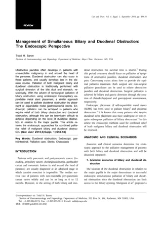
- 1. Gut and Liver, Vol. 4, Suppl. 1, September 2010, pp. S50-56 Review Management of Simultaneous Biliary and Duodenal Obstruction: The Endoscopic Perspective Todd H. Baron Division of Gastroenterology and Hepatology, Department of Medicine, Mayo Clinic, Rochester, MN, USA 1 Obstructive jaundice often develops in patients with denal obstruction the survival time is shorter. During unresectable malignancy in and around the head of this period treatment should focus on palliation of symp- the pancreas. Duodenal obstruction can also occur in toms of obstructive jaundice, duodenal obstruction and these patients, and usually develops late in the dis- pain. Controversy exists about how to provide the opti- ease course. Palliation of both malignant biliary and mal palliative treatment. Both surgical and non-surgical duodenal obstruction is traditionally performed with palliative procedures can be used to relieve obstructive surgical diversion of the bile duct and stomach, re- jaundice and duodenal obstruction. Surgical palliation is spectively. With the advent of nonsurgical palliation of achieved by biliary and gastric diversion through the crea- biliary obstruction using endoscopic transpapillary ex- pandable metal stent placement, a similar approach tion of choledochojejunal and gastrojejunal anastomoses, can be used to palliate duodenal obstruction by place- respectively. ment of expandable metal gastroduodenal stents. En- Endoscopic placement of self-expandable metal stents 2 doscopic palliation can be achieved in patients who (SEMS) has been used to palliate biliary and duodenal 3 require relief of both biliary obstruction and duodenal obstruction. It is known that many patients who require obstruction, although this can be technically difficult to duodenal stent placement also have undergone or will re- achieve depending on the level of duodenal obstruc- 4 quire subsequent palliation of biliary obstruction. In this tion in relation to the major papilla. This article re- article the endoscopic methods used for combined relief views the endoscopic approaches for combined pallia- of both malignant biliary and duodenal obstruction will tive relief of malignant biliary and duodenal obstruc- be reviewed. tion. (Gut Liver 2010;4(Suppl. 1):S50-56) Key Words: Duodenal obstruction; Endoscopy, gas- ANATOMIC AND CLINICAL SCENARIOS trointestinal; Palliative care; Stents; Cholestasis Anatomic and clinical scenarios determine the endo- scopic approach to the palliative management of patients INTRODUCTION with both biliary and duodenal obstruction and will be discussed separately. Patients with pancreatic and peri-pancreatic cancer (in- 1. Anatomic scenarios of biliary and duodenal ob- cluding, ampullary cancer, cholangiocarcinoma, gallbladder struction cancer and metastatic lesions in and around the head of pancreas) are usually diagnosed at an advanced stage in The location of the duodenal obstruction in relation to which curative resection is impossible. The median sur- the major papilla is the major determinant to successful vival rate of patients with non-resectable peri-pancreatic endoscopic simultaneous palliation of biliary and duode- cancer varies widely and can be as long as 6 to 12 nal obstruction since the duodenal obstruction can limit 1 months. However, in the setting of both biliary and duo- access to the biliary opening. Mutignani et al. proposed a Correspondence to: Todd H. Baron Division of Gastroenterology and Hepatology, Department of Medicine, 200 First St. SW, Rochester, MN 55905, USA Tel: +1-507-284-2174, Fax: +1-507-255-7612, E-mail: todd@mayo.edu DOI: 10.5009/gnl.2010.4.S1.S50
- 2. Baron TH: Management of Simultaneous Biliary and Duodenal Obstruction: The Endoscopic Perspective S51 classification system for the three anatomic scenarios of duodenal palliation is least in patients with type III duo- duodenal obstruction in relation to the major papilla that denal stenosis while type I is intermediate and type II is determine the endoscopic approach and technical success the most technically difficult. to combined palliation of biliary and duodenal obstruc- The approach to type I cases is to pass the duodeno- tion. The classification system is as follows (Fig. 1): scope through the duodenal stricture to the major papilla, Type I stenosis occurs at the level of the duodenal bulb if possible. This may require balloon dilation of the stric- or upper duodenal genu but without involvement of the ture to a diameter of 15-18 mm (Fig. 2A) and/or passing papilla. Type II stenosis affects the second part of the du- the balloon to the third duodenum to be used as an an- 5 odenum with involvement of the major papilla. Type III chor to pull the endoscope across the stricture. Once the stenosis involves the third part of the duodenum distal to major papilla is reached the biliary tree is cannulated and and without involvement of the major papilla. an expandable metal biliary stent is placed (Fig. 2B). A Of the three types of biliary-duodenal stenoses, techni- guidewire is then advanced through the channel of the cal difficulty to achieve successful combined biliary and endoscope and passed into the fourth portion of the duo- denum; the duodenoscope is withdrawn into the stomach and a duodenal stent which passes through the working channel of the endoscope is deployed across the duodenal 6 stricture as previously described (Fig. 2C). It is usually necessary to place the proximal end of the stent into the stomach to allow enough stent coverage of the duodenal stricture since type I strictures tend to be in the proximal duodenal bulb. If the duodenoscope cannot be advanced through the stricture despite balloon dilation a duodenal stent is placed across the stricture. Since the luminal di- ameter of commercially available duodenal stents is 20-22 mm and duodenoscopes are approximately 11 mm, the endoscope can usually be passed through the stent during the same procedure to allow the biliary system to be ac- cessed with placement of a biliary SEMS. However, this may require balloon dilation of the duodenal stent. It is important that the duodenal stent be placed with the dis- tal end positioned proximal to the level of the major pap- illa to allow the bile duct to be accessed (Fig. 3). The lo- cation of the major papilla can be estimated easily fluo- roscopically in the presence of a prior biliary stent or by passing a smaller caliber forward-viewing endoscope to the papilla and a obtaining a radiographic image with the endoscope positioned at the level of the papilla for later reference. If the duodenoscope cannot be passed through the duodenal stent lumen because of inadequate stent ex- pansion despite balloon dilation there are three options. The first option is to repeat the endoscopic retrograde cholangiopancreatography (ERCP) after waiting at least 48 to 72 hours at which time the duodenal stent is al- most always fully expanded and will allow passage of the duodenoscope to the second duodenum. Other options to Fig. 1. Classification of duodenal stenosis type in relation to palliate biliary obstruction include percutaneous access to the major papilla as proposed by Mutignani et al. Endoscopy the biliary tree and endoscopic ultrasound (EUS)-guided 1 2007;39:440-447. : (I) at the level of the duodenum proximal entry into the biliary tree (both of these approached will to and without involvement of the papilla, (II) affecting the be discussed in more detail below under management of second part of the duodenum with involvement of the major type II duodenal obstruction). papilla, and (III) involving the third part of the duodenum distal to and without involvement of the major papilla. The approach to type II cases is most difficult. The sec-
- 3. S52 Gut and Liver, Vol. 4, Suppl. 1, September 2010 Fig. 2. Palliation of biliary and duodenal obstruction in type I stenosis. (A) The duodenal stricture is balloon dilated to allow passage of the duodenoscope to the level of the major papilla (note the prior placement of a plastic biliary stent). (B) After the endoscope is passed to the major papilla, the plastic stent is removed and an expandable metal biliary stent is deployed. (C) After deployment of the biliary stent, an expandable metal duodenal stent is placed. Fig. 3. Type I duodenal stenosis with duodenal stent placement to allow access to the bile duct. (A) The duodenal stent is positioned with the distal end proximal to the major papilla (arrow) as visualized using an upper endoscope. (B) In the same patient, the endoscope has been passed through the duodenal stent (arrowheads) lumen and an expandable metal biliary stent (arrows) is placed. ond duodenum is strictured and involves the major major papilla can be identified through the interstices of papilla. Unless the patient has had a prior transpapillary the duodenal stent and the bile duct accessed with place- biliary stent placed, bile duct cannulation is often not ment of a biliary SEMS. If the bile duct cannot be ac- successful since the major papilla is not endoscopically cessed through a transpapillary approach after duodenal identifiable due to extensive tumor infiltration. In addi- stent placement then biliary access can be achieved using 7 tion, the lumen is narrowed and there is little working a percutaneous or EUS approach. Typically, in either of room between the lens of the endoscope and the major the two approaches the bile duct is accessed through the papilla. Nonetheless, one should first attempt placement liver (when EUS is used a transgastric approach is most of an expandable metal biliary stent. If successful, the du- often used). In addition, in either approach there are two odenal stent is then placed across the stricture and over- options: a rendezvous approach or completion alone using lies the biliary stent. If identification and/or cannulation a percutaneous or EUS approach. When the rendezvous of the major papilla cannot be achieved then a duodenal approach is used, a guidewire is passed into the biliary stent is placed across the stricture. Unfortunately, the tree across the stricture and through the interstices of the stent will invariably further impair endoscopic visual- stent into the stent lumen (Fig. 4A, B); if the EUS ap- ization of the major papilla. In some cases, however, the proach is used, the echoendoscope is removed. A duode-
- 4. Baron TH: Management of Simultaneous Biliary and Duodenal Obstruction: The Endoscopic Perspective S53 Fig. 4. Successful simultaneous biliary and duodenal stent placement using the endoscopic ultrasound (EUS) rendezvous technique in type II duodenal stenosis. (A) After placement of a duodenal self-expandable metal stent (SEMS), the bile duct could not be cannulated. A transgastric EUS-guided puncture was performed. (B) A guide wire was passed into the lumen of the duodenal stent. (C) After the echoendoscope was withdrawn, a duodenoscope was advanced into the duodenal stent lumen, the guide wire grasped, and a biliary SEMS deployed. noscope is advanced into the lumen of the duodenal stent into the duodenum. The latter EUS-guided approach is and the guidewire wire is grasped with a snare and with- similar to the percutaneous approach in which the guide- drawn into the channel of the endoscope. A biliary stent wire is passed antegrade into the duodenum across the advanced through the endoscope channel over the guide- duodenal stricture. The stent is then passed through the wire, into the biliary tree and deployed. The distal end of echoendoscope and deployed across the stricture with the 8 the biliary stent resides within the lumen of the duodenal distal end into the duodenum. Finally, in a recent case stent (Fig. 4C). When the biliary stent is placed entirely report an EUS-guided approach was performed where the using the percutaneous approach, the stent is passed an- echoendoscope was passed into the lumen of a previously tegrade and the distal end is positioned into the duodenal placed duodenal stent in a type II stenosis in which the lumen as described above (Fig. 5). When the EUS ap- papilla could not otherwise be endoscopically visualized. proach is undertaken, the distal end of the biliary stent The bile duct opening was identified and a transpapillary 9 can reside within the biliary tree proximal to the biliary biliary SEMS placed. stricture and with the proximal end deployed into the Type III cases are the least common and often result gastric lumen to create a hepaticogastric anastomosis. from pancreatic cancer that arises from the uncinate However, this approach is associated with a higher rate of process. The tumor encases the bile duct causing biliary bile leakage than when the stent crosses the papilla and obstruction and extends inferiorly to causing duodenal
- 5. S54 Gut and Liver, Vol. 4, Suppl. 1, September 2010 Fig. 5. Successful combined biliary and duodenal stent placement using a percutaneous approach in type II duodenal stenosis. A duodenal stent was endoscopically placed and tumor-precluding endoscopic bile-duct cannulation was performed in the duodenum. (A) Percutaneous cholangiography demonstrates a tight distal bile-duct stricture. (B) A guide wire was passed into the lumen of the duodenal stent. (C) After balloon dilation of the stent interstices, a biliary self-expandable metal stent (SEMS) was deployed percutaneously as a one-step procedure. Fig. 6. Successful combined endoscopic biliary and duodenal stent placement in type III duodenal stenosis. (A) The bile duct with a distal stricture is cannulated. (B) After placement of the biliary self-expandable metal stent (SEMS), the duodenal stricture is delineated with contrast agent. (C) The duodenal SEMS is deployed across the duodenal stricture with the proximal end just distal to the biliary SEMS.
- 6. Baron TH: Management of Simultaneous Biliary and Duodenal Obstruction: The Endoscopic Perspective S55 obstruction below the level of the major papilla. These cases are the least technically difficult since the duodeno- scope can be passed to the major papilla and to the level of the stricture. In addition, it is not necessary to pass the endoscope beyond the duodenal stricture and thus balloon dilation of the stricture is not necessary. The se- quence of SEMS placement (biliary stent first then duode- nal stent or vice versa) is usually not critical when the distance between the major papilla and the proximal por- tion of the biliary stricture are not in close proximity (Fig. 6). However, if the proximal level of the duodenal obstruction is very close to the major papilla it is best to place the biliary stent first since the proximal end of the duodenal stent may need to be placed across the level of the major papilla to allow adequate coverage of the duo- denal stricture. Whatever sequence of stent placement is chosen, it is best to avoid placing the duodenal stent Fig. 7. Endoscopic photograph demonstrates successful cannulation of the bile duct in a type I patient in whom a across the biliary opening so that biliary access is pre- gastroduodenal self-expandable metal stent (SEMS) was served both at the time of the initial procedure as well as placed across the papilla 3 days earlier. A biliary SEMS was in the future should biliary stent occlusion occur. placed. 2. Clinical scenarios In addition to the relationship of the duodenal ob- placed across the papilla. If the stent does not cross the struction to the major papilla other factors influence the papilla, the endoscope can be passed through the metal endoscopic approach. These include prior surgical pallia- stent and ERCP performed as usual. If the stent has tion (gastrojejunal bypass) and clinical scenarios. The crossed the papilla in a type I duodenal stenosis, it may most common clinical scenario is the initial development be possible to identify a normal papilla through the inter- of biliary obstruction followed by later onset of duodenal stices of the stent followed by successful cannulation of obstruction. Many patients with duodenal obstruction the bile duct (Fig. 7) or to create a window within the have already undergone a palliative intervention for relief stent at the level of the papilla using rat-toothed forceps 1,10 of biliary obstruction - endoscopic, percutaneous, or sur- or argon plasma coagulation. If these maneuvers fail, gical. If a transpapillary stent was previously placed then then either a percutaneous or EUS-guided approach to the type and timing of prior stent placement needs to be placement of a metal biliary stent is usually required, as determined. For example, if a plastic biliary stent had described above. been previously placed it is likely occluded or will be- Finally, when considering endoscopic management of come occluded and needs to be replaced. In such patients duodenal obstruction in the patient without clinically it is recommended that a metal biliary stent be placed at overt biliary obstruction, prophylactic placement of a bili- the time of duodenal stent placement, especially in type II ary SEMS should be considered, especially if there is any patients (Fig. 2) since the duodenal stent must cross the evidence of biliary ductal dilation by non-invasive imaging level of the major papilla making subsequent biliary stent or in the presence of abnormal liver function tests that placement difficult, if not impossible since it may not be cannot be explained by other processes such as medi- accessible through the interstices of the duodenal stent. cations or presence of liver metastases. Another scenario is simultaneous presentation of gas- 3. Results tric and duodenal obstruction without prior intervention. In this scenario it is recommended that placement of a There are several series describing successful combined 1,11-13 biliary SEMS be performed during the same procedure as biliary and endoscopic drainage. In an early study11 placement of the duodenal SEMS, if possible. 18 patients underwent simultaneous biliary and duodenal An additional clinical scenario is duodenal obstruction self-expandable metal stent. Ten patients had prior plastic followed by later biliary obstruction. This is uncommon biliary stents in place. Combined metal stenting was tech- and could be difficult to treat endoscopically since the nically successful in 17 patients. All the patients had re- papilla is usually inaccessible if the duodenal stent was lief of biliary obstruction and 16 had a relief of gastric
- 7. S56 Gut and Liver, Vol. 4, Suppl. 1, September 2010 outlet obstructive symptoms. No immediate stent-related 2. Moss AC, Morris E, Leyden J, MacMathuna P. Do the ben- complications occurred. Median survival time was 78 efits of metal stents justify the costs? A systematic review and meta-analysis of trials comparing endoscopic stents for days. The authors who devised the duodenal stenosis 1 malignant biliary obstruction. Eur J Gastroenterol Hepatol classification (Fig. 1) achieved technically successful 2007;19:1119-1124. combined biliary and duodenal stent placement in the 3. Jeurnink SM, Steyerberg EW, van Hooft JE, et al. Surgical vast majority of patients in all three types of duodenal gastrojejunostomy or endoscopic stent placement for the stenosis, though the majority of patients (46/64) had bili- palliation of malignant gastric outlet obstruction (SUS- TENT study): a multicenter randomized trial. Gastrointest ary stents placed at a mean interval of 107 days prior to Endosc 2010;71:490-499. duodenal stent placement; prior biliary stent placement 4. Adler DG, Baron TH. Endoscopic palliation of malignant greatly facilitates successful combined stenting in type II gastric outlet obstruction using self-expanding metal stents: duodenal stenoses. However, some of these patients re- experience in 36 patients. Am J Gastroenterol 2002;97: quired rendezvous procedures. Early complications oc- 72-78. 5. Kikuyama M, Itoi T, Sasada Y, Sofuni A, Ota Y, Itokawa F. curred in 6% of patients and late complications occurred Large-balloon technique for one-step endoscopic biliary in 16%. The median survival after combined stenting was stenting in patients with an inaccessible major papilla ow- 81 days. In another series 23 patients who had both bili- ing to difficult duodenal stricture (with video). Gastro- ary and duodenal stenoses, successful combined stenting intest Endosc 2009;70:568-572. 12 was achieved in 91% of cases. More recently, the use of 6. Baron TH. Optimizing endoscopic placement of expandable stents throughout the GI tract. Expert Rev Gastroenterol a dedicated duodenal stent with a central portion de- Hepatol 2008;2:399-409. signed to facilitate passage of a biliary stent through the 7. Shami VM, Kahaleh M. Endoscopic ultrasound-guided 13 interstices was reported in a small number of patients. cholangiopancreatography and rendezvous techniques. Dig Endoscopic placement of duodenal SEMS was achieved in Liver Dis 2010;42:419-424. 8. Nguyen-Tang T, Binmoeller KF, Sanchez-Yague A, Shah all and a self-expandable metal biliary stent through the JN. Endoscopic ultrasound (EUS)-guided transhepatic ante- mesh of the duodenal stent was technically successful in rograde self-expandable metal stent (SEMS) placement 7 (87.5%) of 8 patients. However, 2/3 patients with type across malignant biliary obstruction. Endoscopy 2010;42: II duodenal strictures failed bile duct cannulation and re- 232-236. quired a rendezvous procedure. Early complications oc- 9. Belletrutti PJ, Gerdes H, Schattner MA. Successful endo- scopic ultrasound-guided transduodenal biliary drainage curred in 1 patient. Median survival after combined stent- through a pre-existing duodenal stent. JOP 2010;11:234- ing was 91 days (range, 36-314 days). 236. In summary, the results of these series suggest that in 10. Topazian M, Baron TH. Endoscopic fenestration of duode- experienced centers combined biliary and duodenal stent nal stents using argon plasma to facilitate ERCP. Gas- placement for palliation can be achieved in the majority trointest Endosc 2009;69:166-169. 11. Kaw M, Singh S, Gagneja H. Clinical outcome of simulta- of patients though a rendezvous approach is required neous self-expandable metal stents for palliation of malig- more often in the type II patients. Overall survival from nant biliary and duodenal obstruction. Surg Endosc 2003; the time of combined biliary and duodenal stent place- 17:457-461. ment is relatively short. 12. Maire F, Hammel P, Ponsot P, et al. Long-term outcome of biliary and duodenal stents in palliative treatment of pa- tients with unresectable adenocarcinoma of the head of REFERENCES pancreas. Am J Gastroenterol 2006;101:735-742. 13. Moon JH, Choi HJ, Ko BM, et al. Combined endoscopic 1. Mutignani M, Tringali A, Shah SG, et al. Combined endo- stent-in-stent placement for malignant biliary and duodenal scopic stent insertion in malignant biliary and duodenal obstruction by using a new duodenal metal stent (with obstruction. Endoscopy 2007;39:440-447. videos). Gastrointest Endosc 2009;70:772-777.