Avascular necrosis
•Transferir como PPTX, PDF•
15 gostaram•830 visualizações

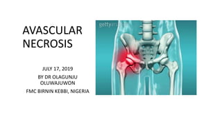
Avascular necrosis of femoral head

Recomendados
Mais conteúdo relacionado
Mais procurados
Mais procurados (20)
Semelhante a Avascular necrosis
Semelhante a Avascular necrosis (20)
Último
Último (20)
Avascular necrosis
- 1. AVASCULAR NECROSIS JULY 17, 2019 BY DR OLAGUNJU OLUWAJUWON FMC BIRNIN KEBBI, NIGERIA
- 2. OUTLINE • INTRODUCTION • EPIDEMIOLOGY • ANATOMY • ETIOLOGY • PATHOPHYSIOLOGY • CLINICAL PRESENTATION
- 3. • STAGING • DIFFERENTIAL DIAGNOSIS • TREATMENT • COMPLICATIONS • PROGNOSIS • REFERENCES
- 4. INTRODUCTION Avascular necrosis (AVN) is defined as cellular death of bone components due to interruption of the blood supply; the bone structures then collapse, resulting in pain, loss of joint function and long-term joint damage.
- 5. • AVN usually involves the epiphysis (end part of a long bone), such as the femoral and humeral heads and the femoral condyles, but small bones can also be affected.
- 9. • In clinical practice, AVN is most commonly encountered in the hip. • Most available data regarding the natural history, pathology, pathogenesis, and treatment of AVN pertains to femoral head necrosis.
- 11. • Avascular necrosis is also called osteonecrosis, aseptic necrosis, and ischemic bone necrosis.
- 12. EPIDEMIOLOGY • The frequency of AVN depends on the site involved. • The most common site is the hip; • other locations include the • carpals, talus, femur, metatarsal, mandible, and humerus.
- 13. • In the United States, approximately 15,000 new cases of AVN are reported each year. • AVN accounts for more than 10% of total hip replacement surgeries performed in the United States.
- 14. • Osteonecrosis of the jaw associated with bisphosphonate has also been well studied and reported. • Most patients with osteonecrosis of the jaw also had an ongoing malignancy and/or had undergone a recent dental procedure.
- 15. • In most countries, the incidence and prevalence of AVN are not well reported.
- 16. • AVN has no racial predilection except for cases associated with sickle cell disease and hemoglobin S and SC disease, which predominantly occur in people of African and Mediterranean descent.
- 17. • With the exception of AVN associated with systemic lupus erythematosus, AVN is more common in men, with an overall male- to-female ratio of 8:1. • AVN is a disease of middle age that most often occurs during the fourth or fifth decade of life and is bilateral in more than half of cases.
- 18. • When it occurs in children at the femoral head, it is known as Legg- Calve-Perthes syndrome. • The use of high-dose corticosteroids carries a reported 3-20% incidence of AVN.
- 19. ANATOMY
- 25. ETIOLOGY • AVN may be primary or idiopathic. • For AVN that is secondary or associated with an underlying condition or exposure, the following factors have been identified: • Systemic corticosteroid use or Cushing disease [12] • Trauma • Alcohol abuse
- 26. • Systemic lupus erythematosus (with or without antiphospholipid syndrome), as well as other connective-tissue diseases • Hemoglobinopathies or hemophilic disorders (eg, sickle cell disease, hemophilia A or B) • Osteoporosis medications (ie, bisphosphonates, denosumab)
- 27. • Hyperlipidemia • Bone disorders (slipped capital femoral epiphysis, congenital dysplasia of the hip, Legg-Calve-Perthes disease) • HIV infection • Renal Transplantation
- 28. • Allogeneic bone marrow transplantation • Radiation therapy • Gaucher disease • Malignancy (marrow infiltration, malignant fibrous histiocytoma)
- 29. • Caisson disease (uncommon diving-related decompression illness) • Pregnancy • Psoriasis • Inflammatory bowel disease
- 30. • Prolonged, repeated exposure to high pressure (as experienced by commercial and military divers) has been linked to AVN, though the relationship is not too well understood.
- 31. PATHOPHYSIOLOGY • Although the pathophysiology of AVN is not fully understood, the final common pathway is interruption of blood flow to the bone. • AVN often affects bones with a single terminal blood supply, such as the femoral head, carpals, talus, and humerus.
- 32. • The earliest pathologic characteristics of osteonecrosis are necrosis of hematopoietic cells and adipocytes followed by interstitial marrow edema. • Osteocyte necrosis occurs after approximately 3 hours of anoxia, but histological signs of osteocyte death do not appear until approximately 24 to 72 hours after oxygen deprivation.
- 33. • Interruption of the vascular supply and resultant necrosis of marrow, medullary bone, and cortex are theorized to be caused by the mechanisms listed below. • However, individual patients usually have more than one risk factor; this indicates that the pathogenesis of AVN is likely multifactorial.
- 34. • Vascular occlusion: • This is characterized by the interruption of the extraosseous blood supply via factors such as direct trauma (eg, fracture, dislocation), nontraumatic stress, and stress fracture.
- 35. • Altered lipid metabolism: • Animal studies have led to the hypothesis that increased levels of serum lipids leads to lipid deposition in the femoral head, causing femoral hypertension and ischemia. • Lipid-level–lowering drugs in animals reverse this process. Corticosteroid administration was associated with fat emboli in the femoral heads of rabbits.
- 36. • Intravascular coagulation: • Disorders of the coagulation system have been implicated in the pathogenesis of AVN. • Typically, it is a secondary event triggered by a familial thrombophilia, hypercholesterolemia, allograft organ rejection, other disorders (e.g, infection, malignancy), or pregnancy.
- 37. • Healing process: • Necrotic bone triggers a process of repair that includes osteoclasts, osteoblasts, histiocytes, and vascular elements. • Osteoblasts build new bone on top of the dead bone, leading to a thick scar that prevents revascularization of the necrotic bone, with resultant abnormal joint remodelling and joint dysfunction.
- 38. • Primary cell death: • Osteocyte death without other features of AVN has been seen in renal transplant patients, as well as in patients receiving steroids and those who consume significant amounts of alcohol.
- 40. CLINICAL PRESENTATION • Avascular necrosis (AVN) may be asymptomatic and is occasionally discovered incidentally on radiographs. • Symptoms depend on the affected joint. • Medullary infarcts are usually silent, and infarcts of the small bones of the hands and feet are often symptomatic.
- 41. • Pain in the affected joint is typically the presenting symptom of AVN, regardless of the location. • Patients with AVN of the femoral head often report groin or anterior thigh pain that is exacerbated by weight bearing.
- 42. • The pain may initially be mild but progressively worsens over time and subsequently may be present at rest or at night. • Large infarcts, such as those due to Gaucher disease and hemoglobinopathies, are associated with very severe pain.
- 43. • Initially, the physical examination findings of AVN may be unrevealing. • Abnormal physical findings depend on the location and severity of disease. • With progression of AVN of the hip, joint function deteriorates and the patient may walk with a limp.
- 44. • AVN of smaller, non–weight-bearing joints typically does not cause significant disability. • Findings may include the following: • Patients with AVN may have tenderness around the affected bone. • Both active and passive joint movements may be restricted and painful.
- 45. • A neurologic deficit may be present if a nerve is affected (compressed) because of necrosis and compression deformity of affected bones. • Advanced AVN can result in joint deformity and muscle wasting.
- 46. STAGING • The French orthopedic surgeon Paul (RP) Ficat (1917-1986) in association with Professor Jacques Arlet devised a system of staging idiopathic avascular necrosis of femoral head in the late 1970s based on two fundamental concepts :
- 47. • a standard radiograph shows only the shadow of the mineralized portion of a bone • bone necrosis is the end result of severe and prolonged ischemia
- 53. • stage 0 • plain radiograph: normal • MRI: normal • clinical symptoms: nil
- 54. • stage I • plain radiograph: normal or minor osteopenia • MRI: edema • bone scan: increased uptake • clinical symptoms: pain typically in the groin
- 55. • stage II • plain radiograph: mixed osteopenia and/or sclerosis and/or subchondral cysts, without any subchondral lucency (crescent sign: see below) • MRI: geographic defect • bone scan: increased uptake • clinical symptoms: pain and stiffness
- 56. • stage III • plain radiograph: crescent sign and eventual cortical collapse • MRI: same as plain radiograph • clinical symptoms: pain and stiffness +/- radiation to knee and limp
- 57. • stage IV • plain radiograph: end-stage with evidence of secondary degenerative change • MRI: same as plain radiograph • clinical symptoms: pain and limp
- 58. DDX • Inflammatory synovitis • Complex regional pain syndrome • Osteoarthritis • Soft tissue trauma (e.g., Labral tear) • Osteomyelitis • Neoplastic bone conditions • Osteoporosis
- 59. DIAGNOSTICS • No laboratory test findings specifically suggest or confirm the presence of avascular necrosis (AVN). • Plain radiographic findings are unremarkable in early stages of AVN.
- 60. • Nevertheless, the American College of Radiology (ACR) considers x- ray of the pelvis and hips the most appropriate initial imaging study in patients at risk for AVN who present with hip pain.
- 62. • If radiographs are normal or show femoral head lucencies suspicious for osteonecrosis, magnetic resonance imaging (MRI) of the hips without contrast is most appropriate.
- 66. • The ACR advises that MRI is the most sensitive and specific imaging modality for diagnosis and provides optimal evaluation of the likelihood of articular collapse.
- 67. • Additional ACR recommendations include the following : • Contrast-enhanced MRI may be needed to detect early osteonecrosis of the hip in pediatric patients, which is indicated by hypoperfusion • In patients with a contraindication for MRI, alternative imaging modalities are computed tomography (CT) or bone scintigraphy with single-photon emission CT (SPECT).
- 68. • Histology is the criterion standard for diagnosis of AVN. • However, bone biopsy is not routinely performed because of the availability of sensitive noninvasive tests such as MRI.
- 69. TREATMENT • MEDICAL • Medical management of avascular necrosis (AVN) primarily depends on the location and severity of disease, as well as the patient's age and general health. • Treatment outcomes correlate directly with the stage of the disease. • No medical treatment has proven effective in preventing or arresting the disease process.
- 70. • Conservative measures include • limited weight bearing with crutches and pain medications. • This may be beneficial and is a reasonable initial course of action if the involved segment is smaller than 15% and far from the weight-bearing region. Immobilization may be helpful in some cases (eg, AVN of the distal femur or tibia). In advanced AVN, the disease course is unaffected by activity and will eventually require surgery.
- 71. • Treatment with bisphosphonates may be helpful. • Iloprost, a vasoactive prostaglandin analog that is approved for inhalational treatment of pulmonary hypertension, has shown clinical and radiographic benefits in early-stage AVN when administered intravenously.
- 72. • Statin therapy to prevent corticosteroid-induced AVN may be helpful. • Extracorporeal shockwave therapy (ESWT) has shown beneficial effects in early AVN of the femoral head. • ESWT may relieve pain and improve hip function of the hip, and induce regression of AVN. • ESWT may be combined with alendronate therapy.
- 73. • SURGICAL MANAGEMENT • Several surgical procedures have been used in an attempt to treat AVN, with variable success. • No surgical procedure is the consensual best among surgeons in the treatment of AVN.
- 74. • In early stages of AVN (precollapse), core decompression with or without bone graft is typically considered the most appropriate treatment. • In late stages, characterized by collapse, femoral head deformity, and secondary osteoarthritis, total hip arthroplasty is the most appropriate treatment.
- 75. Core decompression • Researchers postulate that core decompression improves circulation by decreasing intramedullary pressure and preventing further ischemia and progressive joint destruction. • The best results are obtained when treating patients with early AVN (precollapse). • Core decompression is also effective for pain control.
- 77. Bone graft • Bone graft options include structural cortical or medullary bone graft and vascularized bone graft with either a muscle-pedicle bone graft or free vascularized fibular graft.
- 78. • Bone grafting is combined with the following: • Core decompression, which may interrupt the cycle of ischemia • Excision of sequestrum, which may inhibit revascularization of the femoral head • Period of limited weight bearing
- 81. OSTEOTOMY • Several osteotomy procedures have been tried with variable success. • Intertrochanteric osteotomies have been performed in patients with posttraumatic AVN.
- 83. Total hip arthroplasty • Most patients with advanced disease (stage III and above) require total hip arthroplasty. • Total hip arthroplasty provides excellent pain relief for many years, although most young patients require repeat surgery. • high failure rates (10-50% after 5 years), patients with AVN will probably need a second total hip arthroplasty during their lifetime.
- 85. Cell therapy • The use of stem cells implanted via core decompression has been studied for the treatment of early-stage (precollapse) AVN of the femoral head.
- 86. • Systematic reviews of studies with patient-reported outcomes have demonstrated clinical benefit with the use of stem cells for hip AVN, with a low rate of complications, but have highlighted the lack of standardization with this technique.
- 87. COMPLICATIONS • The natural history of AVN involves subchondral necrosis, subchondral fracture and collapse of bone, deformity of the articular surface, and osteoarthritis. • In later stages, sclerosis and total destruction of the joint may occur. • Nonunion of fracture and secondary muscle wasting are potential complications.
- 88. PROGNOSIS • The prognosis of AVN depends on the disease stage at the time of diagnosis and the presence of any underlying conditions. • More than 50% of patients with AVN require surgical treatment within 3 years of diagnosis. • Half of patients with subchondral collapse of the femoral head develop AVN in the contralateral hip.
- 89. • Poor prognostic factors include the following: • Age older than 50 years • Advanced disease (stage 3 or worse) at the time of diagnosis • Necrosis of more than one third of the weight-bearing area of the femoral head on MRI
- 90. • Lateral involvement of femoral head (compared with medial lesions) • Non-modifiable risk factors such as cumulative dose of corticosteroids (corticosteroid-induced AVN)
- 91. CONCLUSION • Clinical and MRI characteristics need to be evaluated for the critical diagnosis of ONFH. • The progression of ONFH has not been well established, therefore it is difficult to evaluate whether a specific treatment modality changes the natural course of the disease.
- 92. • Medical treatment outcomes correlate directly with the stage of the disease. • No medical treatment has proven effective in preventing or arresting the disease process.
- 93. • Most patients with advanced disease (stage III and above) require total hip arthroplasty.
- 94. REFERENCES • Bose VC, Baruah BD. Resurfacing arthroplasty of the hip for avascular necrosis of the femoral head: a minimum follow-up of four years. J Bone Joint Surg Br. 2010 Jul. 92(7):922-8. [Medline]. • Steffen RT, Athanasou NA, Gill HS, Murray DW. Avascular necrosis associated with fracture of the femoral neck after hip resurfacing: histological assessment of femoral bone from retrieval specimens. J Bone Joint Surg Br. 2010 Jun. 92(6):787-93. [Medline]. • Woo SB, Hellstein JW, Kalmar JR. Narrative [corrected] review: bisphosphonates and osteonecrosis of the jaws. Ann Intern Med. 2006 May 16. 144(10):753-61. [Medline]. • Qi WX, Tang LN, He AN, Yao Y, Shen Z. Risk of osteonecrosis of the jaw in cancer patients receiving denosumab: a meta-analysis of seven randomized controlled trials. Int J Clin Oncol. 2014 Apr. 19(2):403- 10. [Medline]. • Shah KN, Racine J, Jones LC, Aaron RK. Pathophysiology and Risk Factors for Osteonecrosis. Current Reviews in Musculoskeletal Medicine. Sept 2015. 8(3): 201–209:[Medline]. [Full Text]. • Kawai K, Tamaki A, Hirohata K. Steroid-induced accumulation of lipid in the osteocytes of the rabbit femoral head. A histochemical and electron microscopic study. J Bone Joint Surg Am. 1985 Jun. 67(5):755- 63. [Medline]. • Wang GJ, Sweet DE, Reger SI, et al. Fat-cell changes as a mechanism of avascular necrosis of the femoral head in cortisone-treated rabbits. J Bone Joint Surg Am. 1977 Sep. 59(6):729-35. [Medline].
- 95. • Bagan JV, Murillo J, Jimenez Y, et al. Avascular jaw osteonecrosis in association with cancer chemotherapy: series of 10 cases. J Oral Pathol Med. 2005 Feb. 34(2):120-3. [Medline]. • Khan AA, Sándor GK, Dore E, Morrison AD, Alsahli M, Amin F, et al. Bisphosphonate associated osteonecrosis of the jaw. J Rheumatol. 2009 Mar. 36(3):478-90. [Medline]. • Ninomiya S. An epidemiological survey of idiopathic avascular necrosis of the femoral head in Japan. Annual Report of Japanese Investigation Committee for Intractable Disease. 1989. • Chiu HY, Wang IT, Huang WF, Tsai YW, Shiu MN, Tsai TF. Increased risk of avascular necrosis in patients with psoriatic disease: A nationwide population-based matched cohort study. J Am Acad Dermatol. 2017 May. 76 (5):903-910.e1. [Medline]. • Aaron RK, Voisinet A, Racine J, Ali Y, Feller ER. Corticosteroid-associated avascular necrosis: dose relationships and early diagnosis. Ann N Y Acad Sci. 2011 Dec. 1240(1):38-46. [Medline]. • Lawson-Ayayi S, Bonnet F, Bernardin E, et al. Avascular necrosis in HIV-infected patients: a case-control study from the Aquitaine Cohort, 1997-2002, France. Clin Infect Dis. 2005 Apr 15. 40(8):1188-93. [Medline]. • Mehta P, Nelson M, Brand A, Boag F. Avascular necrosis in HIV. Rheumatol Int. 2013 Jan. 33(1):235- 8. [Medline]. • Enright H, Haake R, Weisdorf D. Avascular necrosis of bone: a common serious complication of allogeneic bone marrow transplantation. Am J Med. 1990 Dec. 89(6):733-8. [Medline].
- 96. • Fink JC, Leisenring WM, Sullivan KM, et al. Avascular necrosis following bone marrow transplantation: a case-control study. Bone. 1998 Jan. 22(1):67-71. [Medline] • Rolston VS, Patel AV, Learch TJ, Li D, Karayev D, Williams C, et al. Prevalence and Associations of Avascular Necrosis of the Hip in a Large Well-characterized Cohort of Patients With Inflammatory Bowel Disease. J Clin Rheumatol. 2018 May 24. [Medline]. • [Guideline] Murphey MD, Roberts CC, Bencardino JT, Appel M, Arnold E, Chang EY, et al. ACR Appropriateness Criteria Osteonecrosis of the Hip. J Am Coll Radiol. 2016 Feb. 13 (2):147-55. [Medline]. • Staging of Avascular Necrosis. www.OrthopaedicsOne.com. Available at http://www.orthopaedicsone.com/display/Main/Staging+of+Avascular+Necrosis. Feb 2009; Accessed: October 2, 2018. • Luo RB, Lin T, Zhong HM, Yan SG, Wang JA. Evidence for using alendronate to treat adult avascular necrosis of the femoral head: a systematic review. Med Sci Monit. 2014 Nov 26. 20:2439-47. [Medline]. • Pilge H, Bittersohl B, Schneppendahl J, Hesper T, Zilkens C, Ruppert M, et al. Bone Marrow Aspirate Concentrate in Combination With Intravenous Iloprost Increases Bone Healing in Patients With Avascular Necrosis of the Femoral Head: A Matched Pair Analysis. Orthop Rev (Pavia). 2016 Nov 17. 8 (4):6902. [Medline]. [Full Text]. • Claßen T, Becker A, Landgraeber S, Haversath M, Li X, Zilkens C, et al. Long-term Clinical Results after Iloprost Treatment for Bone Marrow Edema and Avascular Necrosis. Orthop Rev (Pavia). 2016 Mar 21. 8 (1):6150. [Medline]. [Full Text].
- 97. • Pritchett JW. Statin therapy decreases the risk of osteonecrosis in patients receiving steroids. Clin Orthop. 2001 May. (386):173-8. [Medline]. • Wang CJ, Cheng JH, Huang CC, Yip HK, Russo S. Extracorporeal shockwave therapy for avascular necrosis of femoral head. Int J Surg. 2015 Dec. 24 (Pt B):184- 7. [Medline]. • Gao F, Sun W, Li Z, Guo W, Wang W, Cheng L, et al. High-Energy Extracorporeal Shock Wave for Early Stage Osteonecrosis of the Femoral Head: A Single-Center Case Series. Evid Based Complement Alternat Med. 2015. 2015:468090. [Medline]. • Van Laere C, Mulier M, Simon JP, et al. Core decompression for avascular necrosis of the femoral head. Acta Orthop Belg. 1998 Sep. 64(3):269-72. [Medline]. • Aldridge JM 3rd, Urbaniak JR. Avascular necrosis of the femoral head: role of vascularized bone grafts. Orthop Clin North Am. 2007 Jan. 38(1):13-22, v. [Medline].
- 98. THANK YOU FOR LISTENING