Gastric exammination
•Transferir como PPT, PDF•
3 gostaram•1,041 visualizações

Radiology of the GIT

Recomendados
Recomendados
Mais conteúdo relacionado
Mais procurados
Mais procurados (20)
Semelhante a Gastric exammination
Semelhante a Gastric exammination (20)
Último
Último (20)
Gastric exammination
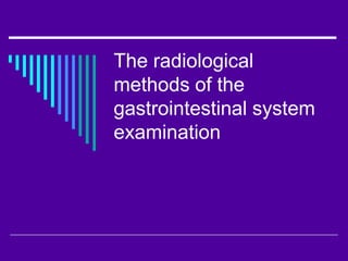
- 1. The radiological methods of the gastrointestinal system examination
- 2. The main methods of examination - Roentgenoscopy (fluoroscopy) - Roentgenography - Fluorography
- 3. The additional methods - Roentgen polygraphy - Roentgen kymography - Tomography - Roentgenoscopy with primary magnification
- 4. The special contrast methods - Double contrasting - Pneumoparietography of the stomach
- 5. The roentgen anatomy and examination of esophagus - Width – from 1,5-2 см to 2 см; - Length – 25-26 см; - The parts of esophagus: А) cervical; Б) thoracic; В) abdominal.
- 6. - Three physiologic narrowings: А) at the level of cricoid cartilage; Б) at the level of aortic arc; В) at the level of cardia. - The folds of mucosa – 2-3 см, parallel, longitudinal; - The velocity of contrast movement is 4-6 sec.
- 7. folds
- 8. peristalsis narrowing (arc) cardia
- 9. The roentgen anatomy and examination of the stomach 1. The survey roentgenoscopy. 2. The first stage of examination – the patient drinks 1-2 swallows of contrast (we can evaluate the folds of mucosa of the stomach). 3. The second stage of examination – tight filling. We can determine the shape, size and position of stomach.
- 10. 4. The stomach peristalsis: - Superficial; - Medium depth; - Deep; - Segmental; - The velocity of peristaltic wave is 21 sec.
- 11. trendelenburg
- 13. parietography
- 14. 5. Parts of the stomach: - Fundus; - Cardiac part; - Body; - Sinus (or angle); - Antrum; - Pyloric part; - Minor and major curvature.
- 16. The roentgen anatomy and examination of duodenum and small intestine 1. The duodenum comes after stomach and it has three parts: - Superior (bulb of triangular shape); - Descending (covers the head of pancreas); - Inferior.
- 17. 2. Small intestine: - duodenum; - jejunum; - ileum; - Kerckring's folds of the mucosal layer.
- 18. N
- 20. The roentgen anatomy and examination of large intestine 1. The methods of examination: - Per os; - Irrigoscopy – contrast enema.
- 21. 2. Roentgen anatomy and parts of the large intestine: - Caecum; - C. ascendens; - C. transversum; - C. descendens; - C. sygmoideum; - Rectum.
- 22. 3. Irrigoscopy: - The first stage of examination – tight filling of the bowel. А) shape; Б) size; В) localization. - The second stage of examination– the evaluation of the mucosa folds (after depletion of patient); - The third stage of examination– double contrasting (inflation of large intestine with barium sulfate by Bobrov device). The elasticity of the walls is determined.
- 23. N
- 27. The pathology of the gastrointestinal tract 1. Esophagus: - Esophageal diverticuli: А) pulsational; Б) tractional; В) functional. - Achalasia of esophagus;
- 32. Balon dilatation Achalasia before after
- 33. - Cancer of esophagus: А) scirrhous; Б) bowl-shaped (saucer-shaped); В) medullar shape.
- 34. The symptoms of cancer: - Stenosis of esophagus; - Filling defect; - Irregular borders; - The delay of contrast media above the level of stenosis; - Deformation and absence of the folds; - The absence of peristalsis at the level of defect.
- 35. cr cr
- 36. - Burn of esophagus: The first examination is possible after 2-3 weeks. Symptoms: • Circular stenosis; • Flat contours; • Deformation – cone-, funnel-, ampullar. - Foreign substances Method by Ivanova-Podobed.
- 37. burn
- 39. 2. Stomach: - Gastritis: А) acute; Б) chronic; В) chronic hyperthrophic gastritis; Г) rigid antral gastritis.
- 40. - The ulcer of stomach and duodenum: Symptoms: • “niche”; • inflammative elevation of mucosa; • folds convergention; • the symptom of “pointing finger”. Complications: • hemorrhage; • perforation; • penetration; • malignisation (transformation into cancer).
- 41. Acute ulcer
- 42. Chronic ulcer
- 43. ulcus
- 45. ulcus
- 46. ulcus
- 47. - Cancer of stomach: • polypous; • bowl-shape; • ulcerative cancer; • diffuse; • cancerous ulcer; • cancer from polyp.
- 48. Symptoms: • filling defect; • the absence of the folds of mucosa; • the absence of peristalsis at the defect localization; • stenosis of the lumen.
- 49. cr
- 50. cr
- 51. polyposis
- 52. Malignant polyps of stomach
- 53. 3. Large intestine: - Inflammations: А) colitis; Б) chronic colitis; В) chronic spastic colitis; Г) unspecific ulcerative colitis. Symptoms: • spasm of intestine; • smoothness of haustrum; • smoothness of the folds of mucosa.
- 54. colitis artefact
- 55. n
- 56. - Cancer of the large intestine Symptoms: • filling defect; • irregular contours; • circular stenosis; • the absence of the folds of mucosa; • evacuation disorders.
- 57. Cr sygm
- 58. The roentgenologic picture of the acute abdomen 1. Bowel obstruction. - high; - low. Symptoms: - Kloyber cups(at the background of swallen bowel there is the presence of horisontal level of fluid).
- 59. B. obstruction
- 60. 2. Perforate ulcer. - Symptom of sickle (the presence of air under the right cupola of the diaphragm).