
Recomendados
Mais conteúdo relacionado
Mais procurados
Mais procurados (20)
Semelhante a Ridge split in implantology
Semelhante a Ridge split in implantology (20)
Mais de Nishu Priya
Mais de Nishu Priya (19)
Último
Último (20)
Ridge split in implantology
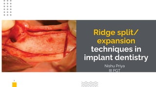
- 1. 1 Ridge split/ expansion techniques in implant dentistry Nishu Priya III PGT
- 2. Introduction ● The fast pace of soaring popularity of implants in dentistry by clinicians and patients alike is substantiated by the existence of a booming share of implants in the global dental market. ● The local conditions may not always be favorable for implant placement as in cases of deficient bone volume after extraction. ● Sufficient horizontal as well as vertical bone dimensions are a prerequisite to warranty the success of implants. ● Hence, often there is the need to augment the available atrophic bone to make it amenable to implant placement in suitable prosthetic positions with desirable stability and aesthetics. ● To feed this need for augmentation, a plethora of agents and techniques are available with volumes of literature; yet there is an absence of consensus for the efficacy of any of these surgical techniques.
- 3. 3
- 4. 4
- 5. Rationale ● Split ridge and expansion techniques are effective for the correction of moderately resorbed edentulous ridges in selected cases. ● Transverse expansion is based on osseous plasticity obtained by corticotomy. It progressively allows for an adequate transversal intercortical diameter large enough to insert one or several dental implants. ● The gap created by sagittal osteotomy expansion undergoes spontaneous ossification, following a mechanism similar to that occurring in fractures. 5
- 6. Products and devices ● Various ridge splitting/expanding chisels (from 3 mm to 6 mm) are designed to define the appropriate shape and size of the osteotomy. 6
- 7. 7
- 8. 8 Surgical procedure Full‐thickness crestal incision with vertical releasing incisions one tooth further than the site being treated. Elevation of a mucoperiosteal flap palatally and buccally to expose the bone ridge. Lamellar cortical splitting initiated by one horizontal crestal osteotomy using a diamond disk, a burr or, preferably, a piezosurgery tip. The longitudinal split can be limited by placing transverse cuts in the bone. Two additional vertical cuts, 2 mm distal to the site of implantation and 1 mm mesial to the adjacent teeth, can be performed to facilitate the expansion. Longitudinal splitting of the alveolar ridge performed using a set of chisels of increasing width. They are optimally used by pressing the instrument manually into the bone or by gently tapping the chisels with a hammer. Normally the chisels are extended to a depth of 5–7 mm, but the penetration depends on the width of the ridge. Facial‐lingual movement should be avoided so as not to further deform the buccal plate.
- 9. 9
- 10. 1 0 The gap created by splitting can either be left empty or filled with different materials, such as collagen sponge, autogenous bone chips or bone substitute. The split ridge can be covered with membranes. •• Dental implant placement at the same time or later, according to a standard procedure. There is a lack of evidence for delayed implant placement when using the split ridge technique. When placed immediately, primary implant stability is achieved by placing the implants at the most apical non‐fractured portion of the jawbone. To improve primary stability of the implants and to prevent fracture of the buccal plate, the use of osteotomes to prepare the implant bed is recommended. •• Wound closure with vertical mattress sutures. •• Dental rehabilitation may be started three to six months afterwards.
- 11. 11
- 12. 12
- 13. 1 3 Indications and advantages • The split ridge/ridge expansion technique is indicated in selected situations where atrophy of the edentulous ridge has developed horizontally and cancellous bone is present between the oral and facial cortical plates, and adequate residual height exists. This technique is mainly indicated in the maxilla. • The procedure results in a significant increase (range 87.5– 100%) in the width of the alveolar ridge (Donos et al., 2008) and allows a gain of 4–5.5 mm of thickness. Survival rates of implants placed at sites augmented using these techniques are similar to those of implants inserted in native bone; that is ranging from 98% to 100% at eight months post‐loading (Chen et al. 2009).
- 14. 1 4 Contraindication and limitations Unfavourable bone angularity Excessive facial inclination of the alveolar ridge may contraindicate this procedure, as it may worsen the initial situation from a prosthetic and aesthetic point of view. When excessive buccal inclination of the implants creates problems, guided bone regeneration (GBR) or bone grafting techniques seem more suitable. Severe horizontal atrophy The technique can only be applied when the buccal and palate/ lingual plates are separated by spongy bone. Therefore, the indications are more limited compared to onlay bone grafts and GBR, which can also be applied in cases presenting severe horizontal atrophy. Ridge expansion in the mandible Although possible, ridge expansion in the mandible is frequently difficult due to the rigidity of the bone. The risk of fracture of the osteotomised fragment is higher than at the maxilla.
- 15. 1 5 Complications • Basal greenstick fracture of the segments during widening has not been controllable to date. Thus, fracture of the buccal plate is the most common complication. Care must be taken in the presence of undercuts that may increase the risk of bone fracture. A minimum width of 2–3 mm of the coronal alveolar crest is necessary to avoid bone fracture. • Loosening or fracture of microscrews may happen. • A labyrinthine concussion may occur during tapping of an osteotome. • The patient may experience a benign positional vertigo.
- 16. 1 6 1 Three consecutive patients (2 males, with a mean age of 49 years, and 1 female, aged 51 years) with thin, narrow alveolar ridges were selected for split-crest ridge augmentation followed immediately by implant placement. Criteria for patient selection included the following: • Good general state of health • Nonsmoker or light smoker status • Class IV alveolar atrophy according to Cawood and Howell • Correct interarch relationships • Patient consent to treatment
- 17. ● A mid crestal full-thickness incision was made, and a partial-thickness flap was dissected and elevated from the palate and reflected to the buccal portion of the alveolar crest. ● Periosteum preservation was intended to reduce bone resorption and prevent free fracture of the split ridge. ● Implant site osteotomies were performed according to the manufacturer’s surgical guide with the use of spiral drills of increasing diameter. ● Finger pressure on the buccal and palatal bone plates during drilling procedures helped to stabilize the buccal bone plate.
- 18. 18 After the implant osteotomies had been completed, the outline of a sagittal osteotomy was scored in the bone with a blade (No. 64 Beaver blade, BD Beaver, Waltham, Mass). The blade then was used as a chisel and was tapped with a surgical mallet in small increments until a 1- to 3-mm-deep furrow was created along the length of the ridge. This same procedure was performed vertically within 2 mm of the adjoining teeth.
- 19. 19 Once the crestal furrow and adjacent vertical bone releases were defined, a bone chisel was progressively driven more deeply into the furrow, and implant osteotomy sites were prepared further to their full dimensions via an osteotome technique (bone condenser) (DENTSPLY Friadent, Mannheim, Germany). Tapered, multithreaded implants were gently placed.
- 20. 20 Mariotti,an orthodontic ligature wire was used to stabilize the bone plates. The present procedure altered the original technique by replacing the ligature wire with absorbable ligatures.
- 21. 21 The furrow between the bone plates was grafted (Biogen equine spongy granular 0.5 gr, Bioteck, Vicenza, Italy) if the gap was more than 2 mm deep. Before suturing, a collagen membrane was layered over the cover screw. The flap was reapproximated buccally and palatally, and primary closure was achieved with 4–0 sutures.
- 22. 22 All patients received 2 gr of amoxicillin and clavunate per day starting approximately 1 hour before surgery and continuing for 6 days after surgery, and a nonsteroidal analgesic was given postoperatively. Postoperative instructions included a soft diet for 2 weeks and appropriate oral hygiene with 0.2% chlorhexidine mouth rinses. Sutures were removed 10 days postoperatively. Six months after implant placement, abutments were connected and the prosthetic rehabilitation was initiated.
- 23. Discussion 23 The split-crest procedure may be indicated for sharp mandibular and maxillary ridges in patients whose bone quantity is inadequate for primary implant stabilization or in those for whom immediate placement of implants is desired. Bone plate stability is fundamental for implant stability and for avoidance of bone sequestration. Rather than a metallic ligature, the present technique used an absorbable suture to prevent its penetration through the soft tissue during healing. Complete suture absorption generally occurs at between 60 and 90 days, when the tissue is normally healed. The split-crest procedure that included GBR with a resorbable collagen membrane was a viable therapeutic alternative for implant placement into areas that otherwise would not be suitable for implants.
- 24. 24 2 A case study series was designed to analyze the surgical/prosthetic treatment of patients undergoing the ARST. The study was developed in the Oral and Maxillofacial Surgery Unit and the Oral Implantology Unit at the Universidad de La Frontera (Temuco, Chile) in conjunction with the Facial Surgery Center. For this study a descriptive analysis of the installed implants was designed, considering intraoperative complications both in the expansion and installation of the implants, stability of the implant and bone loss.
- 25. ● All the procedures were performed under local anesthesia and without sedation. ● Atrophic mandibular and maxillary sectors were used for the development of the treatment sequence. All the patients were prepared initially with study models that permitted surgical and prosthetic planning as well as the non-strict surgical guide for intraoperative use. 2 5
- 26. ● With a direct view of the alveolar crest, if the ridge presented a width close to 1.5 mm, the ARST was performed immediately; if it presented less than 1.5 mm, then it was worn down with a diamond bur (low speed motor at 1800 rpm) to reduce the height, obtaining an improved alveolar crest width up to approximately 1.5 mm. ● With this condition the bone splitting technique could be performed using the piezoelectric system (Piezotome2¨, Satelec Action, France), calibrating the device on the D1 setting and using profuse saline irrigation.
- 27. ● The conclusion of the expansion sequence was followed by preparation for implant installation, which began with a lance drill to planned depth, followed by the drill sequence with normal instrumentation in most cases; in some cases sub instrumentation was needed. ● Conical implants were used with internal connections) and due to the design needed for the low-stress installation in the expanded segment and the need for apical fixation, using 3.5 to 4 mm wide and 10 to 13 mm high implants 27
- 28. ● Post-operative checks took place at weeks 1, 2 and 4. When necessary, a removable prosthesis was used 3 days after the procedure. ● After 5 months, the implants were evaluated by x-ray and a second surgery was performed to install their healing abutment; after two weeks the prosthetic rehabilitation phase began following regular development parameters as per to the previous planning. 2 8
- 29. 29
- 30. Results 30 Eleven patients with an average age of 52 years, 4 male and 7 female, underwent surgery. No systemic complications were observed, nor any other type of alteration to the original protocol offered. All the implants were inserted up to cervical level, as indicated by the manufacturer or lower according to the presence of adjacent bone plates. In the second surgery the loss of two implants was observed (5.88%) in relation to the prosthetic load. The bone remodeling observed was adequate and in agreed with normal sites.
- 31. Discussion ● The alveolar ridge splitting technique (ARST) was proposed in the 1990s by Simion et al. to install implants in sites with broad bone atrophy, sufficient height, less treatment time and less morbidity. Our results have shown good success and low morbidity with the technique, performing the osteotomy exclusively at alveolar crest level, enabled mainly by the piezoelectric system insert scale. ● The use of the piezoelectric system gives a fundamental qualitative advance to this technique. It allows control and safety in the osteotomy as well as adequate visibility in the intraoperative stage (Olate et al., 2013). ● Our experience is exclusively with the piezoelectric system insert scale, without the use of a chisel and with low intraoperative noise, which improves the patient acceptance of the technique. 31
- 32. Conclusion • There are many methods for augmentation for implant placement in deficient alveolar ridges of which ridge split or spreading are advocated in cases where ridge width is >3.5 mm. The most important factor for successful ridge split cases is careful patient selection and bone evaluation. • Generally, the site heals similar to fracture repair of bone wherein, the gap is filled by clot that organizes over a period and is replaced with woven bone and later by load bearing lamellar bone at the interphase. • Although, this surgical approach may be used in both jaws, it is better suited for the maxilla. Thus, to satisfy the ideal goals of implant dentistry augmentation of deficient alveolar ridges is an important aspect of dental implant therapy with the end goal to provide functional restoration that is in harmony with the adjacent natural dentition 3 2
- 33. References 33 ● Goyal M, Mittal N, Gupta GK, Singhal M. Ridge augmentation in implant dentistry. J Int Clin Dent Res Organ 2015;7:94-112. ● OLATE, S.; MARêN, M.; OPORTO, G.; FARIAS, D. & CANTIN, M. Alveolar ridge splitting for implant installation in atrophic sites. Analysis of a case series. Int. J. Odontostomat., 9(2):249-254, 2015. ● Implant dentistry at a glance, 2nd edition ● Ê Basa, S.; Varol, A. & Turker, N. Alternative bone expansion technique for immediate placement of implants in the edentulous posterior mandibular ridge: a clinical report. Int. J. Oral Maxillofac. Implants, 19(4):554-8, 2004. ● Demetriades N, Park JI, Laskarides C. Alternative bone expansion technique for implant placement in atrophic edentulous maxilla and mandible, Journal of Oral Implantology, 201; 37(4)463-471.