SlideShare uma empresa Scribd logo
1 de 39
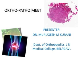
ORTHO-PATHO MEET
PRESENTER:
DR. MURUGESH M KURANI
Dept. of Orthopaedics, J N
Medical College, BELAGAVI.
CLINICAL HISTORY
• Name: xyz
• Age: 53 years.
• Sex: Female.
• Occupation: Housewife.
CHIEF COMPLAINTS
C/O swelling in the right ring finger since 3
months
C/O pain in the swelling since 3 months
HISTORY OF PRESENTING ILLNESS
Patient was apparently alright 3 months back,
later she noticed a small swelling in the right ring
finger which was associated with mild
intermittent pain. Swelling gradually progressed
to attain present size of around 1.5x1 cm.
Since last 15days pain increased in severity and is
continuous in nature.
Pain is more during night time.
Pain increases on movements and relieves
upon rest
No h/o trauma/ fall
No h/o fever/ weight loss/ decreased appetite
No h/o intake of any medication
PAST HISTORY:
• No history of similar complaints in the past.
• No h/o -ASTHAMA/TB / brucellosis
• No h/o DM/HTN
FAMILY HISTORY:
No h/o similar complaints in the family
PERSONAL HISTORY
• Diet : vegeterian
• Appetite : Adequate
• Sleep : decreased because of pain
• Bowel & Bladder : Normal and regular
• No h/o of smoking /tobacco/alcohol intake
GENERAL PHYSICAL EXAMINATION
An elderly female patient , moderately built & nourished,
conscious and co-operative & well oriented to time, place &
person.
VITALS:
• PR: 82/min
• BP: 134/86 mm of Hg in supine
• RR: 19 /min
• Temp- afebrile 98*F
Pallor: absent
Icterus: absent
Cyanosis: absent
Clubbing: absent
Lymphadenopathy: absent
Edema: absent
SYSTEMIC EXAMINATION
CVS: S1 S2 + ,no murmur
RS : B/L Normal vascular breath sounds present
B/L Air entry equal.
PA : Soft, Non tender,
Bowel sounds present, no organomegaly.
CNS: No focal neurological deficit.
LOCAL EXAMINATION
INSPECTION:
Localised ovoid swelling of
around 1.5X1 cm over middle
phalynx of right ring finger on
volar aspect. (parallel to long
axis of bone)
Skin over the swelling is
stretched.
No scar/ sinus/ redness.
No visible pulsations/ dilated
veins.
PALPATION
local rise of temperature.
Tenderness present over swelling.
Size: 1.5x1 cm, hard, fixed to underlying bone.
No distal neuro-vascular deficit.
MOVEMENTS:
Flexion at PIP joint terminally restricted.
INVESTIGATIONS:
Hb : 11.4g%
WBC : 6800 cells/cumm, N62, L 30, E 06, M 02
PLATELET COUNT : 3.05L/cumm
RBS : 116 mg%
UREA : 29mg%
CREATININE: 1.1 mg%
HIV, HBsAg, HCV : NON REACTIVE
X RAY OF RIGHT HAND
Sharply marginated,
lobulated,eccentrically
located lucent defect
in metaphysio-
epiphysial region of
middle phalynx of ring
finger.
MRI OF RIGHT HAND
IMPRESSION:
Well defined signal
abnormality along
palmar aspect of ring
finger in the region of
middle phalynx s/o
GCT of tendon
sheath d/d ganglion
cyst.
DIFFERENTIAL DIAGNOSIS
GANGLION CYST
GCT OF TENDON SHEATH
ENCHONDROMA
PERIOSTEAL CHONDROMA
SIMPLE BONE CYST
ANEURYSMAL BONE CYST
CHONDROBLASTOMA
CHONDROMYXOID FIBROMA
CHONDROSARCOMA
GCT TENDON SHEATH
A giant cell tumor of tendon sheath consists of
multinucleated giant cells, inflammatory cells, histiocytes,
and fibroblasts.
It develops in or near synovial joints, bursae, and tendon
sheaths and may represent a reactive inflammatory
process or a benign neoplasm.
AGE- It most frequently develops in people between 30 and 50
years of age.
SITE- It commonly appears in the hand and less commonly in
the foot, ankle, and knee.
Physical examination reveals a firm, small, lobulated mass
fixed to the underlying tissues or tendon sheaths.
Occasionally it can erode bone. This tumor may grow
slowly and recur following surgical excision.
GANGLION CYST
Ganglion cysts are unilocular or multilocular collections of translucent
fluid or gelatinous myxoid tissue surrounded by fibrous tissue.
AGE- They can occur in patients of any age and are located in a superficial
location adjacent to synovial joints or tendon sheaths.
SITE - They are commonly found near the wrist, hand, and knee.
Occasionally those that develop near the knee grow to a large size and
dissect through the surrounding soft tissues.
Enlargement of the limb or swelling caused by these unusual ganglia may
suggest the presence of a neoplasm. Although aspiration can remove the
fluid from a ganglion cyst, surgical resection is currently the most
predictable method of eradicating symptomatic lesions.
ENCHONDROMA
An enchondroma is a benign hyaline cartilage
lesion located in the medullary cavity of
otherwise normal bones.
It frequently occurs in the bones of the
hands and feet but may appear in any
bone including the femur, tibia, and
humerus.
It is generally considered an asymptomatic,
indolent lesion.
Plain radiographs reveal a central, well-
circumscribed lucent region that may be
mineralized. Enchondromas resemble
bone infarcts. Further imaging studies are
not necessary, but enchondromas normally
show increased activity on a bone scan.
During skeletal growth the lesions may slowly enlarge.
Following completion of normal growth, they cease to
enlarge and the cartilage component calcifies to give a
stippled radiographic appearance.
In extremely rare cases, a chondrosarcoma can develop
from an enchondroma. Because enchondromas are usually
asymptomatic and do not enlarge after skeletal maturity, a
lesion that causes pain or enlarges in an adult strongly
suggests the possibility of malignant transformation.
Enchondromas generally do not require surgical treatment.
PERIOSTEAL CHONDROMA
A periosteal chondroma is a rare, subperiosteal lesion consisting of
hyaline cartilage.
It forms between the cortical bone and overlying periosteum, often
creating an indentation in the bone surface and a smooth bulge of
periosteum-covered cartilage that projects into the soft tissues.
AGE- Most patients are young or middle-aged adults.
Presumably periosteal chondromas develop from proliferation of
cartilage-forming periosteal cells.
SITE- They occur most frequently in the proximal humeral metaphysis,
phalanges, metacarpals, and metatarsals.
They usually present as a solitary, painful mass or as an
incidental radiographic finding.
Radiographs show a scalloped depression in the bone
cortex and may show the faint image of a soft tissue mass
containing speckled regions of calcification.
Periosteal chondromas can slowly enlarge, but they have
not been shown to be aggressive.
For symptomatic or enlarging lesions, surgical resection
provides definitive local control.
SIMPLE BONE CYST
Simple, or unicameral, bone cysts consist of fluid-filled
cavities within bone lined by a thin layer of fibrous tissue.
Age- They occur most commonly in children less than 15
years of age.
Site- Approximately 50% occur in the proximal humeral
metaphysis. Other common sites include the proximal
femur and iliac wing.
They may cause slight expansion of bone and thinning of
the cortex. As a result patients often present with a
pathologic fracture through the cyst.
Radiographically, simple bone cysts are centrally
located, lucent lesions of the metaphysis. As new bone
in skeletally immature patients grows away from the
cyst, the lesion may eventually reside in the diaphysis.
The current recommended treatment of simple cysts
includes observation with restriction of activity and
steroid injections. Intralesional curettage and bone
grafting is generally reserved for large cysts at high risk
for fracture in the proximal femur.
ANEURYSMAL BONE CYST(ABC)
Aneurysmal bone cysts (ABCs) consist of blood-filled cavities lined by
fibrous septae that include giant cells and areas of osteoid but no true
endothelial cells.
AGE- Approximately 85% of patients with ABCs present before age 20.
The most common symptoms are pain, swelling, and tenderness on
palpation of the involved bone.
SITE- ABCs commonly involve the metaphysis of long bones, posterior
elements of the vertebrae, pelvis, or scapula, but they can occur
throughout the skeleton.
ABCs can grow rapidly and frequently cause pathologic fractures. When
located in the spine, they can cause neurologic compromise.
Plain radiographs show a lytic lesion causing marked
expansion of the involved bone and occasional
periosteal new bone formation. An MRI scan often
reveals fluid-fluid levels.
They may stop expanding and begin to ossify after
reaching a certain size or they may regress
spontaneously.
Standard treatment is a confirmatory biopsy followed
by intralesional curettage and bone grafting.
CHONDROBLASTOMA
A chondroblastoma is a benign cartilage tumor
consisting of regions or “islands” of densely
packed polyhedral cells called chondroblasts
admixed with fibrous tissue and chondrocytes
forming a cartilage matrix.
Site- epiphysis of long bones in patients with
open physes. They occur most commonly in the
proximal humerus, distal femur, proximal tibia,
and proximal femur.
Radiographs typically show an eccentric,
epiphyseal lucency with punctate calcifications. A
sclerotic rim surrounds the lucent area.
The lesions rarely involve more than half of the
epiphysis and only occasionally extend into the
metaphysis.
Intralesional curettage and bone grafting is
indicated for a chondroblastoma.
TREATMENT
Excision and curettage done under regional
block.
HISTOPATHOLOGY REPORT
GROSS EXAMINATION:
Single, grey white, firm tissue,
measuring 1.2x0.8x0.7cm. Cut section
shows grey white appearance.
MICROSCOPIC EXAMINATION:
Mass lined by lobules of chondrocytes
surrounded by fibroblasts along with
central areas of ossification. No
evidence of malignancy in the sections
studied.
IMPRESSION:
CHONDROMYXOID FIBROMA- MIDDLE
PHALYNX OF RIGHT RING FINGER.
CHONDROMYXOID FIBROMA
Rare, slow growing, benign cartilagenous tumor <1%, malignant degeneration is rare.
Charecterised by GRM1 gene fusion or promoter swapping.
Associated with a translocation at t(1;5)
Described in 1943 by Jaffe and Lichtenstein, who differentiated the histologic
findings from chondrosarcoma, before 1943 it is considered a giant cell varient.
Site: metaphysis of long bones – distal femur, proximal tibia (60%)
flat bones – ileum, ribs, skull bone
hand bones.
Age – ranges from 3-70 years, 60% are late adolescents and young adults.
Sex – M>F
X - RAY- Sharply marginated,
lobulated,eccentrically located
lucent defect in the
metaphysis, may extend into
epiphysis.
Macroscopy: sharp
circumscribed, often lobulated,
firm, gray white or blue gray.
Microscopy: pseudolobular
architecture, spindle shaped or
stellate cells, abondant myxoid
stroma to chondroid stroma.
TREATMENT:
Intralesional curettage or resection. Resulting bony defect has to filled
using iliac bone graft and screw fixation. Recurrence rate 25%.
Wide en block excision may lower the recurrence, but add unnecessary
morbidity.
Local adjunct treatment agents, such as phenol, methylmethacrylate
and liquid nitrogen have not been shown to decrease the
recurrence rate.
Radiotherapy may be used in tumors that are considered unresectable.
CHONDROSARCOMA
A chondrosarcoma is a malignant tumor, may develop from an
enchondroma or osteochondroma but occurs more commonly in bones
with no known preceding cartilage lesion.
AGE- Chondrosarcomas occur in adults and the elderly.
SITE- most commonly in the pelvis, scapula, humerus, femur, and tibia.
Patients with multiple hereditary exostoses, Ollier's disease and Maffucci's
syndrome have a higher risk for malignant transformation of a
cartilaginous lesion.
Chondrosarcomas vary considerably in their behavior: some enlarge
rapidly, aggressively invade normal tissue, metastasize, and cause death,
but most are low grade tumors that enlarge slowly, cause little damage to
adjacent tissues, and metastasize only after many years.
Plain radiographs reveal bone destruction
as well as mineralization within the
tumor.
HISTOPATHO: Chondrosarcoma of low
grade may mimic Chondromyxoid
fibroma except for the lack of a
myxoid element.
Low grade chondrosarcoma: few
mitotic figures with a bland histologic
appearance, enlarged chondrocytes
with plump multinucleated lacunae.
High grade chondrosarcoma:
hypercellular stroma consisting of
charecteristic “blue balls” of a
cartilage lesion which permeate the
bone trabeculae.
Wide surgical excision is necessary and offers
the best possibility of cure, as radiation and
chemotherapy have not proven to be effective
methods of treatment for chondrosarcoma.
Locally recurrent disease is common and may
be difficult to treat.
THANK YOU

Mais conteúdo relacionado

Mais procurados

Inra medullary nailing - basic concepts
Inra medullary nailing - basic conceptsInra medullary nailing - basic concepts
Inra medullary nailing - basic conceptsharivenkat1990
 
ULNAR NERVE PALSY AND TENDON TRANSFERS
ULNAR NERVE PALSY AND TENDON TRANSFERSULNAR NERVE PALSY AND TENDON TRANSFERS
ULNAR NERVE PALSY AND TENDON TRANSFERSBenthungo Tungoe
 
Patella dislocation by DR.NAVEEN RATHOR
Patella dislocation by DR.NAVEEN RATHORPatella dislocation by DR.NAVEEN RATHOR
Patella dislocation by DR.NAVEEN RATHORDR.Naveen Rathor
 
Calcaneal fractures
Calcaneal fracturesCalcaneal fractures
Calcaneal fracturesRohit Vikas
 
Delayed Union & Nonunion of Fractures
Delayed Union & Nonunion of FracturesDelayed Union & Nonunion of Fractures
Delayed Union & Nonunion of FracturesDr. Armaan Singh
 
CORA (center of rotation of angulation)
CORA (center of rotation of angulation)CORA (center of rotation of angulation)
CORA (center of rotation of angulation)Morshed Abir
 
Knee stiffness dr anil k jain
Knee stiffness dr anil k jainKnee stiffness dr anil k jain
Knee stiffness dr anil k jainvaruntandra
 
Fracture of Distal End Humerus.
Fracture of Distal End Humerus.Fracture of Distal End Humerus.
Fracture of Distal End Humerus.Dr.Anshu Sharma
 
Osteopetrosis(albergs schoberg disease
Osteopetrosis(albergs schoberg diseaseOsteopetrosis(albergs schoberg disease
Osteopetrosis(albergs schoberg diseaseJamil Kifayatullah
 
Three column fixation for complex PROXIMAL TIBIA FRACTURES
Three column fixation for complex PROXIMAL TIBIA FRACTURESThree column fixation for complex PROXIMAL TIBIA FRACTURES
Three column fixation for complex PROXIMAL TIBIA FRACTURESLokesh Sharoff
 
Acetabular Fracture Radiology: Xrays, CT scan & 3D printing
Acetabular Fracture Radiology: Xrays, CT scan & 3D printingAcetabular Fracture Radiology: Xrays, CT scan & 3D printing
Acetabular Fracture Radiology: Xrays, CT scan & 3D printingVaibhav Bagaria
 
Subodh acetabulum ppt
Subodh acetabulum pptSubodh acetabulum ppt
Subodh acetabulum pptSubodh Pathak
 

Mais procurados (20)

Bone graft
Bone graftBone graft
Bone graft
 
Ankle fracture
Ankle fractureAnkle fracture
Ankle fracture
 
Inra medullary nailing - basic concepts
Inra medullary nailing - basic conceptsInra medullary nailing - basic concepts
Inra medullary nailing - basic concepts
 
ULNAR NERVE PALSY AND TENDON TRANSFERS
ULNAR NERVE PALSY AND TENDON TRANSFERSULNAR NERVE PALSY AND TENDON TRANSFERS
ULNAR NERVE PALSY AND TENDON TRANSFERS
 
Patella dislocation by DR.NAVEEN RATHOR
Patella dislocation by DR.NAVEEN RATHORPatella dislocation by DR.NAVEEN RATHOR
Patella dislocation by DR.NAVEEN RATHOR
 
Calcaneal fractures
Calcaneal fracturesCalcaneal fractures
Calcaneal fractures
 
Delayed Union & Nonunion of Fractures
Delayed Union & Nonunion of FracturesDelayed Union & Nonunion of Fractures
Delayed Union & Nonunion of Fractures
 
Fractures of the olecranon
Fractures of the olecranonFractures of the olecranon
Fractures of the olecranon
 
CORA (center of rotation of angulation)
CORA (center of rotation of angulation)CORA (center of rotation of angulation)
CORA (center of rotation of angulation)
 
Knee stiffness dr anil k jain
Knee stiffness dr anil k jainKnee stiffness dr anil k jain
Knee stiffness dr anil k jain
 
The deltopectoral approach
The deltopectoral approachThe deltopectoral approach
The deltopectoral approach
 
Monteggia ppt
Monteggia pptMonteggia ppt
Monteggia ppt
 
Fracture of Distal End Humerus.
Fracture of Distal End Humerus.Fracture of Distal End Humerus.
Fracture of Distal End Humerus.
 
Osteopetrosis(albergs schoberg disease
Osteopetrosis(albergs schoberg diseaseOsteopetrosis(albergs schoberg disease
Osteopetrosis(albergs schoberg disease
 
Protrusio acetabuli
Protrusio acetabuliProtrusio acetabuli
Protrusio acetabuli
 
Three column fixation for complex PROXIMAL TIBIA FRACTURES
Three column fixation for complex PROXIMAL TIBIA FRACTURESThree column fixation for complex PROXIMAL TIBIA FRACTURES
Three column fixation for complex PROXIMAL TIBIA FRACTURES
 
Triplane Fracture
Triplane FractureTriplane Fracture
Triplane Fracture
 
Acetabular Fracture Radiology: Xrays, CT scan & 3D printing
Acetabular Fracture Radiology: Xrays, CT scan & 3D printingAcetabular Fracture Radiology: Xrays, CT scan & 3D printing
Acetabular Fracture Radiology: Xrays, CT scan & 3D printing
 
Subodh acetabulum ppt
Subodh acetabulum pptSubodh acetabulum ppt
Subodh acetabulum ppt
 
Kienbock's disease
Kienbock's diseaseKienbock's disease
Kienbock's disease
 

Semelhante a CHONDROMYXOID FIBROMA

Benign bone tumors - Dr. Sachin M
Benign bone tumors - Dr. Sachin MBenign bone tumors - Dr. Sachin M
Benign bone tumors - Dr. Sachin MSachinMalayaiah1
 
Bone forming tumors rabins
Bone forming tumors rabinsBone forming tumors rabins
Bone forming tumors rabinsRobins Shah
 
Aneurysmal bone cyst
Aneurysmal bone cystAneurysmal bone cyst
Aneurysmal bone cystRobins Shah
 
Common benign and malignant bone tumors
Common benign and malignant bone tumorsCommon benign and malignant bone tumors
Common benign and malignant bone tumorsahm732
 
Simple and aneurysmal Bone cyst - Definition, Classfication, Investigations, ...
Simple and aneurysmal Bone cyst - Definition, Classfication, Investigations, ...Simple and aneurysmal Bone cyst - Definition, Classfication, Investigations, ...
Simple and aneurysmal Bone cyst - Definition, Classfication, Investigations, ...orthoslides
 
Benign bone tumours
Benign bone tumoursBenign bone tumours
Benign bone tumoursArif S
 
Aneurysmal Bone Cyst.pptx
Aneurysmal Bone Cyst.pptxAneurysmal Bone Cyst.pptx
Aneurysmal Bone Cyst.pptxWilliamsMusa1
 
Aneurysmal bone cyst.pptx
Aneurysmal bone cyst.pptxAneurysmal bone cyst.pptx
Aneurysmal bone cyst.pptxSalman Syed
 
farrukh-aneurysmalbonecyst-190530010628.pptx
farrukh-aneurysmalbonecyst-190530010628.pptxfarrukh-aneurysmalbonecyst-190530010628.pptx
farrukh-aneurysmalbonecyst-190530010628.pptxAbrahamEmes
 

Semelhante a CHONDROMYXOID FIBROMA (20)

Osteochondroma
OsteochondromaOsteochondroma
Osteochondroma
 
Benign bone tumors - Dr. Sachin M
Benign bone tumors - Dr. Sachin MBenign bone tumors - Dr. Sachin M
Benign bone tumors - Dr. Sachin M
 
Benign bone tumor ppt
Benign bone tumor pptBenign bone tumor ppt
Benign bone tumor ppt
 
Abc case powerpoint
Abc case powerpointAbc case powerpoint
Abc case powerpoint
 
PPT ABC.pptx
PPT ABC.pptxPPT ABC.pptx
PPT ABC.pptx
 
PPT ABC.pptx
PPT ABC.pptxPPT ABC.pptx
PPT ABC.pptx
 
Bone forming tumors rabins
Bone forming tumors rabinsBone forming tumors rabins
Bone forming tumors rabins
 
Aneurysmal bone cyst
Aneurysmal bone cystAneurysmal bone cyst
Aneurysmal bone cyst
 
Common benign and malignant bone tumors
Common benign and malignant bone tumorsCommon benign and malignant bone tumors
Common benign and malignant bone tumors
 
Simple and aneurysmal Bone cyst - Definition, Classfication, Investigations, ...
Simple and aneurysmal Bone cyst - Definition, Classfication, Investigations, ...Simple and aneurysmal Bone cyst - Definition, Classfication, Investigations, ...
Simple and aneurysmal Bone cyst - Definition, Classfication, Investigations, ...
 
Bone tumor
Bone tumorBone tumor
Bone tumor
 
Osteoid osteoma
Osteoid osteomaOsteoid osteoma
Osteoid osteoma
 
Benign bt
Benign btBenign bt
Benign bt
 
Diskitis
DiskitisDiskitis
Diskitis
 
Benign bone tumours
Benign bone tumoursBenign bone tumours
Benign bone tumours
 
Aneurysmal Bone Cyst.pptx
Aneurysmal Bone Cyst.pptxAneurysmal Bone Cyst.pptx
Aneurysmal Bone Cyst.pptx
 
Osteochondroma (dr. mahesh)
Osteochondroma (dr. mahesh)Osteochondroma (dr. mahesh)
Osteochondroma (dr. mahesh)
 
Aneurysmal Bone Cyst
Aneurysmal Bone CystAneurysmal Bone Cyst
Aneurysmal Bone Cyst
 
Aneurysmal bone cyst.pptx
Aneurysmal bone cyst.pptxAneurysmal bone cyst.pptx
Aneurysmal bone cyst.pptx
 
farrukh-aneurysmalbonecyst-190530010628.pptx
farrukh-aneurysmalbonecyst-190530010628.pptxfarrukh-aneurysmalbonecyst-190530010628.pptx
farrukh-aneurysmalbonecyst-190530010628.pptx
 

Mais de Murugesh M Kurani

Scapula surgical approaches ppt
Scapula surgical approaches pptScapula surgical approaches ppt
Scapula surgical approaches pptMurugesh M Kurani
 
Posterolateral corner knee injuries
Posterolateral corner knee injuriesPosterolateral corner knee injuries
Posterolateral corner knee injuriesMurugesh M Kurani
 
INFECTED FRACTURES, INFIRM PATIENT, INDOMITABLE SURGEON HANDLING NON-UNION AN...
INFECTED FRACTURES, INFIRM PATIENT, INDOMITABLE SURGEON HANDLING NON-UNION AN...INFECTED FRACTURES, INFIRM PATIENT, INDOMITABLE SURGEON HANDLING NON-UNION AN...
INFECTED FRACTURES, INFIRM PATIENT, INDOMITABLE SURGEON HANDLING NON-UNION AN...Murugesh M Kurani
 
Herbert screw fixation and bone graft in nonunited scaphoid
Herbert screw fixation and bone graft in nonunited scaphoidHerbert screw fixation and bone graft in nonunited scaphoid
Herbert screw fixation and bone graft in nonunited scaphoidMurugesh M Kurani
 
Fractures and fracture dislocations of the tarsometatarsal joint
Fractures and fracture dislocations of the tarsometatarsal jointFractures and fracture dislocations of the tarsometatarsal joint
Fractures and fracture dislocations of the tarsometatarsal jointMurugesh M Kurani
 
Genu varum, Genu valgum, Genu recurvatum
Genu varum, Genu valgum, Genu recurvatumGenu varum, Genu valgum, Genu recurvatum
Genu varum, Genu valgum, Genu recurvatumMurugesh M Kurani
 

Mais de Murugesh M Kurani (7)

Scapula surgical approaches ppt
Scapula surgical approaches pptScapula surgical approaches ppt
Scapula surgical approaches ppt
 
Posterolateral corner knee injuries
Posterolateral corner knee injuriesPosterolateral corner knee injuries
Posterolateral corner knee injuries
 
INFECTED FRACTURES, INFIRM PATIENT, INDOMITABLE SURGEON HANDLING NON-UNION AN...
INFECTED FRACTURES, INFIRM PATIENT, INDOMITABLE SURGEON HANDLING NON-UNION AN...INFECTED FRACTURES, INFIRM PATIENT, INDOMITABLE SURGEON HANDLING NON-UNION AN...
INFECTED FRACTURES, INFIRM PATIENT, INDOMITABLE SURGEON HANDLING NON-UNION AN...
 
Herbert screw fixation and bone graft in nonunited scaphoid
Herbert screw fixation and bone graft in nonunited scaphoidHerbert screw fixation and bone graft in nonunited scaphoid
Herbert screw fixation and bone graft in nonunited scaphoid
 
Fractures and fracture dislocations of the tarsometatarsal joint
Fractures and fracture dislocations of the tarsometatarsal jointFractures and fracture dislocations of the tarsometatarsal joint
Fractures and fracture dislocations of the tarsometatarsal joint
 
Evidence based medicine
Evidence based medicineEvidence based medicine
Evidence based medicine
 
Genu varum, Genu valgum, Genu recurvatum
Genu varum, Genu valgum, Genu recurvatumGenu varum, Genu valgum, Genu recurvatum
Genu varum, Genu valgum, Genu recurvatum
 

Último

Call Girls in Lucknow Just Call 👉👉8630512678 Top Class Call Girl Service Avai...
Call Girls in Lucknow Just Call 👉👉8630512678 Top Class Call Girl Service Avai...Call Girls in Lucknow Just Call 👉👉8630512678 Top Class Call Girl Service Avai...
Call Girls in Lucknow Just Call 👉👉8630512678 Top Class Call Girl Service Avai...soniyagrag336
 
7 steps How to prevent Thalassemia : Dr Sharda Jain & Vandana Gupta
7 steps How to prevent Thalassemia : Dr Sharda Jain & Vandana Gupta7 steps How to prevent Thalassemia : Dr Sharda Jain & Vandana Gupta
7 steps How to prevent Thalassemia : Dr Sharda Jain & Vandana GuptaLifecare Centre
 
ANATOMY AND PHYSIOLOGY OF REPRODUCTIVE SYSTEM.pptx
ANATOMY AND PHYSIOLOGY OF REPRODUCTIVE SYSTEM.pptxANATOMY AND PHYSIOLOGY OF REPRODUCTIVE SYSTEM.pptx
ANATOMY AND PHYSIOLOGY OF REPRODUCTIVE SYSTEM.pptxSwetaba Besh
 
Ahmedabad Call Girls Book Now 9630942363 Top Class Ahmedabad Escort Service A...
Ahmedabad Call Girls Book Now 9630942363 Top Class Ahmedabad Escort Service A...Ahmedabad Call Girls Book Now 9630942363 Top Class Ahmedabad Escort Service A...
Ahmedabad Call Girls Book Now 9630942363 Top Class Ahmedabad Escort Service A...GENUINE ESCORT AGENCY
 
Cheap Rate Call Girls Bangalore {9179660964} ❤️VVIP BEBO Call Girls in Bangal...
Cheap Rate Call Girls Bangalore {9179660964} ❤️VVIP BEBO Call Girls in Bangal...Cheap Rate Call Girls Bangalore {9179660964} ❤️VVIP BEBO Call Girls in Bangal...
Cheap Rate Call Girls Bangalore {9179660964} ❤️VVIP BEBO Call Girls in Bangal...Sheetaleventcompany
 
Call Girls Mussoorie Just Call 8854095900 Top Class Call Girl Service Available
Call Girls Mussoorie Just Call 8854095900 Top Class Call Girl Service AvailableCall Girls Mussoorie Just Call 8854095900 Top Class Call Girl Service Available
Call Girls Mussoorie Just Call 8854095900 Top Class Call Girl Service AvailableJanvi Singh
 
Chandigarh Call Girls Service ❤️🍑 9809698092 👄🫦Independent Escort Service Cha...
Chandigarh Call Girls Service ❤️🍑 9809698092 👄🫦Independent Escort Service Cha...Chandigarh Call Girls Service ❤️🍑 9809698092 👄🫦Independent Escort Service Cha...
Chandigarh Call Girls Service ❤️🍑 9809698092 👄🫦Independent Escort Service Cha...Sheetaleventcompany
 
Gastric Cancer: Сlinical Implementation of Artificial Intelligence, Synergeti...
Gastric Cancer: Сlinical Implementation of Artificial Intelligence, Synergeti...Gastric Cancer: Сlinical Implementation of Artificial Intelligence, Synergeti...
Gastric Cancer: Сlinical Implementation of Artificial Intelligence, Synergeti...Oleg Kshivets
 
Call Girl In Indore 📞9235973566📞 Just📲 Call Inaaya Indore Call Girls Service ...
Call Girl In Indore 📞9235973566📞 Just📲 Call Inaaya Indore Call Girls Service ...Call Girl In Indore 📞9235973566📞 Just📲 Call Inaaya Indore Call Girls Service ...
Call Girl In Indore 📞9235973566📞 Just📲 Call Inaaya Indore Call Girls Service ...Sheetaleventcompany
 
Race Course Road } Book Call Girls in Bangalore | Whatsapp No 6378878445 VIP ...
Race Course Road } Book Call Girls in Bangalore | Whatsapp No 6378878445 VIP ...Race Course Road } Book Call Girls in Bangalore | Whatsapp No 6378878445 VIP ...
Race Course Road } Book Call Girls in Bangalore | Whatsapp No 6378878445 VIP ...dishamehta3332
 
Pune Call Girl Service 📞9xx000xx09📞Just Call Divya📲 Call Girl In Pune No💰Adva...
Pune Call Girl Service 📞9xx000xx09📞Just Call Divya📲 Call Girl In Pune No💰Adva...Pune Call Girl Service 📞9xx000xx09📞Just Call Divya📲 Call Girl In Pune No💰Adva...
Pune Call Girl Service 📞9xx000xx09📞Just Call Divya📲 Call Girl In Pune No💰Adva...Sheetaleventcompany
 
❤️Amritsar Escorts Service☎️9815674956☎️ Call Girl service in Amritsar☎️ Amri...
❤️Amritsar Escorts Service☎️9815674956☎️ Call Girl service in Amritsar☎️ Amri...❤️Amritsar Escorts Service☎️9815674956☎️ Call Girl service in Amritsar☎️ Amri...
❤️Amritsar Escorts Service☎️9815674956☎️ Call Girl service in Amritsar☎️ Amri...Sheetaleventcompany
 
Call Girls Kathua Just Call 8250077686 Top Class Call Girl Service Available
Call Girls Kathua Just Call 8250077686 Top Class Call Girl Service AvailableCall Girls Kathua Just Call 8250077686 Top Class Call Girl Service Available
Call Girls Kathua Just Call 8250077686 Top Class Call Girl Service AvailableDipal Arora
 
Call girls Service Phullen / 9332606886 Genuine Call girls with real Photos a...
Call girls Service Phullen / 9332606886 Genuine Call girls with real Photos a...Call girls Service Phullen / 9332606886 Genuine Call girls with real Photos a...
Call girls Service Phullen / 9332606886 Genuine Call girls with real Photos a...call girls hydrabad
 
💰Call Girl In Bangalore☎️63788-78445💰 Call Girl service in Bangalore☎️Bangalo...
💰Call Girl In Bangalore☎️63788-78445💰 Call Girl service in Bangalore☎️Bangalo...💰Call Girl In Bangalore☎️63788-78445💰 Call Girl service in Bangalore☎️Bangalo...
💰Call Girl In Bangalore☎️63788-78445💰 Call Girl service in Bangalore☎️Bangalo...gragneelam30
 
Cara Menggugurkan Kandungan Dengan Cepat Selesai Dalam 24 Jam Secara Alami Bu...
Cara Menggugurkan Kandungan Dengan Cepat Selesai Dalam 24 Jam Secara Alami Bu...Cara Menggugurkan Kandungan Dengan Cepat Selesai Dalam 24 Jam Secara Alami Bu...
Cara Menggugurkan Kandungan Dengan Cepat Selesai Dalam 24 Jam Secara Alami Bu...Cara Menggugurkan Kandungan 087776558899
 
Premium Call Girls Dehradun {8854095900} ❤️VVIP ANJU Call Girls in Dehradun U...
Premium Call Girls Dehradun {8854095900} ❤️VVIP ANJU Call Girls in Dehradun U...Premium Call Girls Dehradun {8854095900} ❤️VVIP ANJU Call Girls in Dehradun U...
Premium Call Girls Dehradun {8854095900} ❤️VVIP ANJU Call Girls in Dehradun U...Sheetaleventcompany
 
Exclusive Call Girls Bangalore {7304373326} ❤️VVIP POOJA Call Girls in Bangal...
Exclusive Call Girls Bangalore {7304373326} ❤️VVIP POOJA Call Girls in Bangal...Exclusive Call Girls Bangalore {7304373326} ❤️VVIP POOJA Call Girls in Bangal...
Exclusive Call Girls Bangalore {7304373326} ❤️VVIP POOJA Call Girls in Bangal...Sheetaleventcompany
 
💰Call Girl In Bangalore☎️7304373326💰 Call Girl service in Bangalore☎️Bangalor...
💰Call Girl In Bangalore☎️7304373326💰 Call Girl service in Bangalore☎️Bangalor...💰Call Girl In Bangalore☎️7304373326💰 Call Girl service in Bangalore☎️Bangalor...
💰Call Girl In Bangalore☎️7304373326💰 Call Girl service in Bangalore☎️Bangalor...Sheetaleventcompany
 
Circulatory Shock, types and stages, compensatory mechanisms
Circulatory Shock, types and stages, compensatory mechanismsCirculatory Shock, types and stages, compensatory mechanisms
Circulatory Shock, types and stages, compensatory mechanismsMedicoseAcademics
 

Último (20)

Call Girls in Lucknow Just Call 👉👉8630512678 Top Class Call Girl Service Avai...
Call Girls in Lucknow Just Call 👉👉8630512678 Top Class Call Girl Service Avai...Call Girls in Lucknow Just Call 👉👉8630512678 Top Class Call Girl Service Avai...
Call Girls in Lucknow Just Call 👉👉8630512678 Top Class Call Girl Service Avai...
 
7 steps How to prevent Thalassemia : Dr Sharda Jain & Vandana Gupta
7 steps How to prevent Thalassemia : Dr Sharda Jain & Vandana Gupta7 steps How to prevent Thalassemia : Dr Sharda Jain & Vandana Gupta
7 steps How to prevent Thalassemia : Dr Sharda Jain & Vandana Gupta
 
ANATOMY AND PHYSIOLOGY OF REPRODUCTIVE SYSTEM.pptx
ANATOMY AND PHYSIOLOGY OF REPRODUCTIVE SYSTEM.pptxANATOMY AND PHYSIOLOGY OF REPRODUCTIVE SYSTEM.pptx
ANATOMY AND PHYSIOLOGY OF REPRODUCTIVE SYSTEM.pptx
 
Ahmedabad Call Girls Book Now 9630942363 Top Class Ahmedabad Escort Service A...
Ahmedabad Call Girls Book Now 9630942363 Top Class Ahmedabad Escort Service A...Ahmedabad Call Girls Book Now 9630942363 Top Class Ahmedabad Escort Service A...
Ahmedabad Call Girls Book Now 9630942363 Top Class Ahmedabad Escort Service A...
 
Cheap Rate Call Girls Bangalore {9179660964} ❤️VVIP BEBO Call Girls in Bangal...
Cheap Rate Call Girls Bangalore {9179660964} ❤️VVIP BEBO Call Girls in Bangal...Cheap Rate Call Girls Bangalore {9179660964} ❤️VVIP BEBO Call Girls in Bangal...
Cheap Rate Call Girls Bangalore {9179660964} ❤️VVIP BEBO Call Girls in Bangal...
 
Call Girls Mussoorie Just Call 8854095900 Top Class Call Girl Service Available
Call Girls Mussoorie Just Call 8854095900 Top Class Call Girl Service AvailableCall Girls Mussoorie Just Call 8854095900 Top Class Call Girl Service Available
Call Girls Mussoorie Just Call 8854095900 Top Class Call Girl Service Available
 
Chandigarh Call Girls Service ❤️🍑 9809698092 👄🫦Independent Escort Service Cha...
Chandigarh Call Girls Service ❤️🍑 9809698092 👄🫦Independent Escort Service Cha...Chandigarh Call Girls Service ❤️🍑 9809698092 👄🫦Independent Escort Service Cha...
Chandigarh Call Girls Service ❤️🍑 9809698092 👄🫦Independent Escort Service Cha...
 
Gastric Cancer: Сlinical Implementation of Artificial Intelligence, Synergeti...
Gastric Cancer: Сlinical Implementation of Artificial Intelligence, Synergeti...Gastric Cancer: Сlinical Implementation of Artificial Intelligence, Synergeti...
Gastric Cancer: Сlinical Implementation of Artificial Intelligence, Synergeti...
 
Call Girl In Indore 📞9235973566📞 Just📲 Call Inaaya Indore Call Girls Service ...
Call Girl In Indore 📞9235973566📞 Just📲 Call Inaaya Indore Call Girls Service ...Call Girl In Indore 📞9235973566📞 Just📲 Call Inaaya Indore Call Girls Service ...
Call Girl In Indore 📞9235973566📞 Just📲 Call Inaaya Indore Call Girls Service ...
 
Race Course Road } Book Call Girls in Bangalore | Whatsapp No 6378878445 VIP ...
Race Course Road } Book Call Girls in Bangalore | Whatsapp No 6378878445 VIP ...Race Course Road } Book Call Girls in Bangalore | Whatsapp No 6378878445 VIP ...
Race Course Road } Book Call Girls in Bangalore | Whatsapp No 6378878445 VIP ...
 
Pune Call Girl Service 📞9xx000xx09📞Just Call Divya📲 Call Girl In Pune No💰Adva...
Pune Call Girl Service 📞9xx000xx09📞Just Call Divya📲 Call Girl In Pune No💰Adva...Pune Call Girl Service 📞9xx000xx09📞Just Call Divya📲 Call Girl In Pune No💰Adva...
Pune Call Girl Service 📞9xx000xx09📞Just Call Divya📲 Call Girl In Pune No💰Adva...
 
❤️Amritsar Escorts Service☎️9815674956☎️ Call Girl service in Amritsar☎️ Amri...
❤️Amritsar Escorts Service☎️9815674956☎️ Call Girl service in Amritsar☎️ Amri...❤️Amritsar Escorts Service☎️9815674956☎️ Call Girl service in Amritsar☎️ Amri...
❤️Amritsar Escorts Service☎️9815674956☎️ Call Girl service in Amritsar☎️ Amri...
 
Call Girls Kathua Just Call 8250077686 Top Class Call Girl Service Available
Call Girls Kathua Just Call 8250077686 Top Class Call Girl Service AvailableCall Girls Kathua Just Call 8250077686 Top Class Call Girl Service Available
Call Girls Kathua Just Call 8250077686 Top Class Call Girl Service Available
 
Call girls Service Phullen / 9332606886 Genuine Call girls with real Photos a...
Call girls Service Phullen / 9332606886 Genuine Call girls with real Photos a...Call girls Service Phullen / 9332606886 Genuine Call girls with real Photos a...
Call girls Service Phullen / 9332606886 Genuine Call girls with real Photos a...
 
💰Call Girl In Bangalore☎️63788-78445💰 Call Girl service in Bangalore☎️Bangalo...
💰Call Girl In Bangalore☎️63788-78445💰 Call Girl service in Bangalore☎️Bangalo...💰Call Girl In Bangalore☎️63788-78445💰 Call Girl service in Bangalore☎️Bangalo...
💰Call Girl In Bangalore☎️63788-78445💰 Call Girl service in Bangalore☎️Bangalo...
 
Cara Menggugurkan Kandungan Dengan Cepat Selesai Dalam 24 Jam Secara Alami Bu...
Cara Menggugurkan Kandungan Dengan Cepat Selesai Dalam 24 Jam Secara Alami Bu...Cara Menggugurkan Kandungan Dengan Cepat Selesai Dalam 24 Jam Secara Alami Bu...
Cara Menggugurkan Kandungan Dengan Cepat Selesai Dalam 24 Jam Secara Alami Bu...
 
Premium Call Girls Dehradun {8854095900} ❤️VVIP ANJU Call Girls in Dehradun U...
Premium Call Girls Dehradun {8854095900} ❤️VVIP ANJU Call Girls in Dehradun U...Premium Call Girls Dehradun {8854095900} ❤️VVIP ANJU Call Girls in Dehradun U...
Premium Call Girls Dehradun {8854095900} ❤️VVIP ANJU Call Girls in Dehradun U...
 
Exclusive Call Girls Bangalore {7304373326} ❤️VVIP POOJA Call Girls in Bangal...
Exclusive Call Girls Bangalore {7304373326} ❤️VVIP POOJA Call Girls in Bangal...Exclusive Call Girls Bangalore {7304373326} ❤️VVIP POOJA Call Girls in Bangal...
Exclusive Call Girls Bangalore {7304373326} ❤️VVIP POOJA Call Girls in Bangal...
 
💰Call Girl In Bangalore☎️7304373326💰 Call Girl service in Bangalore☎️Bangalor...
💰Call Girl In Bangalore☎️7304373326💰 Call Girl service in Bangalore☎️Bangalor...💰Call Girl In Bangalore☎️7304373326💰 Call Girl service in Bangalore☎️Bangalor...
💰Call Girl In Bangalore☎️7304373326💰 Call Girl service in Bangalore☎️Bangalor...
 
Circulatory Shock, types and stages, compensatory mechanisms
Circulatory Shock, types and stages, compensatory mechanismsCirculatory Shock, types and stages, compensatory mechanisms
Circulatory Shock, types and stages, compensatory mechanisms
 

CHONDROMYXOID FIBROMA

  • 1. ORTHO-PATHO MEET PRESENTER: DR. MURUGESH M KURANI Dept. of Orthopaedics, J N Medical College, BELAGAVI.
  • 2. CLINICAL HISTORY • Name: xyz • Age: 53 years. • Sex: Female. • Occupation: Housewife.
  • 3. CHIEF COMPLAINTS C/O swelling in the right ring finger since 3 months C/O pain in the swelling since 3 months
  • 4. HISTORY OF PRESENTING ILLNESS Patient was apparently alright 3 months back, later she noticed a small swelling in the right ring finger which was associated with mild intermittent pain. Swelling gradually progressed to attain present size of around 1.5x1 cm. Since last 15days pain increased in severity and is continuous in nature. Pain is more during night time.
  • 5. Pain increases on movements and relieves upon rest No h/o trauma/ fall No h/o fever/ weight loss/ decreased appetite No h/o intake of any medication
  • 6. PAST HISTORY: • No history of similar complaints in the past. • No h/o -ASTHAMA/TB / brucellosis • No h/o DM/HTN FAMILY HISTORY: No h/o similar complaints in the family
  • 7. PERSONAL HISTORY • Diet : vegeterian • Appetite : Adequate • Sleep : decreased because of pain • Bowel & Bladder : Normal and regular • No h/o of smoking /tobacco/alcohol intake
  • 8. GENERAL PHYSICAL EXAMINATION An elderly female patient , moderately built & nourished, conscious and co-operative & well oriented to time, place & person. VITALS: • PR: 82/min • BP: 134/86 mm of Hg in supine • RR: 19 /min • Temp- afebrile 98*F
  • 9. Pallor: absent Icterus: absent Cyanosis: absent Clubbing: absent Lymphadenopathy: absent Edema: absent
  • 10. SYSTEMIC EXAMINATION CVS: S1 S2 + ,no murmur RS : B/L Normal vascular breath sounds present B/L Air entry equal. PA : Soft, Non tender, Bowel sounds present, no organomegaly. CNS: No focal neurological deficit.
  • 11. LOCAL EXAMINATION INSPECTION: Localised ovoid swelling of around 1.5X1 cm over middle phalynx of right ring finger on volar aspect. (parallel to long axis of bone) Skin over the swelling is stretched. No scar/ sinus/ redness. No visible pulsations/ dilated veins.
  • 12. PALPATION local rise of temperature. Tenderness present over swelling. Size: 1.5x1 cm, hard, fixed to underlying bone. No distal neuro-vascular deficit.
  • 13. MOVEMENTS: Flexion at PIP joint terminally restricted.
  • 14. INVESTIGATIONS: Hb : 11.4g% WBC : 6800 cells/cumm, N62, L 30, E 06, M 02 PLATELET COUNT : 3.05L/cumm RBS : 116 mg% UREA : 29mg% CREATININE: 1.1 mg% HIV, HBsAg, HCV : NON REACTIVE
  • 15. X RAY OF RIGHT HAND Sharply marginated, lobulated,eccentrically located lucent defect in metaphysio- epiphysial region of middle phalynx of ring finger.
  • 16. MRI OF RIGHT HAND IMPRESSION: Well defined signal abnormality along palmar aspect of ring finger in the region of middle phalynx s/o GCT of tendon sheath d/d ganglion cyst.
  • 17. DIFFERENTIAL DIAGNOSIS GANGLION CYST GCT OF TENDON SHEATH ENCHONDROMA PERIOSTEAL CHONDROMA SIMPLE BONE CYST ANEURYSMAL BONE CYST CHONDROBLASTOMA CHONDROMYXOID FIBROMA CHONDROSARCOMA
  • 18. GCT TENDON SHEATH A giant cell tumor of tendon sheath consists of multinucleated giant cells, inflammatory cells, histiocytes, and fibroblasts. It develops in or near synovial joints, bursae, and tendon sheaths and may represent a reactive inflammatory process or a benign neoplasm. AGE- It most frequently develops in people between 30 and 50 years of age. SITE- It commonly appears in the hand and less commonly in the foot, ankle, and knee. Physical examination reveals a firm, small, lobulated mass fixed to the underlying tissues or tendon sheaths. Occasionally it can erode bone. This tumor may grow slowly and recur following surgical excision.
  • 19. GANGLION CYST Ganglion cysts are unilocular or multilocular collections of translucent fluid or gelatinous myxoid tissue surrounded by fibrous tissue. AGE- They can occur in patients of any age and are located in a superficial location adjacent to synovial joints or tendon sheaths. SITE - They are commonly found near the wrist, hand, and knee. Occasionally those that develop near the knee grow to a large size and dissect through the surrounding soft tissues. Enlargement of the limb or swelling caused by these unusual ganglia may suggest the presence of a neoplasm. Although aspiration can remove the fluid from a ganglion cyst, surgical resection is currently the most predictable method of eradicating symptomatic lesions.
  • 20.
  • 21. ENCHONDROMA An enchondroma is a benign hyaline cartilage lesion located in the medullary cavity of otherwise normal bones. It frequently occurs in the bones of the hands and feet but may appear in any bone including the femur, tibia, and humerus. It is generally considered an asymptomatic, indolent lesion. Plain radiographs reveal a central, well- circumscribed lucent region that may be mineralized. Enchondromas resemble bone infarcts. Further imaging studies are not necessary, but enchondromas normally show increased activity on a bone scan.
  • 22. During skeletal growth the lesions may slowly enlarge. Following completion of normal growth, they cease to enlarge and the cartilage component calcifies to give a stippled radiographic appearance. In extremely rare cases, a chondrosarcoma can develop from an enchondroma. Because enchondromas are usually asymptomatic and do not enlarge after skeletal maturity, a lesion that causes pain or enlarges in an adult strongly suggests the possibility of malignant transformation. Enchondromas generally do not require surgical treatment.
  • 23. PERIOSTEAL CHONDROMA A periosteal chondroma is a rare, subperiosteal lesion consisting of hyaline cartilage. It forms between the cortical bone and overlying periosteum, often creating an indentation in the bone surface and a smooth bulge of periosteum-covered cartilage that projects into the soft tissues. AGE- Most patients are young or middle-aged adults. Presumably periosteal chondromas develop from proliferation of cartilage-forming periosteal cells. SITE- They occur most frequently in the proximal humeral metaphysis, phalanges, metacarpals, and metatarsals.
  • 24. They usually present as a solitary, painful mass or as an incidental radiographic finding. Radiographs show a scalloped depression in the bone cortex and may show the faint image of a soft tissue mass containing speckled regions of calcification. Periosteal chondromas can slowly enlarge, but they have not been shown to be aggressive. For symptomatic or enlarging lesions, surgical resection provides definitive local control.
  • 25. SIMPLE BONE CYST Simple, or unicameral, bone cysts consist of fluid-filled cavities within bone lined by a thin layer of fibrous tissue. Age- They occur most commonly in children less than 15 years of age. Site- Approximately 50% occur in the proximal humeral metaphysis. Other common sites include the proximal femur and iliac wing. They may cause slight expansion of bone and thinning of the cortex. As a result patients often present with a pathologic fracture through the cyst.
  • 26. Radiographically, simple bone cysts are centrally located, lucent lesions of the metaphysis. As new bone in skeletally immature patients grows away from the cyst, the lesion may eventually reside in the diaphysis. The current recommended treatment of simple cysts includes observation with restriction of activity and steroid injections. Intralesional curettage and bone grafting is generally reserved for large cysts at high risk for fracture in the proximal femur.
  • 27. ANEURYSMAL BONE CYST(ABC) Aneurysmal bone cysts (ABCs) consist of blood-filled cavities lined by fibrous septae that include giant cells and areas of osteoid but no true endothelial cells. AGE- Approximately 85% of patients with ABCs present before age 20. The most common symptoms are pain, swelling, and tenderness on palpation of the involved bone. SITE- ABCs commonly involve the metaphysis of long bones, posterior elements of the vertebrae, pelvis, or scapula, but they can occur throughout the skeleton. ABCs can grow rapidly and frequently cause pathologic fractures. When located in the spine, they can cause neurologic compromise.
  • 28. Plain radiographs show a lytic lesion causing marked expansion of the involved bone and occasional periosteal new bone formation. An MRI scan often reveals fluid-fluid levels. They may stop expanding and begin to ossify after reaching a certain size or they may regress spontaneously. Standard treatment is a confirmatory biopsy followed by intralesional curettage and bone grafting.
  • 29. CHONDROBLASTOMA A chondroblastoma is a benign cartilage tumor consisting of regions or “islands” of densely packed polyhedral cells called chondroblasts admixed with fibrous tissue and chondrocytes forming a cartilage matrix. Site- epiphysis of long bones in patients with open physes. They occur most commonly in the proximal humerus, distal femur, proximal tibia, and proximal femur.
  • 30. Radiographs typically show an eccentric, epiphyseal lucency with punctate calcifications. A sclerotic rim surrounds the lucent area. The lesions rarely involve more than half of the epiphysis and only occasionally extend into the metaphysis. Intralesional curettage and bone grafting is indicated for a chondroblastoma.
  • 31. TREATMENT Excision and curettage done under regional block.
  • 32. HISTOPATHOLOGY REPORT GROSS EXAMINATION: Single, grey white, firm tissue, measuring 1.2x0.8x0.7cm. Cut section shows grey white appearance. MICROSCOPIC EXAMINATION: Mass lined by lobules of chondrocytes surrounded by fibroblasts along with central areas of ossification. No evidence of malignancy in the sections studied. IMPRESSION: CHONDROMYXOID FIBROMA- MIDDLE PHALYNX OF RIGHT RING FINGER.
  • 33. CHONDROMYXOID FIBROMA Rare, slow growing, benign cartilagenous tumor <1%, malignant degeneration is rare. Charecterised by GRM1 gene fusion or promoter swapping. Associated with a translocation at t(1;5) Described in 1943 by Jaffe and Lichtenstein, who differentiated the histologic findings from chondrosarcoma, before 1943 it is considered a giant cell varient. Site: metaphysis of long bones – distal femur, proximal tibia (60%) flat bones – ileum, ribs, skull bone hand bones. Age – ranges from 3-70 years, 60% are late adolescents and young adults. Sex – M>F
  • 34. X - RAY- Sharply marginated, lobulated,eccentrically located lucent defect in the metaphysis, may extend into epiphysis. Macroscopy: sharp circumscribed, often lobulated, firm, gray white or blue gray. Microscopy: pseudolobular architecture, spindle shaped or stellate cells, abondant myxoid stroma to chondroid stroma.
  • 35. TREATMENT: Intralesional curettage or resection. Resulting bony defect has to filled using iliac bone graft and screw fixation. Recurrence rate 25%. Wide en block excision may lower the recurrence, but add unnecessary morbidity. Local adjunct treatment agents, such as phenol, methylmethacrylate and liquid nitrogen have not been shown to decrease the recurrence rate. Radiotherapy may be used in tumors that are considered unresectable.
  • 36. CHONDROSARCOMA A chondrosarcoma is a malignant tumor, may develop from an enchondroma or osteochondroma but occurs more commonly in bones with no known preceding cartilage lesion. AGE- Chondrosarcomas occur in adults and the elderly. SITE- most commonly in the pelvis, scapula, humerus, femur, and tibia. Patients with multiple hereditary exostoses, Ollier's disease and Maffucci's syndrome have a higher risk for malignant transformation of a cartilaginous lesion. Chondrosarcomas vary considerably in their behavior: some enlarge rapidly, aggressively invade normal tissue, metastasize, and cause death, but most are low grade tumors that enlarge slowly, cause little damage to adjacent tissues, and metastasize only after many years.
  • 37. Plain radiographs reveal bone destruction as well as mineralization within the tumor. HISTOPATHO: Chondrosarcoma of low grade may mimic Chondromyxoid fibroma except for the lack of a myxoid element. Low grade chondrosarcoma: few mitotic figures with a bland histologic appearance, enlarged chondrocytes with plump multinucleated lacunae. High grade chondrosarcoma: hypercellular stroma consisting of charecteristic “blue balls” of a cartilage lesion which permeate the bone trabeculae.
  • 38. Wide surgical excision is necessary and offers the best possibility of cure, as radiation and chemotherapy have not proven to be effective methods of treatment for chondrosarcoma. Locally recurrent disease is common and may be difficult to treat.