The Extracellular matrix
•Transferir como PPTX, PDF•
15 gostaram•21,982 visualizações

Extracelluar Matrix

Recomendados
Mais conteúdo relacionado
Mais procurados
Mais procurados (20)
Semelhante a The Extracellular matrix
Semelhante a The Extracellular matrix (20)
Mais de Muhammadasif909
Mais de Muhammadasif909 (20)
Último
Último (20)
The Extracellular matrix
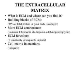
- 1. THE EXTRACELLULAR MATRIX • What is ECM and where can you find it? • Building blocks of ECM: (25% of total protein in your body is collagen) • More ECM components: (Laminin, Fibronectin etc. heparan-sulphate proteoglycan) • ECM functions: (it is not only to keep cells in place) • Cell-matrix interactions. (Integrins)
- 2. EXTRACELLULAR MATRIX • In biology, the extracellular matrix (ECM) is a collection of extracellular molecules secreted by cells that provide structural and biochemical support to the surrounding cells. • The extracellular matrix (ECM) is the non-cellular component present within all tissues and organs, and provides not only essential physical scaffolding for the cellular constituents but also initiates crucial biochemical and biomechanical cues that are required for tissue morphogenesis, differentiation and homeostasis.
- 3. The importance of the ECM is vividly illustrated by the wide range of syndromes, which can be anything from minor to severe, that arise from genetic abnormalities in ECM proteins (Jarvelainen et al., 2009). •Complex arrangements of molecules filling in spaces between the cells. •Not an amorphous jelly or glue but highly organized structure. •Mostly found in connective tissues, such as tendon, cartilage, bone or dermis of the skin. •Diverse structures created by different amounts and organization of ECM components •ECM is a local product for local cells. Cells secrete ECM that is finally assembled outside the cell.
- 6. SOME FUNCTIONS OF ECM • Due to its diverse nature and composition, the ECM can serve many functions, such as: Providing support Segregating tissues from one another, & Regulating intercellular communication. The extracellular matrix regulates a cell's dynamic behavior.
- 7. Although, fundamentally, the ECM is composed of water, proteins and polysaccharides, each tissue has an ECM with a unique composition and topology that is generated during tissue development through a dynamic and reciprocal, biochemical and biophysical dialogue between the various cellular components (e.g. epithelial, fibroblast, adipocyte, endothelial elements) and the evolving cellular and protein micro-environment. Indeed, the physical, topological, and biochemical composition of the ECM is not only tissue-specific, but is also markedly heterogeneous. Cell adhesion to the ECM is mediated by ECM receptors, such as integrins, discoidin domain receptors and syndecans (Harburger and Calderwood, 2009; Humphries et al., 2006; Leitinger and Hohenester, 2007; Xian et al., 2010). Adhesion mediates cytoskeletal coupling to the ECM and is involved in cell migration through the ECM (Schmidt and Friedl, 2010).
- 8. Moreover, the ECM is a highly dynamic structure that is constantly being remodeled, either enzymatically or non-enzymatically, and its molecular components are subjected to a myriad of post-translational modifications. Through these physical and biochemical characteristics the ECM generates the biochemical and mechanical properties of each organ, such as its tensile and compressive strength and elasticity, and also mediates protection by a buffering action that maintains extracellular homeostasis and water retention. In addition, the ECM directs essential morphological organization and physiological function by binding growth factors (GFs) and interacting with cell-surface receptors to elicit signal transduction and regulate gene transcription. The biochemical and biomechanical, protective & organizational properties of the ECM in a given tissue can vary tremendously from one tissue to another (e.g. lungs versus skin versus bone) and even within one tissue (e.g. renal cortex versus renal medulla), as well as from one physiological state to another (normal versus cancerous).
- 9. MAJOR TYPES OF ECM MOLECULES • Glycosaminoglycans: polysaccharide chains usually found attached to proteins to form proteoglycans • Fibrillar proteins such as collagens (mainly structural role) or fibronectin (adhesive glycoprotein)
- 12. CONNECTIVE TISSUES • Tissue that connects, supports, binds, or separates other tissues or organs, typically having relatively few cells embedded in an amorphous matrix, often with collagen or other fibres, and including cartilaginous, fatty, and elastic tissues.
- 13. Connective tissue Connective tissue (CT) is one of the four types of biological tissue in animal body that supports, connects or separates different types of tissues and organs.
- 14. Connective tissue It develops from the mesoderm. The other three types are epithelial, muscle, and nervous tissue. Connective tissue is found in between other tissues everywhere in the body, including the nervous system. In the central nervous system, the three outer membranes (the meninges) that envelop the brain and spinal cord are composed of connective tissue. All connective tissue consists of three main components: fibers (elastic and collagenous fibers), ground substance and cells. Not all authorities include blood or lymph as connective tissue because they lack the fiber component. All are immersed in the body water. The cells of connective tissue include fibroblasts, adipocytes, macrophages, mast cells and leucocytes.
- 15. Collagen Collagen is the most abundant protein in our bodies, especially type 1 collagen. It's found in muscles, bones, skin, blood vessels, digestive system and tendons. It's what helps give our skin strength and elasticity, along with replacing dead skin cells. Tropocollagen molecule: procollagens (red, green, blue) join to form a triple helical tropocollagen.
- 16. THE COLLAGEN FAMILY • Triple helical domain • Repeated Gly - X - Y amino acid sequence, where X is often proline and Y hydroxyproline • 19 different collagen types (+ possibly 4 more) containing polypeptides encoded by at least 38 genes Proline hydroxyproline
- 17. COMMON THEMES IN ECM SYNTHESIS • Extensive post-translational modification • Route: ER - Golgi - Secretory vesicles • During this journey protein are glycosylated or decorated with long GAG chains • Amino acid recidues can be modified (in collagens proline -> hydroxyproline) Chondroitin sulfate nb R1, R2, R3 Hyaluronan (-4GlcUAβ1-3GlcNAcβ1-)n Glycosaminoglycans[(GAGs) or mucopolysaccharides[ are long unbranched polysaccharides consisting of a repeating disaccharide unit. The repeating unit (except for keratan) consists of an amino sugar (N-acetylglucosamine or N- acetylgalactosamine) along with a uronic sugar (glucuronic acid or iduronic acid) or galactose. Glycosaminoglycans are highly polar and attract water. They are therefore useful to the body as a lubricant or as a shock absorber.
- 18. Aggrecan (ACAN), also known as cartilage-specific proteoglycan core protein (CSPCP) or chondroitin sulfate proteoglycan 1, is a protein that in humans is encoded by the ACAN gene. This gene is a member of the lectican(chondroitin sulfate proteoglycan) family. The encoded protein is an integral part of the extracellular matrix in cartilaginous tissue and it withstands compression in cartilage. Aggrecan (ACAN)
- 20. T cells may recognize these complexes using their T cell receptors (TCRs). These cells process antigens and present them to T-cells. Almost all cell types can serve as some form of APC. They are found in a variety of tissue types.
- 21. Non-collagenous domains • Triple-helical collagen rods are not the only functional domains • Example: Type XVIII collagen that is found in many tissues is associated with basal lamina. – Endostatin is a 22kDa polypeptide that is proteolytically cleaved from the C-terminus of type XVIII collagen – Endostatin found in blood vessel walls and basement membranes – Endostatin is a potent inhibitor of angiogenesis and tumour growth !!! – Endostatin is currently being tested in clinical trials – Similar domains in other collagen family members
- 22. Collagens in disease • Inherited diseases with mutations in collagen genes • Osteogenesis Imperfecta (Osteogenesis imperfecta (OI), also known as brittle bone disease, is a group of genetic disorders that mainly affect the bones. It results in bones that break easily. The severity may be mild to severe.[1] Other symptoms may include a blue tinge to the whites of the eye, short height, loose joints, hearing loss, breathing problems, and problems with the teeth • Fibrotic diseases with accumulation of ECM • Liver Chirrosis • Lung Fibrosis
- 23. Collagens in disease • Osteogenesis Imperfecta - Brittle bone disease (not to be confused with osteoporosis) • Variable from mild to embryonic lethal • Often a point mutation in one of type I collagen genes can cause disease • Glycine substitutions to another amino acid more severe than mutations of X or Y in Gly - X - Y triplet. • Dominant negative effect of some mutations. • Predisposing mutations (e.g. Type II collagen in osteoarthrosis)
- 24. Collagens in disease • Fibrotic diseases such as liver cirrhosis are characterized by accumulation of ECM • Collagen synthesis is mainly regulated by the level of gene activity. • Some growth factors such as TGF-b signal to increase collagen synthesis. • Enzymes in the collagen synthesis are investigated as drug targets to treat fibrotic diseases TGFα is upregulated in some human cancers. It is produced in macrophages, brain cells, and keratinocytes, and induces epithelial development. TGFβ exists in three known subtypes in humans, TGFβ1, TGFβ2, and TGFβ3. These are upregulated in Marfan's syndrome and some human cancers, and play crucial roles in tissue regeneration, cell differentiation, embryonic development, and regulation of the immune system. Isoforms of transforming growth factor-beta (TGF-β1) are also thought to be involved in the pathogenesis of pre-eclampsia. TGFβ receptors are single pass serine/threonine kinase receptors.
- 25. Collagens: • All collagens contain a repeating Gly-X-Y sequence and fold into a characteristic triple- helical structure • Collagens assemble to fibrils or networks • Procollagen chains are modified in ER where they also assemble into a triple helix • Type I collagen is the most abundant type; it is a major structural protein of bone, tendon and dermis • Mutations in collagen chains can render the fibrils unstable
- 26. Fibronectin • Large extracellular glycoprotein • Name = fibro + nectere (to bind) • Multiple domains with different binding sites for other ECM proteins or for receptors on cell surface • Present in tissues and in blood plasma Fibronectin is a high-molecular weight (~440kDa) glycoprotein of the extracellular matrix that binds to membrane-spanning receptor proteins called integrins. Similar to integrins, fibronectin binds extracellular matrix components such as collagen, fibrin, and heparan sulfate proteoglycans (e.g. syndecans).
- 27. Fibronectin is essential for embryonic development • Gene targeting => complete lack of fibronectin Embryonic lethal. • Gross malformations, notochord and somites* missing, heart malformation • Problems in cell adhesion, migration and differentiation • *A somite is a division of the body of an animal or embryo. Somites are bilaterally paired blocks of paraxial mesoderm that form along the head-to-tail axis of the developing embryo in segmented animals.
- 28. Proteoglycan biosynthesis • Signal peptide directs the nascent polypeptide to ER • Protein modifications starts in late ER. GAG side chains elongation and modification takes place in Golgi. • Several specific enzymes to add disaccharide units and to modify them (e.g. sulphation). For example over 30 enzymes are needed in synthesis of aggrecan, a cartilage matrix proteoglycan.
- 29. Syndecans and glypicans • Syndecans are transmembrane proteins. Four family members. Short cytoplasmic tail contains highly conserved sequences that bind to adaptor proteins. Variable part of syndecan-4 cytoplasmic domain binds protein kinase C and affect cell signalling • Glypicans (6 known family members) are lipid anchored to plasma membrane. GPI= glycosylphosphatidylinositol. • Both families: individual family members have distinct expression patterns: e.g. syndecan-1 in epithelia and syndecan-3 in neural cells
- 30. Proteoglycans modulate growth factor activity • Matrix associated PG: sequestration • Membrane-bound PG: presentation • Certain sugar sequences promote FGF signalling and others inhibit • Membrane-bound PGs can be cleaved from cell surface into matrix • Sugar chains can be cleaved by heparanase enzymes to oligosaccharides. Fibroblast growth factors, or FGFs, are a family of growth factors, with members involved in angiogenesis, wound healing, embryonic development and various endocrine signaling pathways.
- 31. Heparin binding proteins • Certain growth factors, especially Fibroblast Growth Factor family (FGF) • Enzymes and their inhibitors, e.g. proteases • Blood coagulation factors • ECM proteins • Note: Proteoglycans can bind several proteins at the same time
- 32. More functions for proteoglycans: - syndecan-3 regulates appetite • Serendipitous discovery in transgenic mice over- expressing syndecan-1 under a viral promoter => maturity-onset obesity. • Heparan-sulphate sugar chains potentiate signalling in hypothalamus that induces over-eating. • In normal mice syndecan-3 is present in hypothalamus (in addition to other neural tissues). • Food deprivation induces syndecan-3 expression several fold and triggers reflex hyperphagia.
- 33. Aggrecan: Example of Matrix Proteoglycans • Aggrecan (ACAN), also known as cartilage- specific proteoglycan core protein (CSPCP) or chondroitin sulfate proteoglycan 1, is a protein that in humans is encoded by the ACAN gene. This gene is a member of the lectican (chondroitin sulfate proteoglycan) family.
- 36. Aggrecan is one of the major macromolecules in the extracellular matrix of articular cartilage and endows the tissue with its characteristic water imbibing properties and its ability to undergo reversible compression. During normal aging of cartilage, Aggrecan undergoes many posttranslational modifications to its protein core and to the number, size, and proportion of the chondroitin sulfate and keratin sulfate chains that are covalently associated with it. These events are mostly catabolic in nature and are a consequence of the relatively long resident time of aggrecan in the matrix. However, it is clear that a number of these age-related changes could only have come about by an altered anabolic response of the chondrocyte
- 37. Proteoglycans in human diseases Proteoglycans are a major component of the animal extracellular matrix, the "filler" substance existing between cells in an organism. Here they form large complexes, both to other proteoglycans, to hyaluronan, and to fibrous matrix proteins, such as collagen. The combination of proteoglycans and collagen form cartilage, a sturdy tissue that is usually heavily hydrated (mostly due to the negatively charged sulfates in the glycosaminoglycan chains of the proteoglycans).[5] They are also involved in binding cations (such as sodium, potassium and calcium) and water, and also regulating the movement of molecules through the matrix. Evidence also shows they can affect the activity and stability of proteins and signalling molecules within the matrix.[citation needed] Individual functions of proteoglycans can be attributed to either the protein core or the attached GAG chain. They can also serve as lubricants. An inability to break down proteoglycans is characteristic of a group of genetic disorders, called mucopolysaccharidoses. The inactivity of specific lysosomal enzymes that normally degrade glycosaminoglycans leads to the accumulation of proteoglycans within cells. This leads to a variety of disease symptoms, depending upon the type of proteoglycan that is not degraded.
- 38. Hyaluronic acid Hyaluronic acid, also called hyaluronan, is an anionic, nonsulfated glycosaminoglycan distributed widely throughout connective, epithelial, and neural tissues. Hyaluronic acid (HA; conjugate base hyaluronate), also called hyaluronan, is an anionic, nonsulfated glycosamino- glycan distributed widely throughout connective, epithelial, and neural tissues. It is unique among glycosaminoglycans in that it is nonsulfated, forms in the plasma membrane instead of the Golgi apparatus, and can be very large, with its molecular weight often reaching the millions. One of the chief components of the extracellular matrix, hyaluronan contributes significantly to cell proliferation and migration, and may also be involved in the progression of some malignant tumors
- 40. Hyaluronic acid Hyaluronic Acid is a glucosaminoglycan consisting of D- glucuronic acid and N-acetyl-D- glucosamine disaccharide units that is a component of connective tissue, skin, vitreous humour, umbilical cord, synovial fluid and the capsule of certain microorganisms contributing to adhesion, elasticity, and viscosity of extracellular substances.
- 41. CD44* • Adhesive glycoprotein • Numerous isoforms from alternative splicing • Originally found as a ‘homing receptor’ in T-lymphocytes • Some splice isoforms are suggested to play a role in tumour metastasis • Cytoplasmic tail of CD44 binds to ERM proteins (ezrin-radixin- moiesin family) that can regulate dynamics of actin cytoskeleton • *Hyaluronic acid (HA), an important component of the extracellular matrix (ECM), is the principal, but by no means the only, ligand of CD44. Other CD44 ligands include the ECM components collagen, fibronectin, laminin, and chondroitin sulfate.
- 42. Basement membrane • Also known as basal lamina • Thin sheet-like network • Epithelial, endothelial, muscle and Schwann cells • Physical support, developmental control, filtering functions • Major constituents: laminins, collagen type IV, perlecan (a proteoglycan)
- 43. Laminins • The laminin molecules are named according to their chain composition. Thus,laminin-511 contains α5, β1, and γ1 chains. ... The trimeric proteins intersect to form a cross- like structure that can bind to other cell membrane and extracellular matrix molecules. Molecular composition of basement membranes is tissue-specific • Laminins: at least 11 heterotrimers – Five alternative alpha chains, – Three alternative beta chains – Two alternative gamma chains – For example: in skin in the BM between epidermis and dermis, Laminin-5 (a3b3g2) is the predominant laminin isoform.
- 44. Structure and function of laminin: anatomy of a multidomain glycoprotein Laminin is a large (900 kDa) mosaic protein composed of many distinct domains with different structures and functions. Globular and rodlike domains are arranged in an extended four-armed, cruciform shape that is well suited for mediating between distant sites on cells and other components of the extracellular matrix. The alpha-helical coiled-coil domain of the long arm is involved in the specific assembly of the three chains (A, B1, B2, and possible variants) of laminin and is the only domain composed of multiple chains. H-D-Tyr-Ile-Gly-Ser-Arg-OH Chain, Laminin M Glycoprotein GP 2 Glycoprotein GP-2 Laminin Laminin M Laminin M Chain M Chain, Laminin Merosin
- 45. Laminin Laminin has active domains for collagen binding, cell adhesion, heparin binding, and neurite outgrowth (PHD) fragment.1Laminin chains are designated A (molecular weight 400kDa), B1 (molecular weight 210 kDa) and B2 (molecular weight 200 kDa). The cohesion between these chains is the result of many inter- and intrachain disulfide bonds. Together, they cause the molecule to look like a crucifix. Laminin is a ubiquitous non-collagenous connective tissue glycoprotein that is a major constituent of basement membranes.
- 46. Name Source Storage Temp Target Cells For Attachment Concentration For Use Cat. No. Laminin, aqueous solution from Engelbreth- Holm-Swarm murine sarcoma basement membrane −20°C Epithelial cells, endothelial cells, muscle cells, tumor cells, hepatocytes, Schwannoma 1 - 2 μg/cm2 L2020-1MG Laminin, liquid from human placenta −70°C Epithelial cells, endothelial cells, muscle cells, tumor cells, hepatocytes, Schwannoma 1 - 2 μg/cm2 L6274-.5MG
- 47. Human proteins containing laminin domains Laminin Domain I LAMA1; LAMA2; LAMA3; LAMA4; LAMA5; Laminin Domain II LAMA1; LAMA2; LAMA3; LAMA4; LAMA5; Laminin B (Domain IV) HSPG2; LAMA1; LAMA2; LAMA3; LAMA5; LAMC1; LAMC2; LAMC3; Laminin EGF-like (Domains III and V) AGRIN; ATRN; ATRNL1; CELSR1; CELSR2; CELSR3; CRELD1; HSPG2; LAMA1; LAMA2; LAMA3; LAMA4; LAMA5; LAMB1; LAMB2; LAMB3; LAMB4; LAMC1; LAMC2; LAMC3; MEGF10; MEGF12; MEGF6; MEGF8; MEGF9; NSR1 ; NTN1; NTN2L; NTN4; NTNG1; NTNG2; RESDA1; SCARF1; SCARF2; SREC; STAB1; USH2A; Laminin G domain AGRIN; CELSR1; CELSR2; CELSR3; CNTNAP1; CNTNAP2; CNTNAP3; CNTNAP3B; CNTNAP4; CNTNA P5; COL11A1; COL11A2; COL12A1; COL14A1; COL15A1; COL16A1; COL18A1; COL19A1; COL20A1; COL21A1; COL22A1; COL24A1; COL27A1; COL5A1; COL5A3; COL9A1; CRB1; CRB2; CSPG4; EGFLA M; EYS; FAT; FAT2; FAT3; FAT4; GAS6; HSPG2; LAMA1; LAMA2; LAMA3; LAMA4; LAMA5; NELL1; N ELL2; NRXN1; NRXN2; NRXN3; PROS1; SLIT1; SLIT2; SLIT3; SPEAR; THBS1; THBS2; THBS3; THBS4; USH2A; Laminin N-terminal (Domain VI) LAMA1; LAMA2; LAMA3; LAMA5; LAMB1; LAMB2; LAMB3; LAMB4; LAMC1; LAMC3; NTN1; NTN2L; NTN4; NT NG1; NTNG2; USH2A
- 48. More Basal Lamina Proteins • Perlecan: a large heparan sulfate proteoglycan. HS chains bind other BM components and contribute to filtering functions • Entactin: interacts with laminin and type IV collagen • Nidogen, a laminin binding protein
- 49. Hemidesmosome: a cell - basement membrane adhesion site • In some epithelia: epidermis, bladder, trachea, breast and amnion • Shares some ultrastructural features with desmosomes: both display dense, membrane- associated cytoplasmic plaques that are connected to intermediate fialments. But molecular composition is different. • Transmembrane glycoproteins connect basement membrane to intracellular plaque
- 50. Basal lamina functions -developmental guidance • Early embryo: keeps 4 and 8 cell stages together • Differentiation of epithelial organs; epithelial - mesenchymal interactions • Neurite outgrowth: guidance of axon growth by ECM containing laminin subunits • (Neurite Outgrowth is a process wherein developing neurons produce new projections as they grow in response to guidance cues. ... Dynamic neurite outgrowth during development results in the formation of a complex neuronal architecture that results in the establishment of the functional nervous system and brain)
- 51. ECM Turnover - MMPs • Matrix metalloproteinases are enzymes that cleave components of ECM • Over 20 different enzymes with different specifications. • Common theme: expressed as an inactive pro- enzyme • Also other substrates than ECM proteins • TIMPs = tissue inhibitors of MMPs
- 52. MMPs - some examples • “Old names” collagenases, gelatinases and stromelysin replaced by numbers (e.g. MMP- 1) • MMP-1 (collagenase-1) cuts triple helical collagens • MMP-9, (Gelatinase-B) chops e.g. type IV collagen and laminins • MT-MMPs are membrane-bound enzymes
- 53. MMPs - some functions • Regulate amount of ECM - degradation and remodelling • Cell migration, wound healing, angiogenesis* • Activate other MMPs • Release or activate growth factors and other bioactive molecules • *Angiogenesis is the formation of new blood vessels. This process involves the migration, growth, and differentiation of endothelial cells, which line the inside wall of blood vessels. The process of angiogenesis is controlled by chemical signals in the body. • (Angiogenesis is an important process that occurs both during health and disease. Blood is important in the body as it carries oxygen and nutrients to all the parts of the body via blood vessels like arteries and brings back the toxins and wastes from these peripheral organs for purification via veins)
- 54. MMPs in diseases • Extensive matrix degradation e.g. in periodontitis*, rheumatoid arthritis • Tumour cell invasion and metastasis: – Carcinoma breaks basement membrane and invades surrounding stroma. • MMP inhibitors tested for therapeutic use *Periodontitis (per-e-o-don-TIE-tis) is a serious gum infection that damages the soft tissue and destroys the bone that supports your teeth
- 55. Integrins • At least 24 different heterodimers from 9 b subunits and 18 a subunits. • Variable pairing: b1 integrin can have 11 different a partners. • Overlapping ECM binding: e.g. 8 different integrins can bind fibronectin • An integrin can bind one or several ECM proteins Integrins are proteins that function mechanically, by attaching the cell cytoskeleton to the extracellular matrix (ECM), and biochemically, by sensing whether adhesion has occurred. The integrin family of proteins consists of alpha and beta subtypes, which form transmembrane heterodimers.
- 57. Integrins and cell behaviour • Clustering of integrins (“velcro effect” in adhesion) • Responses to cell adhesion include spreading, cytoskeletal re-organisation, polarisation, migration, proliferation, activation of specific genes • Cell survival: epithelial cells that are detached commit suicide (this type of apoptosis* is called anoikis). • Also, inside out signalling: integrins can have inactive conformation that does not bind matrix unless first activated. *the death of cells which occurs as a normal and controlled part of an organism's growth or development. Velcro effect: The Velcro Effect is when words and images are associated with sensation and energy in the body.
- 58. SUMMARY • Collagens: Triple helical rod and non-collagenous domains. Important structural role. Extensive post-translational modifications • Fibronectin: adhesive glycoprotein in matrix and plasma • Proteoglycans: GAG-chains attached to core protein. • Laminins: major components of basement membranes • Matrix metalloproteinases degrade and re-model matrix • Integrins: heterodimeric proteins that mediate cell adhesion to extracellular matrix. • Cell-matrix interactions important regulator of cell behaviour
- 59. Integrins - variety in functions Integrins Basal lamina functions -developmental guidance Basal lamina functions -filter Basal lamina functions- structural support Hemidesmosomes and basement membrane- molecular composition Hemidesmosomes and basement membrane- ultrastructural view Type IV collagen Interactions of laminins Laminins Basement membrane