
Recomendados
Mais conteúdo relacionado
Semelhante a Eye
Semelhante a Eye (20)
Mais de MrWestbury
Mais de MrWestbury (17)
Último
Último (20)
Eye
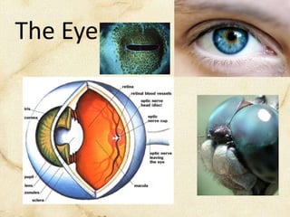
- 1. The Eye
- 3. • Cornea refracts light through the pupil & lens • Lens may change shape for objects at distance • Image is projected INVERTED on the retina • Each eye has a blind spot where the optic nerve & blood vessels enter the eye.
- 4. Retina • A thin layer on the back of the eye where light is converted to an electrical signal and passed through the optic nerve to the eye.
- 5. • Photoreceptors called RODS & CONES sense light • RODS: great at low light sensing, poor resolution & colour • CONES: functions only in bright light & provides information about colour & detail
- 6. CONES are responsible for our colour vision is known as TRI-CHROMATIC vision We see as a combination of 3 colours: RED (630 nm) BLUE (420 nm) GREEN (560 nm) Rods absorb light best at 540 nm Each cone cell contains pigment molecules designed to absorb different wavelengths of light
- 7. Visual Pigments • Each pigment is composed of two molecules: – OPSIN • Proteins that react to specific wavelengths of light • All rods contain a specific type – CHROMOPHORE • RETINAL – a modified form of Vitamin A which changes shape in response to light
- 8. Different firing rates of photoreceptor cells are sent to the brain where the rates are compared. The optic nerves cross at the OPTIC CHIASM and each eye’s signal is sent to the occipital lobe of the brain where the image is ‘assembled’.
- 9. MYOPIA Hyperopia
- 10. ASTIGMATISM