Lymphomas+ Multiple Choice Questions
•Transferir como PPTX, PDF•
15 gostaram•11,034 visualizações

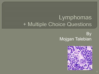
Lymphoma is a cancer of lymphocytes. The most common place for abnormal lymphocytes is in lymph nodes (glands) particularly under the arms, in the neck and in the groin. Lymphoma is solid tumors of the immune system arising from cells of lymphoid tissues; lymphocytes, histiocytes, and reticulum cells. It can happen anywhere in the immune system, but usually in lymph nodes, spleen, marrow, and tonsils. Location and the behavior of lymphomas separate them from leukemia.The malignancy starts and restricted to lymphoid tissues and progress to involve the BM and appears in PB, at this stage it may be named, “lymphosarcoma cell leukemia.

Recomendados
Mais conteúdo relacionado
Mais procurados
Mais procurados (20)
Destaque
Destaque (9)
Semelhante a Lymphomas+ Multiple Choice Questions
Semelhante a Lymphomas+ Multiple Choice Questions (20)
Último
Último (20)
Lymphomas+ Multiple Choice Questions
- 2. Solid tumors of the immune system Arise from cells of lymphoid tissues; lymphocytes, histiocytes, and reticulum cells Anywhere in the immune system, but usually in lymph nodes, spleen, marrow, and tonsils Location and the behavior of lymphomas separate them from leukemias The malignancy starts and restricted to lymphoid tissues and progress to involve the BM and appears in PB, at this stage it may be named, “lymphosarcoma cell leukemia” 2
- 3. A heterogeneous disorder • Hodgkin lymphoma or Hodgkin disease • Lymphocytic lymphomas or referred to as non- Hodgkin’s lymphoma (NHL’s), most cases are fatal 3
- 4. Based on the following categories • Physical examination, measurement of the enlarged lymph node/spleen • Bone marrow examination, Aspirate/biopsy • Radiology studies/CT scans • Hematology studies • Chemistry tests; pr electrophoresis, kidney and liver function tests • Lapratomy to determine the spread of the disease • Lymph node biopsy/histological features 4
- 5. Stage I only 1 lymph node area or lymphoid organ (I) 1 area of a single organ outside of the lymph system (IE) Stage II on the same side of the diaphragm(above or below) in 2 or more groups of lymph nodes ( II) extends from a single group of lymph node(s) into a nearby organ (IIE) Stage III in lymph node areas on both sides of (above and below) the diaphragm It may also have spread into an area or organ next to the lymph nodes (IIIE), into the spleen (IIIS), or both (IIISE). Stage IV spread outside the lymph system into an organ that is not right next to an involved node. spread to the bone marrow(IVM ) , liver ( IVL), brain or spinal cord, or the pleura (thin lining of the lungs 5
- 6. A, asymptomatic B, presence of any or all of the following signs: • Greater than 10% of total body weight loss in the last 6 months • Unexplained fever above 38° C, night sweats 6
- 7. A malignant lymphoma with clonal proliferation Unknown etiology • majority of cases both classical HL(CHL) and nodular lymphocyte predominant HL(NLPHL) are clonal proliferation of B cells • may caused by infectious transmission in the host: Epidemiologic evidence Presence of two peaks period of onset Recovery of the EB Virus DNA from “lesions of approximately 80% of cases of HL” certain RNA tumor viruses CMV, herpes virus 6 Presence of Reed-Sternberg cells accompanied with H-cells (malignant histiocytes), these cells are surrounded by lymphocytes, plasma cells, and eosinophils 7
- 8. The hallmark of the HL Very large cells, 50 -100 µm, irregular with abundant pale acidophilic cytoplasm, usually with 2 lobulated nuclei each contains a gigantic azurophilic nucleolus with a perinuclear halo The usual immunophenotypes: CD45- ,CD30+, CD15+ These cells can be found in other diseases: Rubella, EBV, IMN, Toxoplasmosis, T-cell lymphoma, multiple myeloma 8
- 9. Classical Lacunar Popcorn cells Sarcomatous Each type of these variants is associated with a specific histological type of HL Immunophenotypes: • More types of RS cells are HLA-DR + expressing T-cell null lineage receptors for IL-2(CD25+), CD13 +, CD15+, CD45-,CD30+ Not reactive to B-cell antigens Clonal T-cell rearrangement T zone distribution Lymphocyte predominant form of HD • RS variant typically displays Pan B-cell antigens, tendency of invasion to B-cell area • Some RS variants have feature of monocyte-macrophage lineage; phagocytic and contain lysozyme and alpha -naphthyl esterase 9
- 10. 10 Mononuclear Hodgkin cell L& H (popcorn cell) Characteristic cell in NLPHL Pleomorphic Reed-Sternberg cell Lacunar cell
- 11. Histological classification of HD is based on histological examination of a lymph node Rye classification system is a modification of Lukes and Butler system • Lymphocyte predominant, * WHO recognizes it as a distinct form of HD • Nodular sclerosing • Mixed cellularity • Lymphocyte depleted * Each classification is based on: the amount of Lymphocyte presents, the type of RS cells, and the degree of fibrosis 11
- 12. Lymphocyte predominant Classic HD • Nodular sclerosing • Mixed cellularity • Lymphocyte rich • Lymphocyte depletion 12
- 13. Patients: usually young, asymptomatic and commonly in stage I or II, good prognosis The lymph node: • Small lymphocytes, mature B-cells • Varying number of histiocytes • A few plasma cells • There is no: neutrophils, eosinophils, necrosis and fibrosis • RS variant cells: Popcorn cells/L&H cells in large numbers: small cells with lobulated nuclei and small nucleoli • L&H cells relates to proliferating germinal center (centroblast) cells • Intra-follicular pattern • B cells; CD30+ and CD15- 13
- 14. Two types of LP • Nodular pattern Malignant cells surrounded by lymphocytes Male predominant In younger patients • Diffuse pattern Cluster of malignant cells surrounded by loosely arranged non- malignant reactive T cells 14 NLPHL http://www.pathpedia.com/
- 15. Common type of HD, 60% to 80% cases of HL More common in female patient less than 50 Invasion to mediastinum Good prognosis because of the patient’s fibrogenic defense Lymph node: • Histological pattern: follicular, birefringent collagenous sclerosis • Nodules of H-cells • Variable number of lymphocytes separated by thick bands of collagen • T, B, Null cells; CD30+ and CD15+ • Classic Reed-Sternberg cells • Lacunar RS cells An empty space /lacuna around the nucleus Because of cytoplasm shrinkage during formalin fixation 15
- 16. 16 Nodular Sclerosis HL, Collagen bands between the lymphoid tissue
- 17. • 15% to 30% cases of HL • Patients usually present in stage III or IV, poor prognosis Heterogeneous mixed Cellularity: • Diffuse or follicular histologic pattern • Neoplastic cells outnumber the reactive cells • Numerous classical RS and RS variants (mononuclear) • Lymphocytes, macrophages, eosinophils and plasma cells, histiocytes • Presence of fibrosis but no collagen • T, B and Null cells;CD30+ and CD15+ 17 MCHL
- 18. 5% of HL cases Rich in small lymphocytes • Small numbers of classic RS and H cells • Clinical and pathological features more close to Mixed Cellularity HD 18
- 19. Rarest (<1%) and the most aggressive form of HD In older patients or HIV Positive, median age 35 Presents in stage III or IV, the poorest prognosis Lymph node: • Diffuse and fibrotic pattern • Large numbers of RS (sarcomatous variants) and H cells • Rare lymphocytes in fibrotic stroma • Large and bizarre neoplastic cells • Irregular sclerosis • Unknown cells; CD30+ and CD15+ 19
- 20. In young adult and after the age of 60: • Lymphadenopathy Non-painful lymph node swelling • Weight loss • Fever, night sweats, fatigue • Mediastinal involvement, in 60% of cases • Supraclavicular area invasion; distension of the neck • Pruritus and purpura; less common • Paralysis as a result of spinal/nerve compression • Suppressed cellular immunity because of the disease and therapy; viral, fungal and protozoan infections • Extranodal involvement is common in • and NSHL 20
- 21. Normochromic normocytic, mild anemia, sometimes development of autoimmune hemolytic anemia Slightly increased leukocyte, neutrophilia Lymphopenia is common Increased ESR BM involvement is focal and associated with fibrosis, more seen in advanced stages 21
- 22. Significant percentage of patients treated successfully Chemotherapy • for patient in stage II and above or not cured by radiotherapy Radiation therapy • Best choice for patients with good prognosis; localized lesions, asymptomatic ,and at stage IA Mantle field Inverted Y field Combination of mantle and inverted Y, Total Nodal Irradiation TNI 22
- 23. Most commonly chemotherapeutic regime used: ABVD Adriamycin, inhibiting replication and transcription of DNA Bleomycin, breaking single and double stranded DNA Vinblastine, disrupting mitotic spindles; BM suppression Dacarbazine, interfering with DNA synthesis • 4 cycles of ABVD for patients with early stages • 6 to 8 cycles of ABVD for more advance disease • For lesions greater than 10cm a combination of ABVD and radiotherapy 23
- 24. Solid tumors of the immune system Mostly B-cell lineage (in the follicles of the lymph nodes),85% to 90% of lymphomas T-cell/NK about 10% to 15% lymphomas Histiocytic/dendritic cell lineage about <1% 3-5% of all malignancies in modern countries, seventh most common malignancy Average age 60 yrs-old, more common in men 24
- 25. Three major anatomical regions of a lymph node: Cortex, paracortex, and medulla Cortex: B-cells, macrophages, plasma cells, and reticular cells • Primary follicles, predominant cells are small B cells • in response to antigenic stimulation primary follicles convert to Secondary follicles (germinal center surrounded by a crescent of B cells named mantle zone): Germinal center : centrocytes (resting B cells), centroblasts ( proliferating B cells), dendritic cells, and histiocytes/macrophages Starry sky of the germinal center is because of the pale large macrophages Paracortex: mostly T cells, endothelial venules for entry of circulating lymphocytes into the lymph node parenchyma Medulla: T cells, B cells, macrophages, and plasma cells 25
- 26. 26
- 27. Well differentiated lymphocytes • T or B-cells • Small cells • Small non-cleaved; plasmacytoid B-cells, B- centrocytes • Small and large cleaved cells Poorly differentiated lymphocytes • T or B-cells, immunoblasts • T or B-cells, centroblasts • B-cell large non-cleaved cells Macrophages 27
- 28. Classification is based on: • Histologic pattern • cell size • Nuclear characteristics; cleaved or non-cleaved, convoluted or cerebriform • Immunophenotypes Keil classification • Morphology • Immunophenotyping Luke and Collins system • Immunophenotyping The Rappaport system • Growth pattern • cytological characteristics Revised European/American Lymphoma or REAL classification • Clinical features • Immunophenotypes • Genotypic features WHO classification of tumors of lymphoid tissues • Modified form of the REAL classification • The classification includes NHLs, and all lymphoid neoplasms ; leukemias, HD and plasma cell dyscrasia 28
- 29. B cell Neoplasms • Precursor B cell neoplasm Precursor B lymphoblastic leukemia/lymphoma • Mature B-cell neoplasms • B cell proliferations of uncertain malignant potential T-cell and NK-cell neoplasms • Precursor T cell neoplasms Precursor T lymphoblastic leukemia/lymphoma • Mature T cell and natural killer neoplasms • T cell proliferation of uncertain malignant potential Hodgkin Lymphoma Histiocytic and dendritic cell neoplasms Posttransplantation lymphoproliferative disorders (PTLDs) http://www.ncbi.nlm.nih.gov/pmc/articles/PMC3109529/table/T1/ 29
- 30. The majority of lymphoblastic lymphomas are T cell type (85% to 90%) Solid tumor most commonly relative to T-cell ALL, small percentage as pre-B cells Mostly in older male children, young male adults Lymph node involvement and < 25% blasts in bone marrow Malignant cells at early stage of thymic T-cell differentiation; TdT+, CD7+, CD2+, CD5+, CD4+/CD8+ CD3 expression is lineage specific Non- T-cell Ags expression:CD79a, CD13, and CD33 A high risk disease Mediastinal involvement is common Infiltration to BM and CNS is common Bone lytic lesions is common in B- cell LBL 30
- 31. B cell lymphomas • Precursor B cell lymphomas , uncommon (<1% of NHLs) • Mature B cell lymphomas, 90% of all lymphoid neoplasms Most common in developed countries Predominant in older adults, 60 yrs Some of the subtypes of Mature B cell lymphomas : Follicular lymphoma Diffuse large B cell lymphoma Burkitt lymphoma CLL/Small Lymphocytic lymphoma Marginal Zone lymphomas (MALT lymphomas) 31
- 32. 40% of NHLs and majority of low grade lymphomas (partially) follicular growth pattern with two principal cell types in the germinal center: • Centrocytes (cleaved cells) • Centroblasts (large noncleaved cells) • the malignant follicle the absence of a well- developed mantle zone the absence of tingible body macrophages 32 Secondary follicle (benign) Dark and Light Zone, Mantle Zone Tingible body macrophages (arrows) Follicular Lymphoma Poorly defined mantle zone Lack of polarization into dark and light zones
- 33. Chromosomal Translocation: • t(14;18) in 70% to 95% of cases of follicular lymphoma, involving BCL2 and IgH gene Immunophenotype of follicular lymphoma: • sIg+ • B cell associated Antigens: CD19+, CD20+, BCL- 6+, CD10+, CD5-, CD23-, CD43-, CD11c-, 33
- 34. The most common type of NHL worldwide 30% Starts de novo or as an advanced phase of a pre- existing B-cell lymphoma; cloned B cells of large size Patients average age; 50 yrs old, male/female At the time of diagnosis the disease is mostly limited to one side of the diaphragm, but has the ability to infiltrate to extranodal sites (gastrointestinal tract) Presence of sclerosis in cleaved cell variant Large mononuclear cells • Mostly B cell centroblast; CD5-, CD10- • Large nucleus cleaved or non-cleaved 34 DLBCL a mixture of large B cells: Centroblastic, immunoblastic , and pleomorphic morphology
- 35. Highly aggressive and rapidly fatal but potentially curable the doubling time of the lymphoma is the highest of any tumor chromosomal rearrangements of the c-myc oncogene Associated with AIDS and transplant recipients Median age 7 yrs old, male-to female ratio approximately 2.5 to 1 • Translocation of t(8;14) in majority of cases Two forms of the disease; • Endemic, tropical Africa and New Guinea, cells are positive for EBV genome, accompanied by tumors in the face and jaw, an early B cell origin of the lymphoma cells • Non-endemic/sporadic, median age of 11 yrs old, negative for EBV genome, and in immunocompromised patients (AIDS),tendency to Payer's patches and abdominal areas (abdominal mass), the latter stage of B cell development Burkitt’s lymphocytes: • Moderate size cells with small uniform nuclei and many prominent nucleoli • High mitotic rate • Lipid vacuoles in the cytoplasm on smears or imprint of the tissue • “Starry sky” pattern is because of phagocytosis of apoptotic debris • Morphology of the cell types in Romanowsky staining is ALL L3 • Immunophenotype : CD10+, sIgM+, CD5-, CD23-, Bcl-2-, TdT- 35
- 36. • Solid state version of B cell CLL • B cell lineage with surface IgM; CD5+ (a T-cell Ag), CD19+, CD20+, CD22+, CD23+, CD43+ • B-cell SLL cells with plasmacytoid differentiation lack CD5 • 80% of SLL exhibit clonal cytogenetic abnormality, del 13q14 (50%) *presence of CD5 distinguishes these lymphomas from the follicular type, • Malignant cells spread to other parts of body through bloodstream, BM involvement seen • tendency to turn into more aggressive lymphoma • Growth centers (pseudofollicular pattern) are composed of prolymphocytes and paraimmunoblasts cells 36
- 37. Median age 60 yrs old, more common in females A low grade neoplasm, localized at the time of diagnosis The extranodal MZL; lung, stomach, thyroid, eyes, lungs, salivary glands, stomach; lymphomas of mucosal-associated lymphoid tissue (MALT), associated with chronic Ag stimulation; autoimmune disease (Hashimoto's thyroiditis, and Sjogren's syndrome) or infections like Helicobacter pylori gastritis Lymph node-based MZL: in patients with autoimmune disorders e.g. Sjogren’s syndrome or coexistent extranodal type of marginal zone lymphoma Lymphoepithelial lesions is a constant feature of this lymphoma Follicular colonization, the neoplastic cells infiltrate the reactive follicules B- cells • Marginal zone B-cells, centrocyte-like cells (small cleaved cells) • monocytoid lymphocytes: in lymphoid sinuses and paracortex of some reactive lymph nodes but not normal lymph nodes • Plasma cell differentiation is common • Cells are; sIg+, cIg+, CD10-, CD19+,CD23- and CD43+ 37
- 38. Mostly in elderly male patients Involvement of lymph nodes and spleen Aggressive neoplasm Lymphomatous polyposis: • Presentation of MCL in intestine Small to medium size B- cells • originate at germinal center • Forma a mantle zone around the benign germinal center • sIg+, CD19+, CD5+, and CD43+, CD10- • Translocation of t(11;14)in most cases, involving the BCL1 proto-oncogene and immunoglobulin heavy- chain gene 38 Normal lymph node
- 39. 12% of NHLs worldwide Among the most aggressive of hematolymphoid malignancies Divided to mature and precursor types; precursor T- cell lymphoma is identical to T-cell acute lymphoblastic leukemia Mature T-cell/NK cell lymphomas, the mature T-cells are mostly differentiate into CD4+ or CD8+ • Three patterns of clinical features in mature T/NK cell lymphoma: Nodal presentation e.g.: Peripheral T-cell lymphoma, not otherwise specified Anaplastic large cell lymphoma Extranodal presentation e.g.: Primary cutaneous anaplastic large cell lymphoma Leukemic/disseminated 39
- 40. • 50% of the mature T/NK cell lymphomas • A disease of adults but some cases is in childhood • Predominantly nodal presentation, with extranodal sites involvement • Lymphoma cells Polymorphous mixture of atypical small, medium, and large sized cells mix with variable numbers of reactive cells ( Eos, plasma cells and macrophage) Mature T-helper phenotype:CD3+, TdT-, CD4+, and CD8-, however some phenotypes show CD8+ with CD4+/- 40
- 41. • Mostly in children and young adults • A nodal based lymphoma • Rare CD30+ (Ki-1ag) lymphoma cells • Translocation t(2;5) leads to expression of the ALK- NPM protein • Expression of epithelial membrane antigen (EMA) leads to confusion with carcinoma • Clonal rearrangement of T cell receptor, in 90% of cases • Heterogenous cells with variable number of distinctive kidney/horseshoe-shaped large cells and juxtanuclear eosinophilic inclusion – like zone next to the nucleus 41http://imagebank.hematology.org/
- 42. Dysplastic and malignant T-cell proliferations A tendency for infiltration of the skin Disorders: • Mycosis fungoides and Sezary syndrome: Infiltration of the dermis and epidermis by malignant T- cells with cerebriform nucleus • Lymphomatoid papulosis • Primary cutaneous anaplastic large-cell lymphoma 42
- 43. Three clinical phases: • Premycotic (erythroderma) phase, 6 months to 50 yrs, with eczematous dermatosis because of T-cell infiltrate • Formation of plaques, plaque stage • Tumor nodules, tumor stage Systemic dissemination develops in the later stages As a terminal event transformation to a large cell lymphoma Prognosis is good and > 10 yrs, except for extracutaneous spread with a survival rate <1 year 43
- 44. In the early erythroderma stage, leukemia of the cerebriform T cells (Sezary cells) Tumor stage is unusual in Sezary syndrome 44
- 45. Two types of Sezary cells; 1. Larger than mature small lymphocytes, dense chromatin pattern, cerebriform, is deeply creased or grooved, and lumpy 2. Atypical Sezary cell, large with convoluted nucleus ringed by a string of cytoplasmic vacuoles • Phenotyping; in 90% of cases Pan T +, CD4+, CD8-, in 10% of cases CD8+, monoclonal TCR gene rearrangements • Cytogenetic abnormalities present but not specific 45
- 46. In early stages; patchy, scaly or generalized reddening areas of skin, itching Skin infections (fungal) secondary to scratching and ulceration Tumor like lesions, plaques (raised circular patches of skin) Diagnosis • Histology of the infiltrated skin areas: malignant Sezary cells in clusters known as Pautrier’s microabsecesses • Presence of Sezary cells in PB • Lymph node biopsy in the case of lymphadenopathy 46
- 47. Topical chemotherapy ( nitrogen mustard , steroid creams) Radiation therapy, electron beam irradiations Photo chemotherapy, psoralin (methoxsalen), UVA For advanced stages of the disease; topical therapeutic combined with systematic chemotherapy 47
- 48. Infiltrating the skin, most often head and neck as red and purple nodules Lung Gastrointestinal involvement in lymphomas around abdominal area, mostly in stomach and the small intestine; vomiting, abdominal mass, diarrhea, GI bleeding, and obstruction; diffuse large cell lymphomas Waldeyer’s ring; the oral cavity, maxillary sinuses, nasal cavities Liver involvement Spleen, splenomegaly and as a result pancytopenia BM involvement, but lytic lesions of the bone is not common CNS involvement in high grade lymphomas, in Burkitt’s lymphoma CNS involvement rate is high 48
- 49. Blood cell counts normal Anemia due to replacement of marrow cells, hypersplenism, AHA, GI bleeding, and therapy effects Thrombocytopenia secondary to the above causes Lymphocytopenia depending to the stage of the disease Invasion of lymphoma cells in 10% lymphomas leads to absolute lymphocyte counts of 15.0 to 40.0 X 10⁹ /L; usually in diffuse, small lymphocytic, and the lymphoblastic lymphomas 49
- 50. Positive biopsy of BM indicates stage IV BM involvement mostly in: • Small cleaved cell lymphoma • Mixed small cleaved lymphoma • Large cell lymphoma • Lymphoblastic lymphoma; nodular or focal pattern 50
- 51. Radiotherapy, most of lymphomas are sensitive • Hodgkin disease • NHL • Low grade lymphomas in the early stages • Lymphomas in stage I Chemotherapy • CVP/CHOP ( Cyclophosphamide, Adriamycin, Oncovin, vincristine, and prednisone) 51
- 52. 1. The tissue equivalent of CLL? a. Diffuse large B cell lymphoma b. Small lymphocytic lymphoma c. Lymphoblastic lymphoma, precursor T cell d. Lymphoblastic lymphoma precursor B cell 2.. Monocytoid B cells in the reactive lymph nodes particularly common in: a. The HIV infection b. Toxoplasmosis c. MZL d. All of the above 3. An anterior Mediastinal mass is often present in: 3. NSHL 4. NLPHL 5. MCHL 6. LR 4.The most aggressive form of HL: a. Lymphocyte depletion b. Lymphocyte predominant c. Mixed cellularity d. Nodular sclerosing 5. Identification of reed-Sternberg cell a. Seen in a variety of malignant and benign conditions b. With immunophenotyping techniques is no more a requirement c. Finding the cells suggests the possibility of HL d. All of the above 52
- 53. 6. Leukemia and lymphoma are in different categories because of their: a. Location b. behavior c. a and b d. non of the above 7. Which of the followings are hematologic finding in HD? a. Eosinophilia b. Neutrophilia c. Lymphopenia d. all of the above 8. In MALT lymphomas: a. the malignant cells are T- cells b. the malignant cells are monocytoid lymphocytes c. there is extranodal presentation d. b and C 9. In Burkitt’s lymphoma: a. cell type is that of L3 b. Is EBV positive in sporadic form c. Is HIV positive in endemic form d. a and c 10. Sezary cells: a. seen in PB of mycosis fungoides patients b. Pan T +, CD4+, CD8-, monoclonal TCR gene rearrangements c. Pan T +, CD4+, CD8-, poly clonal TCR gene rearrangements d. a and b 53
- 54. 11. Infection commonly related to Hodgkin’s lymphoma: a. Herpes b. EBV C. H. pylori d. CMV 12. Cell feature in all types of classic Hodgkin’s lymphoma: a. Sezary cell b. Lacunar cell c. Histiocytes d. Reed-Sternberg cell 13. Asymptomatic Mediastinal mass is common in: a. LD b. MC c. LP d. NS 14. Small Lymphocytic Lymphoma is characterized by all of the followings except: a. the tissue equivalent of CLL b. growth centers c. prolymphocytes and paraimmunoblasts are common d. CD19+, CD20+, CD5- 15. BCl1 proto-oncogene AND IgH genes are associated to : a. Mantle cell Lymphoma b. Marginal Zone lymphoma c. SLL d. Burkitt lymphoma 54
- 55. 16.Burkitt’s lymphoma is histological characterized by: a. Starry sky b. high proliferation rate c. uniform nuclei d. a and b and c 17. A lymphoma of monoclonal B cells expressing CD10 and BCL2 gene is from: a. mantle cells b. marginal zone cells c. inter-follicular cells d. follicular center cells 18. Which one of the following is not a T cell disorder: a. mycosis fungoides b. Sezary syndrome c. Burkitt’s lymphoma d. Anaplastic Large cell lymphoma 19. Lymphoblastic Lymphomas are related to all the following except: a. development to ALL b. mostly are B-cell phenotype c. potentially curable in young patients d. Mediastinal mass 55
- 56. 20. The staging system for Hodgkin’s lymphoma and NHLs is: a. Duke’s staging system b. Rye staging system c. Ann Arbor staging system d. Working Formulation 21. Reed- Sternberg cells’ immunophenotyping: a. CD45+, CD30+, CD15+ b. CD45+, CD30+, CD15- c. CD45-, CD30+, CD15+ d. CD45-, CD30-, CD15+ 22. The bone marrow involvement indicates which stage of lymphoma: a. Stage II b. stage IIIE c. stage IIIS d. stage IV 56
- 57. 1. b 2. d 3. a 4. a 5. d 6. c 7. d 8. d 9. a 10. b 11. b 12. d 13.d 14. d 15. a 16. d 17. d 18. c 19. b 20. c 21. c 22. d 57
- 58. 1. D M Harmening. Clinical Hematology and Fundamental of Hemostasis, 5th ed. USA: F.A. Davis; 2009. 2. E Campo, S H. Swerdlow, N L. Harris, S Pileri, H Stein, E S. Jaffe. The 2008 WHO classification of lymphoid neoplasms and beyond: evolving concepts and practical applications. Blood 2011; 19(117): . http://www.bloodjournal.org/content/117/19/5019.full?sso- checked=true (accessed 2016). 3. J H. Carr, B F.Rodak. Clinical Hematology Atlas, 2nd ed. US: Elsevier Inc.; 2004. 4. American Society of Hematology. Image Bank. http://imagebank.hematology.org/ (accessed 14 March 2016). TI Mughal, JM Goldman, ST Mughal, Understanding Leukemias, Lymphomas and Myelomas,, 2nd edition, UK, Taylor and Francis Group LLC.;2013. 58