1. Radiography involves the use of penetrating
gamma- or X-radiation to examine material's and
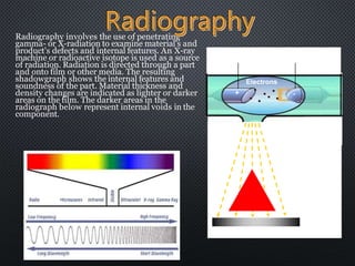
product's defects and internal features. An X-ray
machine or radioactive isotope is used as a source
of radiation. Radiation is directed through a part
and onto film or other media. The resulting
shadowgraph shows the internal features and
soundness of the part. Material thickness and
density changes are indicated as lighter or darker
areas on the film. The darker areas in the
radiograph below represent internal voids in the
component.
High Electrical Potential
Electrons
+ -
X-ray Generator or
Radioactive Source
Creates Radiation
Radiation
Penetrate
the Sample
Exposure Recording Device
2. X-RAYS OR GAMMA RADIATION IS
USED
be calculated with the
equation
E = h = hc/
e.g. the x-ray photon with
wavelength 1Å has energy
12.5 keV
4.1.1 x-ray source
Properties and Generation of X-ray
• X-rays are electromagnetic
radiation with very short
wavelength ( 10-8 -10-12 m)
• The energy of the x-ray can
3. target X-rays
W
Vacuum
X-RAYS ARE PRODUCED
WHENEVER HIGH-
SPEED ELECTRONS
COLLIDE WITH A
METAL TARGET.
A source of electrons – hot
W filament, a high
accelerating voltage
(30-50kV) between the
cathode (W) and the anode
and a metal target.
The anode is a water-cooled
block of Cu containing
desired target metal.
4. Fast moving e- will then be deflected or
decelerated and EM radiation will be
emitted.
The energy of the radiation depends on
the severity of the deceleration, which is
more or less random, and thus has a
continuous distribution.
These radiation is called white
radiation or bremsstrahlung (German
word for „braking radiation‟).
If an incoming electron has sufficient
kinetic energy for knocking out an electron
of the K shell (the inner-most shell), it may
excite the atom to an high-energy state (K
state).
One of the outer electron falls into the K-
shell vacancy, emitting the excess energy as a
x-ray photon -- K-shell emission Radiation.
5. Emitted gamma radiation is one of the three types of natural radioactivity. It
is the most energetic form of electromagnetic radiation, with a very short
wavelength of less than one-tenth of a nano-meter. Gamma rays are
essentially very energetic x-rays emitted by excited nuclei. They often
accompany alpha or beta particles, because a nucleus emitting those
particles may be left in an excited (higher-energy) state.
Man made sources are produced by introducing an extra neutron to atoms
of the source material. As the material rids itself of the neutron, energy is
released in the form of gamma rays. Two of the more common industrial
Gamma-ray sources are Iridium-192 and Colbalt-60. These isotopes emit
radiation in two or three discreet wavelengths. Cobalt 60 will emit a 1.33
and a 1.17 MeV gamma ray, and iridium-192 will emit 0.31, 0.47, and 0.60
MeV gamma rays.
Advantages of gamma ray sources include portability and the ability to
penetrate thick materials in a relativity short time.
Disadvantages include shielding requirements and safety considerations.
6. Top view of developed film
X-ray film
THE PART IS PLACED BETWEEN THE
RADIATION SOURCE AND A PIECE OF
FILM. THE PART WILL STOP SOME OF
THE RADIATION. THICKER AND
MORE DENSE AREA WILL STOP MORE
OF THE RADIATION.
= less exposure
= more exposure
• The film darkness (density) will
vary with the amount of radiation
reaching the film through the
test object.
•Defects, such as voids, cracks,
inclusions, etc., can be detected.
7. Contrast
The first subjective criteria for determining radiographic quality is
radiographic contrast. Essentially, radiographic contrast is the
degree of density difference between adjacent areas on a
radiograph.
It is essential that sufficient
contrast exist between the defect
of interest and the surrounding
area. There is no viewing
technique that can extract
information that does not
already exist in the original
radiograph
low kilovoltage high kilovoltage
8. DEFINITIO
N
Radiographic definition is the abruptness of change in going from
one density to another.
good poor
High definition: the detail portrayed in the radiograph is equivalent to
physical change present in the part. Hence, the imaging system
produced a faithful visual reproduction.
9. CAN BE USED IN ANY SITUATION WHEN ONE
WISHES TO VIEW THE INTERIOR OF AN OBJECT
TO CHECK FOR INTERNAL FAULTS AND
CONSTRUCTION DEFECTS,
e.g. faulty welding
To ‘see’ through what is inside an object
To perform measurements of size, e.g. thickness
measurements of pipes
Standard:
ASTM
– ASTM E94-84a RadiographicTesting
– ASTM E1032-85 Radiographic Examination of Weldments
– ASTM E1030-84 Radiographic Testing of Metallic Castings
10.
11. There is an upper limit of thickness through
which the radiation can penetrate, e.g. -ray
from Co-60 can penetrate up to 150mm of steel
The operator must have access to both sides of
an object
Highly skilled operator is required because of
the potential health hazard of the energetic
radiations
Relative expensive equipment