SlideShare uma empresa Scribd logo
1 de 75
Dr. S. MEENATCHISUNDARAM
ASSOCIATE PROFESSOR
DEPARTMENT OF MICROBIOLOGY
SNMV COLLEGE OF ARTS & SCIENCE, COIMBATORE
https://orcid.org/0000-0002-8691-449X
95496
https://scholar.google.com/citations?user=IkdZ5XsAAAAJ&hl=en
HISTORY AND
SCOPE OF
IMMUNOLOGY
HISTORY AND SCOPE OF IMMUNOLOGY
Immunology started in the last quarter of the
nineteenth century with two major discoveries.
The first of these was Elias Metchnikff's (1845–1916)
identification of phagocytic cells, which engulf and
destroy invading pathogens.
This laid the basis for innate immunity.
The second discovery was Emil Behring's (1854–
1917) and Paul Ehrlich's (1854–1915) identification
of antibodies, which neutralize microbial toxins.
This became the basis for acquired immunity.
Elie Metchnikoff
1880's- Metchnikoff
discovered phagocytic
cells that ingest
microbes and particles
HISTORY AND SCOPE OF IMMUNOLOGY
JOSEPH LISTER (1827–1912) Robert Koch: 1843-1910
He introduced antiseptic techniques in surgery
(1867). He established the guiding principle of
antisepsis for good surgical practice.
Edward Jenner, English country doctor
1798 - Cowpox lesions used to vaccinate against smallpox
Robert Koch created a series of
four generalized principles linking
specific microorganisms to specific
diseases (Koch’s postulates).
Edward Jenner: 1749-1823
FATHER OF VACCINATION
FATHER OF ANTISEPTIC SURGERY
HISTORY AND SCOPE OF IMMUNOLOGY
Louis Pasteur: 1822-1895
1885 - Louis Pasteur, developed the second
human vaccine which was against rabies
JOSEPH LISTER (1827–1912)
He introduced antiseptic techniques
in surgery (1867). He established the
guiding principle of antisepsis for
good surgical practice.
FATHER OF ANTISEPTIC SURGERY
1880’s - Metchnikoff discovered phagocytic cells that ingest microbes and particles
1868 - Louis Pasteur gave the concept of attenuation explaining that exposure to
adverse conditions can change the virulence of pathogens. He also prepared
the first vaccine against cholera, anthrax, and rabies.
1883 - Klebs and Loeffler isolated diphtheria bacillus.
1888 - Landsteiner dominated for 40 years in the field of immunology. He discovered
blood group antigen and antibody. He also introduced the concept of
incomplete antigen (Hapten). He was also awarded the Nobel Prize.
1892 - The first book published on the chemistry of antigen and antibody.
1886 - Julius Bordet ultimately discovered complement and got the Noble Prize
1954 Development of polio vaccines (Salk, Sabin)
HISTORY AND SCOPE OF IMMUNOLOGY
1979 Declaration of smallpox eradication by World Health Organization
1990 Development of first human gene therapy with a retrovirus vector (Anderson, Blaese)
2006 Development of vaccine against human papillomavirus, the first vaccine designed
to prevent human cancer
2011 World Organisation for Animal Health officially declared the eradication of
Rinderpest, a contagious viral disease of cattle disease
HISTORY AND SCOPE OF IMMUNOLOGY
In 1975, Georges Köhler and César Milstein succeeded in making fusions of myeloma cell lines
with B cells to create hybridomas that could produce antibodies, specific to known antigens and
that were immortalized (Monoclonal Antibodies).
Susumu Tonegawa is a Japanese molecular biologist who won the Nobel Prize for Physiology or
Medicine in 1987 for his discovery of "the genetic principle for generation of antibody diversity".
In 1996 Peter C Doherty and Rolf M Zinkernagel got Nobel Prize for their discoveries concerning
“the specificity of the cell mediated immune defence”.
THE BASIS OF DEFENCE MECHANISMS
Immunity [Latin immunis, free of burden] refers to the resistance exhibited by the host
towards injury caused by microorganisms and their products.
The human body has three primary lines of defence to fight against foreign invaders,
including viruses, bacteria, and fungi. The immune system’s three lines of defence include
physical and chemical barriers, non-specific innate responses, and specific adaptive
responses.
THE BASIS OF DEFENCE MECHANISMS
The innate immune system provides the first line of defense, which is divided broadly into two
categories – physical/chemical barriers and nonspecific resistance.
Physical barriers, including the skin and mucosa of the digestive and respiratory tracts, help
eliminate pathogens and prevent tissue and/or blood infections.
Moreover, components that are secreted by the skin or mucosa, such as sweat, saliva, tears,
mucous, help provide a basic barrier against invading pathogens.
The skin is the impermeable physical/mechanical barrier that protects many pathogens from
entering the body.
Similarly, mucosa or mucous membranes that line the immediate internal systems help trap
pathogens by producing mucous.
Hairs inside the nasal cavity, as well as cerumen (earwax), also trap pathogens and
environmental pollutants.
THE BASIS OF DEFENCE MECHANISMS
Some acidic fluids, such as gastric juice, urine, and vaginal secretions, destroy pathogens by creating
low pH conditions.
Also, lysozyme found in tears, sweat, and saliva acts as a vital antimicrobial agent to destroy pathogens.
Pathogens that successfully cross the physical barriers are next encountered by the second line of
defense.
This innate immune response mostly involves immune cells and proteins to nonspecifically recognize
and eliminate any pathogen that enters the body.
Phagocytosis is a crucial phenomenon of the innate immune system that utilizes a special type of
immune cells called phagocytes.
There are two types of phagocytes namely macrophages and neutrophils. These cells are found in the
tissues and blood.
In the beginning, phagocytes recognize and bind pathogens and then use the plasma membrane to
surround and engulf pathogens inside the cell.
THE BASIS OF DEFENCE MECHANISMS
As a result, a separate internal compartment (phagosome)
is generated, which subsequently fuses with another type
of cellular compartment called the lysosome.
The digestive enzymes present inside lysosomes finally
destroy pathogens by breaking them into fragments.
Digestion of pathogens inside a phagosome produces
indigestible materials and antigenic fragments; of which,
indigestible materials are removed by exocytosis.
However, the antigenic fragments are displayed on the
surface of phagocytes, which are subsequently recognized
and destroyed by cytotoxic T cells.
In addition, complement proteins are activated, which in
turn recruit more white blood cells (neutrophils,
eosinophils, and basophils) at the site of infection, leading
to an inflammatory response (swelling, redness, pain).
THE BASIS OF DEFENCE MECHANISMS
The third line defense aims at eliminating specific pathogens that have been encountered by the
immune system previously (adaptive or acquired immune response).
Instead of being restricted to the site of infection, the adaptive immune response occurs throughout the
body.
The adaptive immune system mainly involves two types of white blood cells (lymphocytes) -
B lymphocytes (B cells) and T lymphocytes (T cells).
B cells are involved in antibody-mediated immune responses (humoral immunity), whereas T cells are
involved in cell-mediated immune responses.
In antibody-mediated immunity, B cells are activated when they encounter a ‘known’ antigen. Activated
B cells then engulf and digest the antigen, which is followed by a representation of MHC (major
histocompatibility complex)-bound antigenic fragments on the B cell surface.
The combination of antigen-MHC further activates helper T cells, which in turn secrete cytokines
(interleukins) to trigger the growth and maturation of antigen-presenting B cells into antibody-
producing B cells (plasma cells). At this point, some B cells are transformed into memory cells to keep
the immune system ready for the next attack.
CELL AND ORGANS INVOLVED IN IMMUNE SYSTEM
Cells of the immune system
 B - Cell
 T - Cell
 Natural killer cells
Organs of immune system
Primary lymphoid organs
 Bone marrow
 Thymus
Secondary lymphoid organs
 Lymph nodes
 Spleen
 Payers patches
 MALT,GALT.
MALT- MALT is the mucosa associated Lymphoid tissue
GALT (Gut associated lymphoid tissue)
CELL AND ORGANS INVOLVED IN IMMUNE SYSTEM
CELL OF THE IMMUNE SYSTEM
Two types of lymphocytes namely B-cell and T - cell are critical for
the immune system. In addition, several accessory cells and effector
cells also participate.
B- lymphocytes - the site of development and maturation of B -Cell
occurs in bursa fabricus in birds and bone marrow in mammals.
During the course of immune response B-cell mature into plasma
cell and secrete antibodies [Immunoglobulins].
B-Lymphocytes are intimately associated with humoral
Immunity
T-Lymphocytes- the mononuclear nongranular leucocyte that
matures in thymus and that brings about cell mediated immunity is
called T Lymphocyte.
The T-Lymphocytes are thymus dependent cell. They mature under
the influence of thymus hormones.
The T-Lymphocytes have a large nucleus and a rim of cytoplasm.
They are highly concentrated in the blood and spleen.
CELL AND ORGANS INVOLVED IN IMMUNE SYSTEM
Ts cell [T-Suppressor cells]- Ts cells are a sub population of T cells that suppress the
activity of B cells and other T cells.
They are the regulatory T cells. They inhibit antibody production by B cell . They suppress
the functions of the T killer cells and T helper cells.
T cytotoxic cells [Tc] or T Killer cells [Tk] - The T Killer cells are a sub population of
T Lymphocytes that kill microorganism or body's own cells.
They are also called cytotoxic cells. They are represented by Tc Or Tk.
The T Killer cells are effector cells.
T Helper cells -T helper cells- T helper cells are a sub population of T lymphocytes that
help B cells and other T cells in immune responses.
They are the regulator cells. They help the B cells and T cells in multiple ways .
CELL AND ORGANS INVOLVED IN IMMUNE SYSTEM
Primary lymphoid organ - primary lymphoid organs are
the major site of lymphopoiesis*. The thymus, the Bursa
of fabricius in birds and bone marrow in mammals are
the primary lymphoid organs.
THYMUS
Thymus is a primary lymphoid organ .
In mammals , the thymus is a pharyngeal derivation
arising from the epithelium of the 3rd and 4th pharyngeal
pouches at about the 6th week of gestation.
BONE MARROW
Bone marrow is a primary lymphoid organ.
It is a salt tissue within the cavities of bones .
In marrow is divisible into two regions , namely
1) vascular and adipose region
2) Haemopoietic region
CELL AND ORGANS INVOLVED IN IMMUNE SYSTEM
Secondary lymphoid organ - The secondary lymphoid organs are concerned with immune
reactions. In the secondary lymphoid organs the lymphocytes are made functional.
Lymph nodes - Lymph nodes are secondary Lymphoid organs.
They are complex, cellular spherical or ovoid structures present along the lymphatic ducts.
Spleen - Spleen is a secondary lymphoid organ .
It is a solid, encapsulated organ located in the upper part of the abdominal cavity behind
the stomach and close to the diaphragm.
Peyer's patches- Peyer's patches are secondary lymphoid tissues.
They are MALT. (mucosa associated lymphoid tissue) .
They are collection of lymphoid nodules packed together to from oblong elevation of the
mucous membrane of the small intestine.
CELL AND ORGANS INVOLVED IN IMMUNE SYSTEM
 Growth and maturation of T - lymphocytes takes place in Thymus only.
 It is large at the time of birth (70 g) but with ag , the size keep on reducing and becomes
very small (3g)
 It is a flat, bilobed organ situated above the heart.
 Each Lobe is surrounded by a Capsule and is divided into Lobules, which are separated
from each other by strands of Connective tissue called Trabeculae.
 Each lobule is organized into 2 compartments: the outer compartment, or cortex, is
densely packed with immature T cells, called Thymocytes, where as the inner
compartment, or medulla, is sparsely (dispersed manner) populated with Thymocytes
THYMUS
Bilobed organ - Divided into or having two lobes
Lobes - a curved or rounded projection or division
THYMUS
CELL AND ORGANS INVOLVED IN IMMUNE SYSTEM
 Both the cortex and medulla of the thymus are
crisscrossed (moved or travel around) by a three
- dimensional stromal - cell network composed
of Epithelial cells, Dendritic cells and
Macrophages, which make up the frame work
of the organ and contribute to the growth and
maturation of Thymocytes.
 Some thymic epithelial cells in the outer cortex,
called Nurse cells, have long membrane
extensions that surround as many as 50
Thymocytes, forming large multicellular
complexes.
 Hassall corpuscles are a characteristic
morphologic feature located within the
medullary region of the thymus.
CELL AND ORGANS INVOLVED IN IMMUNE SYSTEM
FUNCTIONS OF THYMUS
 The main function of the Thymus is to release Thymosin hormone that will stimulate
the maturation of T -cells.
 Failure of Thymus development shows dramatic decrease in circulating Lymphocytes of
the T-ce11 lineage and absence of Cell - mediated immunity.
 Aging is accompanied by a decline in Thymic function.
CELL AND ORGANS INVOLVED IN IMMUNE SYSTEM
 Bone marrow is the soft, flexible connective
tissue present within the bone cavities.
 In humans and Mice, bone marrow is the site of B
- cell origin and development.
 Bone marrow forms around 4 % of total body
weight.
 There are two categories of bone marrow tissue :
Red marrow and Yellow marrow. From birth to
early adolescence , the majority of our bone
marrow is red marrow.
 As we grow and mature , increasing amounts of
red marrow is replaced by yellow marrow.
 Bone marrow can generate 200 billions of new
blood cells every day.
BONE MARROW
Bone marrow is a spongy substance found in the center of the bones
CELL AND ORGANS INVOLVED IN IMMUNE SYSTEM
RED BONE MARROW
 Also known a Myeloid tissue.
 Hematopoietic (formation of blood
cell component) in nature and
produces RBC, WBC & Platelets.
 Gets its red color from the
hemoglobin in the erythro1d cells.
 High Vascular supply.
 Function - Helps to remove old cells
from circulation.
YELLOW BONE MARROW
 Also known as Fatty tissue.
 Multipotent Stromal (connective
tissue cell of any organ) in nature and
produce Fat,. Cartilage and Bone.
 Gets its yellow color from the
carotenoids in the fat droplets in the
high number of fat cells.
 Poor Vascular supply
 Function - When blood supply is
extremely low, yellow marrow can be
converted to red marrow in order to
produce more blood cell .
CELL AND ORGANS INVOLVED IN IMMUNE SYSTEM
FUNCTIONS OF BONE MARROW
 Bone marrow is the site of B – Cell development.
 A bone marrow transplant can save the lives of people battling leukemia, lymphoma
and other blood cancers.
 Bone marrow generates RBCs which carry oxygen to the tissues.
 Bone marrow generates Platelets or Thrombocytes help prevent bleeding and aid in
clotting of blood.
 Granulocytes (Neutrophils, Basophils & Eosinophils) and Macrophages fight against
microbial infections. They also remove dead cell and remodel tissue and bones
CELL AND ORGANS INVOLVED IN IMMUNE SYSTEM
 Lymph nodes are a group of small, bean-
shaped organs (2.6 cm in length) found
mainly in the neck and trunk of the
human body.
 They play vital roles in the filtration of
antigens and debris from Lymph
(circulating colourless watery fluid) and
in the generation of immune responses to
pathogens.
 Lymph nodes are often removed from
cancer patients as their filtration function
catches tumor cells metastasized (spread
to other sites in the body) from primary
tumors.
LYMPH NODE
CELL AND ORGANS INVOLVED IN IMMUNE SYSTEM
STRUCTURE OF LYMPH NODE
 The Capsule is made of Collagen and has a sub-capsular Sinus.
 The Lymph flows into the Sinus carrying Lymphocytes, Antigen processing,
macrophages and Dendritic cells to the node Cortex, Paracortex and Medulla.
 Morphologically, Lymph node can be divided into 3 roughly concentric regions:
(1) Cortex (2) Paracortex and (3) Medulla.
 The outer most layer, Cortex contains Lymphocytes - (mostly B cells), Macrophages
and Follicular dendritic cells arranged in Primary follicles.
 The Primary follicles enlarge into Secondary follicles, each containing a Germinal
center.
CELL AND ORGANS INVOLVED IN IMMUNE SYSTEM
 Beneath the cortex is the Paracortex, which is populated largely by T - lymphocytes
and also contains Interdigitating dendritic cells thought to have migrated from tissues
to the node
 The inner most layer of a lymph node, the Medulla is more sparsely populated with
Lymphoid-lineage cells of those present, many are Plasma cells actively secreting
antibody molecules.
 The Medulla in the core of the lymph node mainly processes T- lymphocytes.
CELL AND ORGANS INVOLVED IN IMMUNE SYSTEM
FUNCTIONS OF LYMPH NODE
 Drainage of fluid from blood stream into the tissues.
 Filtration of the lymph at the lymph nodes.
 Filtering blood.
 Raise an immune reaction and fight against microbial infections.
CELL AND ORGANS INVOLVED IN IMMUNE SYSTEM
SPLEEN
 The spleen is the large secondary lymphoid organ located in the Abdominal cavity
under the Diaphragm, the muscular partition between the Abdomen and the Chest.
 Similar to a Lymph node, it acts primarily as a blood filter.
 Old RBCs are recycled in the Spleen.
 Platelets and WBCs are stored in Spleen.
 The spleen also helps to fight against certain kinds of bacteria that cause Pneumonia
and Meningitis.
CELL AND ORGANS INVOLVED IN IMMUNE SYSTEM
Plasma cells, also called
plasma B cells, are white
blood cells that originate in
the Lymphoid organs by B
Lymphocytes and secrete
large quantities of proteins
called antibodies in response
to being presented specific
substances called antigens.
CELL AND ORGANS INVOLVED IN IMMUNE SYSTEM
 The spleen varies in size and
shape between people, but it's
commonly Ovoid shaped and
Reddish brown in colour.
 The spleen, in healthy adult
humans, is approximately 7 cm
(2.8 in) to 14 cm (5.5 in) in
length. It usually weighs
between 150 g and 200 g.
 The spleen is surrounded by a
Capsule that extends a number
of projections (Trabeculae) into
the interior to form a
compartmentalized structure.
 The compartments are of two
types, white pulp and the red
pulp.
CELL AND ORGANS INVOLVED IN IMMUNE SYSTEM
 White pulp: The splenic
white pulp surrounds the
branches of the splenic
artery, forming a
periarteriolar lymphoid
sheath (PALS) populated
mainly by T-lymphocytes.
 Primary lymphoid follicles
are attached to the PALS.
 These follicles are rich in B-
cells and some of them
contain germinal centers
which develop following
antigenic stimulation.
CELL AND ORGANS INVOLVED IN IMMUNE SYSTEM
 Red pulp: The splenic red pulp
consists of a network of
sinusoids.
Functions of spleen
 Functions as the graveyard for
blood cells.
 Mounting immune responses to
antigens in the blood stream.
CELL AND ORGANS INVOLVED IN IMMUNE SYSTEM
 The mucous membranes lining the digestive , respiratory, and urogenital systems have a combined
surface area of about 400 m2 and are the major sites of entry for most pathogens.
 These vulnerable membrane surfaces are defended by a group of organized lymphoid tissues
mentioned earlier and known collectively as Mucosal-associated lymphoid tissue (MALT).
 MALT can be further classified a Gut-associated lymphoid tissue (GALT) or Bronchus-associated
lymphoid tissue (BALT).
 Gut-associated lymphoid tissue (GALT): GALT includes the tonsils, adenoids, and Peyer’s patches
in the intestine.
 Bronchial associated lymphoid tissue (BALT):Also occurs in the respiratory system.
CELL AND ORGANS INVOLVED IN IMMUNE SYSTEM
 Tonsils are collections of Lymphoid tissue facing into the Aerodigestive tract.
 The Tonsils play a role in protecting the body against Respiratory and Gastrointestinal infections.
 Each tonsil consists of a network of crypts (pits) that store cells used to fight infection.
 The tonsils contain B & T- cells, that fights against infections.
 Tonsils also produce Antibodies against Polio,. Streptococcal pneumonia, Influenza, and numerous
infections.
 Tonsillitis occurs when bacterial or viral organisms cause inflammation of the Tonsillar tissue .
This results in fever, difficulty swallowing,. sore throat, ear pain, loss of voice and throat
tenderness.
CELL AND ORGANS INVOLVED IN IMMUNE SYSTEM
 Peyer's patches are small masses of lymphatic tissue found throughout the lleum region of the
Small intestine .
 Peyer's patches are roughly egg- shaped lymphatic tissue nodules that are similar to lymph nodes
in structure , except that they are not surrounded by a connective tissue capsule .
 Important part of the immun system by monitoring intestinal bacteria populations and preventing
the growth of pathogenic bacteria in the intestins.
 Peyer's patches also playing an important role in traping antigens from pathogens and destroying
them.
CELL AND ORGANS INVOLVED IN IMMUNE SYSTEM
Pluripotent hematopoietic stem cells thus differentiate into bone marrow as myeloid
or lymphoid stem cells.
Hematopoietic stem cells (HSC) are the rare, multipotent cells that reside in the bone marrow (BM) and are
responsible for the life-long production of all types of mature blood cells
Haematopoiesis is the formation of blood cellular components.
All cellular blood components are derived from haematopoietic stem cells.
Myeloid progenitor cells are the precursors
of red blood cells, platelets, granulocytes,
monocyte-macrophages, dendritic cells.
Lymphoid progenitor cells (also known as
lymphoblasts) are precursors to other mature
blood cell types, including: T-cells/T-
lymphocytes. B-cells/B-lymphocytes.
Granulocytes are a type of white blood cell
that has small granules. The specific types of
granulocytes are neutrophils, eosinophils,
and basophils.
Dendritic cells are a type of
antigen-presenting cell (APC)
that form an important role in
the adaptive immune system.
Leukocyte - A type of blood cell that is made in the bone marrow and found in the blood and lymph
tissue. Leukocytes are part of the body’s immune system. They help the body fight infection and other
diseases. Types of leukocytes are granulocytes (neutrophils, eosinophils, and basophils), monocytes,
and lymphocytes (T cells and B cells).
Mast cells are immune cells of the myeloid lineage and are present in
connective tissues throughout the body.
Lymphopoiesis is the generation of lymphocytes, one of the five types of white blood cell (WBC).
Types of white blood cells
Monocytes. They have a longer
lifespan than many white blood cells
and help to break down bacteria.
Lymphocytes. They create
antibodies to fight against
potentially harmful invaders.
Neutrophils. They kill and digest
bacteria and fungi. They are the
most numerous type of white blood
cell and the first line of defense
when infection strikes.
Basophils. These small cells seem to
sound an alarm when infectious
agents invade your blood. They
secrete chemicals such as histamine,
a marker of allergic disease, that
help control the body's immune
response.
Eosinophils. They attack and kill
parasites and cancer cells, and help
with allergic responses.
Types of T cells
Based on their surface markers, target cells and functions, the following T cell categories have been
identified.
T cells are classified as regulatory or effector cells.
The regulatory T cells (Treg cells), formerly known as suppressor T cells, are a subpopulation of T cells
that modulate the immune system, maintain tolerance to self-antigens, and prevent autoimmune disease.
Effector T cells are the key players in steering the immune responses to execute immune functions.
cytolysis of cells infected with microbes and tumor cells and lymphokine production
Immunological functions of T-lymphocytes:
 Helps B- cell maturation, expression and antibody production
 Helps in recruitment and activation of mononuclear phagocytic cells.
 Helps in recruitment and activation of specialized cytotoxic T- cells (TCL) in antiviral response.
 Secretes cytokines which is responsible for growth and differentiation of T- cells, monocytes,
macrophages etc
 Helps in regulation of immune reactions.
T cells can be grouped into categories depending primarily on their function.
Helper T cells (CD4+ T cells)
Helper CD4+ T cells or T helper cells are lymphocytes that assist the maturation of other lymphocytes like B
cells to differentiate into plasma cells and memory B cells.
CD4+ T cells are activated by class II MHC molecules that are named after the CD4 receptors present on their
membranes.
Helper T cells account for about 50-60% of total T cells, which then further activate other immune cells to
protect the body from attacks by foreign particles.
The activation of CD4+ T cells results in the differentiation of the cells and secretion of cytokines to regulate
the overall immune response.
These cells can further be differentiated into other subtypes, which depends on the type of cytokines produced.
The two subsets of helper T cells are Th1 and Th2 cells based on the type of toll-like receptors involved in the
activation of the T cells.
The Th1 cells are involved in attacks on pathogens by increasing the phagocytic activity of the macrophages,
whereas the Th2 cells are involved in B cell activation.
Th2 cells are also involved in opsonization, where they coat the surface of pathogens to induce phagocytosis.
T cells can be grouped into categories depending primarily on their function.
Cytotoxic T cells (CD8+ T cells)
Cytotoxic T cells or CD8+ T cells are the T cells activated by the class I MHC molecules on the antigen-
presenting cells.
These are named CD8+ T cells as these contain the CD8 receptors on the surface. The receptor is present in
about 40% of the total T cells.
The CD8+ T cells, after activation, differentiate into the cytotoxic or killer T cells by the process of clonal
expansion.
Cytotoxic T cells usually destroy virus-infected cells or tumor cells as well as cells involved in transplants.
These cells recognize their target cells by binding to short peptides present together with class I MHC
molecules.
Cytotoxic T cells also secrete cytokines like IL-2 and IFN-γ, which regulate the effector functions of other
immune cells.
Memory T cells protect against previously encountered pathogens, but their origins are unclear.
 When the common lymphoid progenitor first arrives
in the thymus it doesn’t express anything on its
surface so it’s considered CD3-, CD4-, and CD8- and
is referred to as a double negative stage or DN stage
cell because it has neither CD4 or CD8 at this point.
 The DN stage can be further broken down into DN1,
DN2, DN3, DN4.
 As the cell moves through the steps to
create a functional T cell receptor it will
then begin to express CD3 in addition to
CD4 and CD8, all on the same cell.
 The dual expression of CD4 and CD8 is
why it’s called the double positive or DP
stage.
 Once the cell completes its expression of
a functional T cell receptor it will
continue to express CD3 but then will
downregulate expression of either CD4
or CD8 and be known as a single
positive T cell or SP.
A key element of T cell development is formation of this T cell receptor,
which is made of two chains, an alpha chain which is like the light chain
of a B cell receptor, and a beta chain which is like the heavy chain of a B
cell receptor.
The alpha and beta chains are made up of gene segments called V for
variable, D for diversity, and J for joining. [VDJ segments]
The beta chain includes 1 V segment, 1 D segment, and 1 J segment; while
the alpha chain only contains 1 V segment and 1 J segment.
Every person inherits multiple copies of the V, D, and J segments, and they
can be rearranged interchangeably to make a unique structure.
For the alpha chain, each person has between 70-80 variable gene
segments and 61 joining J segments. There are more V,D, and J segments
for the beta chain.
So one T cell might have a beta chain on it’s T cell receptor that has a V1-
D3-J5 combination and an alpha chain that is V7-J2, and another T cell
might have a beta chain that has a V44-D10-J1 combination and an alpha
chain that is V2-J3 thus making completely different T cell receptors with
completely different antigen specificities.
Throughout all of these stages the T cell interacts
with epithelial cells of the thymus which release
growth factors as well as molecules like interleukin 7
and 2 which makes these DN1 cells mature.
Two enzymes, Rag-1 and Rag-2, start getting
expressed, and that signifies that the cell is now a
DN2 cell.
To begin forming the beta chain, Rag-1 and Rag-2
help to splice together D and J segments on both
chromosomes, and the chromosome that successfully
rearranges first will then suppress the other
chromosome from rearranging - a process called
allelic exclusion.
If a cell successfully joins a D segment to a J
segment, then it’s considered a DN3 cell.
Next, the DN3 cell has to attach its D-J gene segment
to a V gene segment, with the help of an additional
enzyme called V(D)J recombinase.
Once the VDJ segment are combined, the full beta
chain’s antigen binding site is complete and needs to
be recombined with the constant region of the T cell
receptor. Once that’s complete, it’s considered a DN4
cell.
Allelic exclusion is a process by which only one allele of a
gene is expressed while the other allele is silenced.
The two complexes join together to form a homodimer, and
that sends a signal down the cytoplasmic tails of the receptors
to the cell’s nucleus.
When the signal is sensed within the cell, the cell begins to
divide, and the daughter cells of the rapidly proliferating DN4
cells are called Double positive cells [DP cells], because they
begin to express both CD4 and CD8 on their surface.
The DP cells now begin rearranging the alpha chain, which
means that all the DP cells will have the same beta chain, but
each will have its own distinct alpha chain.
As long as it creates an alpha chain that can bind to the beta
chain and is not self-reactive the DP T cell moves on to the
next step. If it doesn’t, the developing T cell dies.
At this point, it’s time to test out if the beta chain is
functional, by seeing if it can bind to an invariant preTalpha
chain and get expressed on the cell surface.
So the beta chain, the invariant pre-T alpha chain, and CD3
are all expressed on the surface together, and they’re joined
by another beta chain, invariant pre-T alpha chain, and CD3.
The invariant pre T alpha chain is basically something for the
beta chain to practice with until the real alpha chain is
eventually made.
T cells undergo a process called central tolerance, which is a quality control process that makes sure that T cells can
recognize and bind to MHC molecules - a step called positive selection, but that they don’t bind too strongly to
become self-reactive and attack self-tissue and cause disease - a step called negative selection.
 The T cell receptor has a couple of different parts - one part forms the antigen binding groove.
 And surrounding the antigen binding groove is the invariant region which has to bind to the MHC molecule.
 In fact, MHC molecules also have an antigen binding groove and an invariant region.
 So the invariant regions on both the T cell receptors and the MHC molecules bind to one another.
 In positive selection, only the DP cells that bind to an MHC molecule are allowed to survive, those that don’t
are killed off.
The T cell receptor
MHC molecules
In negative selection, to help identify the self-reactive T-cells, there’s an autoimmune regulator gene, called
AIRE for short.
AIRE is expressed in primary lymphoid organs and allows them to express antigens which are normally found
all over the body - the brain, ovaries, pancreas, etcetera.
If a DP cell binds strongly to a self-antigen then it dies swiftly and silently by apoptosis, whereas if a DP cell
recognizes self-MHC but does not recognize the self-antigen presented in the MHC molecule, it will go on to
become a single positive naive T cell by down regulating either the CD4 or CD8 receptor.
Whenever a T cell receptor and MHC molecule bind, CD4 and CD8 are involved in the interaction.
CD4 binds to the MHC class 2 molecule a bit like an arm that reaches out and holds the T cell receptor and MHC
together.
if there’s a double positive (DP) cell, it’s CD4 will bind to a MHC II molecule.
Now, if the strength of the binding between the T cell receptor and MHC class II molecule is somewhat strong then the
cell will downregulate CD8 and become a single positive CD4+ T cell.
On the other hand, if the strength of the binding between the T cell receptor and MHC class II is somewhat weak, then
the cell will downregulate CD4 and become a single positive CD8+ T cell.
At this point, the cell is considered a single positive naive T cell that will travel to the secondary lymphoid organs like
the lymph nodes or the spleen to deal with antigens presented on MHC molecules by antigen presenting cells like
dendritic cells.
A homodimer forms with two copies of the beta chain, the invariant pre-T alpha chain, and CD3
molecules.
At that point, the cells proliferate and each daughter cell rearranges its alpha chain combining a v
segment to a J segment.
The alpha chain will then join the beta chain and undergo positive selection and negative selection to
make sure that they can bind MHC molecules but are not self-reactive.
At this point, it becomes either a naive CD4+ T cell or a naive CD8+ T cell and is released into the
periphery.
The development of plasma cell and memory B cells can be
divided into three broad stages:
1. Generation of mature, immunocompetent B-cells
(maturation)
2. Activation of mature B-cells
3. Differentiation of the activated B-cells, into plasma cells
and memory B cells.
These three stages can be divided into two phases:
Antigen independent phase:
 This takes place in bone marrow.
 It involves the maturation of lymphoid progenitors to
matured naive B cells.
Antigen dependent phase:
 This takes place in lymph node.
 It involves activation of mature B-cells then they
encounter antigen and their differentiation into plasma
cells and memory B-cells.
B Cell Development
Blood Stream
B Cell Travel through blood stream and reach
Germinal centers in lymphnodes
B lymphocyte precursors, pro-B cells, develop in the fetal
liver during embryonic life and in the bone marrow afterwards
continuously throughout life.
Rearrangement of immunoglobulin gene takes place on their
becoming pre-B cells, which synthesise cytoplasmic IgM
In the next stage-immature B cells-IgM is expressed on the
cell surface. These cells migrate to the periphery and undergo
immunoglobulin isotype switching so that instead of IgM
alone, the cell expresses on its surface lgD as well as one of
the other lg classes- lgG, lgA, lgE
By re-assortment of lg genes, B cells develop the capacity to
produce Ig molecules which can react with all the possible
epitopes
B Cell Development
By a process of allelic exclusion each B cell is programmed to form only one class of Ig , with either
kappa and lambda light chain, possessing specificity to a single epitope alone, and to express it on the
cell surface.
Mature B-cell undergoes clonal proliferation on contact with its appropriate antigen. Interaction
between antigen and the membrane-bound antibody on a mature naive B-cell, as well as interactions
with T-cells and macrophages, selectively induces the activation and differentiation of B-cell clones of
corresponding specificity. In this process, the B-cell divides repeatedly and differentiates, generating a
population of plasma cells and memory cells. Plasma cells, synthesize and secrete antibody. Memory
cells circulate until activated by specific antigen.
When a mature B cell encounters
antigen that binds to its B cell
receptor it becomes activated.
It then proliferates and becomes a
blasting B cell.
These B cells form germinal centres.
The germinal centre B cells undergo
class switching, and somatic
hypermutation which which results
in their differentiation into memory
B cells
Plasma cell is the antibody secreting cell. It is oval, about twice the size of a
small lymphocyte, with
an eccentrically placed oval nucleus containing large blocks of chromatin
located peripherally (cartwheel appearance).
The cytoplasm is large and contains abundant endoplasmic reticulum and a
well-developed Golgi apparatus.
It is structurally designed to be an immunoglobulin producing factory.
Plasma cells are end cells and have a short life span of two or three days
A plasma cell makes an antibody of a single specificity, of a single
immunoglobulin class and allotype, and of a single light chain type only.
An exception is seen in the primary antibody response, when a plasma cell
producing IgM initially, may later be switched to IgG production.
After B cells are selected in the germinal centre for those bearing high-affinity
membrane lg for antigen, some B cells differentiate into plasma cells and
others become memory B cells.
These express all isotyes, IgM, IgD ,IgG, IgA, and IgE, as compared to naive
B cells which express only lgM and IgD.
They are characteristically long lived and more readily stimulated than naive
cells and mediate secondary immune response to subsequent encounters with
the same antigen.
Null Cells
A small proportion (5-10%) of lymphocytes that lack distinguishing phenotypic markers
characteristic of T- or B-lymphocytes are called null cells.
Because of their morphology, they are also known as large granular lymphocytes (LGL).
The member of this group is the
a. Natural killer (NK)-cells
b. Antibody dependent cellular cytotoxic (ADCC)-cells
c. Lymphokine-activated killer (LAK)-cells.
The term NK-cell is sometimes used as a common name for all null cells.
Natural Killer (NK) Cells
Natural killer (NK) cells are derived from large granular lymphocytes (LGL). They differ from K-
cells in being independent of antibody.
NK-cells differ Tc cells in other properties as well
 They are not inducible by antigen.
 They lack the classic antigen-specificity of T-cells and B-cells.
 They are not restricted by MHC-encoded proteins.
 NK activity is ‘natural’ or ‘nonimmune’ as it does not require sensitisation by prior antigenic
contact.
 They belong to a different lineage from T- and B-cells.
Functions: They are considered to be important
 Immune surveillance
 Natural defence against virus infected and maliganant mutant cells.
 NK-cells are capable of nonspecific killing of virus-transformed target cells and are involved in
allograft and tumour rejection.
Antibody-dependent Cell-mediated Cytotoxicity (ADCC )
A subpopulation of LGLs possesses surface Receptors for the Fc part of Ig.
They are capable of lysing or killing target cells sensitised with IgG antibodies.
This is an example of a process known as antibody-dependent cell-mediated cytotoxicity
(ADCC).
This antibody dependent cellular cytotoxicity is distinct from the action of cytotoxic T-cells, which
is independent of antibody.
ADCC-cells were formerly called killer (K)-cells but are now classified with NK-cells.
Lymphokine-activated Killer (LAK) Cells
Lymphokine activated killer (LAK) cells are NK-lymphocytes treated with interleukin-2 (lL-2),
which are cytotoxic to a wide range of tumour cells without affecting normal cells. IL-2 also acts as
a growth factor for NK-cells.
Phagocytic Cells
Phagocytic cells are the mononuclear macrophages (of blood and tissues) and the polymorphonuclear
microphages.
Mononuclear cells: The mononuclear phagocytic system consists of monocytes circulating in the
blood and macrophages in the tissues Both types are highly phagocytic and make up the monocyte
macrophage system.
Macrophages: Monocytes leave the circulation and reach various tissues to become transformed into
macrophages, with morphological and functional features characteristic of the tissues. Macrophages
spread throughout the animal body and take up residence in specific tissues where they are given
special names, e.g. alveolar macrophages in the lung, histiocytes in connective tissues, Kupffer cells in
the liver, Mesangial cells in the kidney, microglial cells in the brain and osteoclasts in the bone.
Functions of Macrophages
1. Phagocytosis: The primary function of macrophages is phagocytosis.
2. Antigen presentation to T-cells to initiate immune immune responses.
3. Secretion of cytokines to activate and promote innate immune response.
Macrophages secrete interleukin-1, interleukin-6, tumor necrosis factor, and interleukin-12 in response
to bacterial interaction, which stimulate immune and inflammatory responses, including fever. One of
the most potent activators of macrophages is interferon gamma (IFN-g) secreted by activated TH cells.
Polymorphonuclear Microphages
Microphages are the polymorphonuclear leucocytes of the blood.Because of the irregularshaped
nuclei, granulocytes are also called polymorphonuclear leukocytes or PMNs. Three types of
granulocytes exist: basophils, eosinophils, and neutrophils.
a. Neutrophils: They are actively phagocytic and form the predominant cell type in acute
inflammation. The phagocytic property of neutrophils is nonspecific, except for its augmentation by
opsonins.
b. Eosinophils: They possess phagocytic activity but only to a limited degree. They are found in large
numbers in allergic inflammation, parasitic infections and around antigen antibody complexes.
c. Basophils: Basophil leukocytes are found in the blood and tissues (mast cells). Their cytoplasm has
large numbers of prominent basophilic granules containing heparin, histamine, serotonin and other
hydrolytic enzymes. Degranulation of mast cells, with release of pharmacologically active agents,
constitutes the effector mechanism in anaphylactic and atopic allergy.
Dendritic cells:
While macrophages are the major antigen presenting cells, dendritic cell also performs this function.
Dendritic cells are bone marrow derived cells of a lineage different from the macrophages and T- or B-
lymphocytes. They possess MHC class II antigens.
They are more potent antigen-presenting cells than macrophages and B-cells, both of which need to be
activated before they can function as antigen presenting cells (APCs). Dendritic cells are specially
involved in the presentation of antigens to T-cells during the primary immune response.

Mais conteúdo relacionado

Mais procurados

Cells of the immune system, Overview of immune cells. Immune cells
Cells of the  immune system, Overview of immune cells. Immune cellsCells of the  immune system, Overview of immune cells. Immune cells
Cells of the immune system, Overview of immune cells. Immune cells
manojjeya
 
Humoral immune response
Humoral immune responseHumoral immune response
Humoral immune response
sufihannan
 
B CELL RECEPTOR final.pptx
B CELL RECEPTOR final.pptxB CELL RECEPTOR final.pptx
B CELL RECEPTOR final.pptx
BinteHawah1
 

Mais procurados (20)

Tumour immunology
Tumour immunologyTumour immunology
Tumour immunology
 
Cells of the immune system, Overview of immune cells. Immune cells
Cells of the  immune system, Overview of immune cells. Immune cellsCells of the  immune system, Overview of immune cells. Immune cells
Cells of the immune system, Overview of immune cells. Immune cells
 
T and B cell activation
T  and B cell activationT  and B cell activation
T and B cell activation
 
Brief history of immunology
Brief history of immunologyBrief history of immunology
Brief history of immunology
 
Cell mediated & humoral immunity
Cell mediated & humoral immunityCell mediated & humoral immunity
Cell mediated & humoral immunity
 
Antigen lecture-51 (1)
Antigen lecture-51 (1)Antigen lecture-51 (1)
Antigen lecture-51 (1)
 
Antigens
AntigensAntigens
Antigens
 
B cell generation-activation_and_differentiation
B cell generation-activation_and_differentiationB cell generation-activation_and_differentiation
B cell generation-activation_and_differentiation
 
Adjuvants
AdjuvantsAdjuvants
Adjuvants
 
Isotypes allotypes and idiotypes
Isotypes allotypes and idiotypesIsotypes allotypes and idiotypes
Isotypes allotypes and idiotypes
 
T dependent and t independent antigens
T dependent and t independent antigensT dependent and t independent antigens
T dependent and t independent antigens
 
History of immunology
History of immunologyHistory of immunology
History of immunology
 
Antibodies
AntibodiesAntibodies
Antibodies
 
Organs of the immune system
Organs of the immune systemOrgans of the immune system
Organs of the immune system
 
Antigen processing & presentation
Antigen processing & presentationAntigen processing & presentation
Antigen processing & presentation
 
1 practical applications of immunology
1 practical applications of immunology1 practical applications of immunology
1 practical applications of immunology
 
Humoral immune response
Humoral immune responseHumoral immune response
Humoral immune response
 
B CELL RECEPTOR final.pptx
B CELL RECEPTOR final.pptxB CELL RECEPTOR final.pptx
B CELL RECEPTOR final.pptx
 
Antigens notes
Antigens notes Antigens notes
Antigens notes
 
02.11.09(b): B-Cell Differentiation
02.11.09(b): B-Cell Differentiation02.11.09(b): B-Cell Differentiation
02.11.09(b): B-Cell Differentiation
 

Semelhante a IMMUNOLOGY - HISTORY - BASIC DEFENCE MECHANISM - CELLS AND ORGANS INVOLVED IN IMMUNE SYSTEM

Mechanisms Of Defense Immune System.ppt
Mechanisms Of Defense Immune System.pptMechanisms Of Defense Immune System.ppt
Mechanisms Of Defense Immune System.ppt
Shama
 
The Immune System-An introduction.pptx
The Immune System-An introduction.pptxThe Immune System-An introduction.pptx
The Immune System-An introduction.pptx
malti19
 
The immune system final
The immune system finalThe immune system final
The immune system final
rharrison545
 

Semelhante a IMMUNOLOGY - HISTORY - BASIC DEFENCE MECHANISM - CELLS AND ORGANS INVOLVED IN IMMUNE SYSTEM (20)

Overview of immune response
Overview of immune responseOverview of immune response
Overview of immune response
 
Immunology
ImmunologyImmunology
Immunology
 
Immunology by Tai
Immunology by TaiImmunology by Tai
Immunology by Tai
 
Immunology by tai
Immunology by taiImmunology by tai
Immunology by tai
 
Mechanisms Of Defense Immune System.ppt
Mechanisms Of Defense Immune System.pptMechanisms Of Defense Immune System.ppt
Mechanisms Of Defense Immune System.ppt
 
The Immune System-An introduction.pptx
The Immune System-An introduction.pptxThe Immune System-An introduction.pptx
The Immune System-An introduction.pptx
 
What is immunology.ppt
What is immunology.pptWhat is immunology.ppt
What is immunology.ppt
 
What is immunology and concepts of immunology.ppt
What is immunology and concepts of immunology.pptWhat is immunology and concepts of immunology.ppt
What is immunology and concepts of immunology.ppt
 
Immunity
ImmunityImmunity
Immunity
 
The immune system final
The immune system finalThe immune system final
The immune system final
 
CELLS OF IMMUNE SYSTEM
CELLS OF IMMUNE SYSTEM CELLS OF IMMUNE SYSTEM
CELLS OF IMMUNE SYSTEM
 
Immunity
ImmunityImmunity
Immunity
 
Immune System
Immune SystemImmune System
Immune System
 
Anatomy lab. 8 lymphatics & immune sys.
Anatomy lab. 8 lymphatics & immune sys.Anatomy lab. 8 lymphatics & immune sys.
Anatomy lab. 8 lymphatics & immune sys.
 
Cells and organs of the immune system
Cells and organs of the immune systemCells and organs of the immune system
Cells and organs of the immune system
 
Immune responses
Immune responsesImmune responses
Immune responses
 
Overview of the immune system
Overview of the immune systemOverview of the immune system
Overview of the immune system
 
Immunity
ImmunityImmunity
Immunity
 
Defense Mechanism (2).pptx
Defense Mechanism (2).pptxDefense Mechanism (2).pptx
Defense Mechanism (2).pptx
 
Basic immunology and hypersensitive disorders beba
Basic immunology and hypersensitive disorders bebaBasic immunology and hypersensitive disorders beba
Basic immunology and hypersensitive disorders beba
 

Mais de Meenatchisundaram Subramani

Mais de Meenatchisundaram Subramani (12)

Research Paper Publication
Research Paper PublicationResearch Paper Publication
Research Paper Publication
 
Differential WBC Count
Differential WBC CountDifferential WBC Count
Differential WBC Count
 
Candida albicans
Candida albicansCandida albicans
Candida albicans
 
Pseudomonas aeruginosa
Pseudomonas aeruginosaPseudomonas aeruginosa
Pseudomonas aeruginosa
 
MYCOBACTERIUM TUBERCULOSIS
MYCOBACTERIUM TUBERCULOSISMYCOBACTERIUM TUBERCULOSIS
MYCOBACTERIUM TUBERCULOSIS
 
VIROLOGY - POLIO
VIROLOGY - POLIOVIROLOGY - POLIO
VIROLOGY - POLIO
 
VIROLOGY - DIAGNOSTIC METHODS
VIROLOGY - DIAGNOSTIC METHODSVIROLOGY - DIAGNOSTIC METHODS
VIROLOGY - DIAGNOSTIC METHODS
 
BACILLUS ANTHRACIS
BACILLUS ANTHRACISBACILLUS ANTHRACIS
BACILLUS ANTHRACIS
 
VIROLOGY - INTRODUCTION
VIROLOGY - INTRODUCTIONVIROLOGY - INTRODUCTION
VIROLOGY - INTRODUCTION
 
VIROLOGY - CULTIVATION AND DETECTION OF VIRUS
VIROLOGY - CULTIVATION AND DETECTION OF VIRUSVIROLOGY - CULTIVATION AND DETECTION OF VIRUS
VIROLOGY - CULTIVATION AND DETECTION OF VIRUS
 
VIROLOGY - RABIES VIRUS
VIROLOGY - RABIES VIRUSVIROLOGY - RABIES VIRUS
VIROLOGY - RABIES VIRUS
 
HIV - Human Immunodeficiency Virus
HIV - Human Immunodeficiency VirusHIV - Human Immunodeficiency Virus
HIV - Human Immunodeficiency Virus
 

Último

Making and Justifying Mathematical Decisions.pdf
Making and Justifying Mathematical Decisions.pdfMaking and Justifying Mathematical Decisions.pdf
Making and Justifying Mathematical Decisions.pdf
Chris Hunter
 
Seal of Good Local Governance (SGLG) 2024Final.pptx
Seal of Good Local Governance (SGLG) 2024Final.pptxSeal of Good Local Governance (SGLG) 2024Final.pptx
Seal of Good Local Governance (SGLG) 2024Final.pptx
negromaestrong
 
Gardella_PRCampaignConclusion Pitch Letter
Gardella_PRCampaignConclusion Pitch LetterGardella_PRCampaignConclusion Pitch Letter
Gardella_PRCampaignConclusion Pitch Letter
MateoGardella
 
1029 - Danh muc Sach Giao Khoa 10 . pdf
1029 -  Danh muc Sach Giao Khoa 10 . pdf1029 -  Danh muc Sach Giao Khoa 10 . pdf
1029 - Danh muc Sach Giao Khoa 10 . pdf
QucHHunhnh
 
Gardella_Mateo_IntellectualProperty.pdf.
Gardella_Mateo_IntellectualProperty.pdf.Gardella_Mateo_IntellectualProperty.pdf.
Gardella_Mateo_IntellectualProperty.pdf.
MateoGardella
 
The basics of sentences session 2pptx copy.pptx
The basics of sentences session 2pptx copy.pptxThe basics of sentences session 2pptx copy.pptx
The basics of sentences session 2pptx copy.pptx
heathfieldcps1
 
1029-Danh muc Sach Giao Khoa khoi 6.pdf
1029-Danh muc Sach Giao Khoa khoi  6.pdf1029-Danh muc Sach Giao Khoa khoi  6.pdf
1029-Danh muc Sach Giao Khoa khoi 6.pdf
QucHHunhnh
 
Beyond the EU: DORA and NIS 2 Directive's Global Impact
Beyond the EU: DORA and NIS 2 Directive's Global ImpactBeyond the EU: DORA and NIS 2 Directive's Global Impact
Beyond the EU: DORA and NIS 2 Directive's Global Impact
PECB
 

Último (20)

ICT Role in 21st Century Education & its Challenges.pptx
ICT Role in 21st Century Education & its Challenges.pptxICT Role in 21st Century Education & its Challenges.pptx
ICT Role in 21st Century Education & its Challenges.pptx
 
Paris 2024 Olympic Geographies - an activity
Paris 2024 Olympic Geographies - an activityParis 2024 Olympic Geographies - an activity
Paris 2024 Olympic Geographies - an activity
 
Mattingly "AI & Prompt Design: Structured Data, Assistants, & RAG"
Mattingly "AI & Prompt Design: Structured Data, Assistants, & RAG"Mattingly "AI & Prompt Design: Structured Data, Assistants, & RAG"
Mattingly "AI & Prompt Design: Structured Data, Assistants, & RAG"
 
Making and Justifying Mathematical Decisions.pdf
Making and Justifying Mathematical Decisions.pdfMaking and Justifying Mathematical Decisions.pdf
Making and Justifying Mathematical Decisions.pdf
 
SOCIAL AND HISTORICAL CONTEXT - LFTVD.pptx
SOCIAL AND HISTORICAL CONTEXT - LFTVD.pptxSOCIAL AND HISTORICAL CONTEXT - LFTVD.pptx
SOCIAL AND HISTORICAL CONTEXT - LFTVD.pptx
 
Seal of Good Local Governance (SGLG) 2024Final.pptx
Seal of Good Local Governance (SGLG) 2024Final.pptxSeal of Good Local Governance (SGLG) 2024Final.pptx
Seal of Good Local Governance (SGLG) 2024Final.pptx
 
How to Give a Domain for a Field in Odoo 17
How to Give a Domain for a Field in Odoo 17How to Give a Domain for a Field in Odoo 17
How to Give a Domain for a Field in Odoo 17
 
SECOND SEMESTER TOPIC COVERAGE SY 2023-2024 Trends, Networks, and Critical Th...
SECOND SEMESTER TOPIC COVERAGE SY 2023-2024 Trends, Networks, and Critical Th...SECOND SEMESTER TOPIC COVERAGE SY 2023-2024 Trends, Networks, and Critical Th...
SECOND SEMESTER TOPIC COVERAGE SY 2023-2024 Trends, Networks, and Critical Th...
 
Presentation by Andreas Schleicher Tackling the School Absenteeism Crisis 30 ...
Presentation by Andreas Schleicher Tackling the School Absenteeism Crisis 30 ...Presentation by Andreas Schleicher Tackling the School Absenteeism Crisis 30 ...
Presentation by Andreas Schleicher Tackling the School Absenteeism Crisis 30 ...
 
Application orientated numerical on hev.ppt
Application orientated numerical on hev.pptApplication orientated numerical on hev.ppt
Application orientated numerical on hev.ppt
 
Measures of Central Tendency: Mean, Median and Mode
Measures of Central Tendency: Mean, Median and ModeMeasures of Central Tendency: Mean, Median and Mode
Measures of Central Tendency: Mean, Median and Mode
 
Código Creativo y Arte de Software | Unidad 1
Código Creativo y Arte de Software | Unidad 1Código Creativo y Arte de Software | Unidad 1
Código Creativo y Arte de Software | Unidad 1
 
Gardella_PRCampaignConclusion Pitch Letter
Gardella_PRCampaignConclusion Pitch LetterGardella_PRCampaignConclusion Pitch Letter
Gardella_PRCampaignConclusion Pitch Letter
 
psychiatric nursing HISTORY COLLECTION .docx
psychiatric  nursing HISTORY  COLLECTION  .docxpsychiatric  nursing HISTORY  COLLECTION  .docx
psychiatric nursing HISTORY COLLECTION .docx
 
1029 - Danh muc Sach Giao Khoa 10 . pdf
1029 -  Danh muc Sach Giao Khoa 10 . pdf1029 -  Danh muc Sach Giao Khoa 10 . pdf
1029 - Danh muc Sach Giao Khoa 10 . pdf
 
Gardella_Mateo_IntellectualProperty.pdf.
Gardella_Mateo_IntellectualProperty.pdf.Gardella_Mateo_IntellectualProperty.pdf.
Gardella_Mateo_IntellectualProperty.pdf.
 
Holdier Curriculum Vitae (April 2024).pdf
Holdier Curriculum Vitae (April 2024).pdfHoldier Curriculum Vitae (April 2024).pdf
Holdier Curriculum Vitae (April 2024).pdf
 
The basics of sentences session 2pptx copy.pptx
The basics of sentences session 2pptx copy.pptxThe basics of sentences session 2pptx copy.pptx
The basics of sentences session 2pptx copy.pptx
 
1029-Danh muc Sach Giao Khoa khoi 6.pdf
1029-Danh muc Sach Giao Khoa khoi  6.pdf1029-Danh muc Sach Giao Khoa khoi  6.pdf
1029-Danh muc Sach Giao Khoa khoi 6.pdf
 
Beyond the EU: DORA and NIS 2 Directive's Global Impact
Beyond the EU: DORA and NIS 2 Directive's Global ImpactBeyond the EU: DORA and NIS 2 Directive's Global Impact
Beyond the EU: DORA and NIS 2 Directive's Global Impact
 

IMMUNOLOGY - HISTORY - BASIC DEFENCE MECHANISM - CELLS AND ORGANS INVOLVED IN IMMUNE SYSTEM

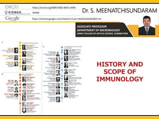
  • 1. Dr. S. MEENATCHISUNDARAM ASSOCIATE PROFESSOR DEPARTMENT OF MICROBIOLOGY SNMV COLLEGE OF ARTS & SCIENCE, COIMBATORE https://orcid.org/0000-0002-8691-449X 95496 https://scholar.google.com/citations?user=IkdZ5XsAAAAJ&hl=en HISTORY AND SCOPE OF IMMUNOLOGY
  • 2. HISTORY AND SCOPE OF IMMUNOLOGY Immunology started in the last quarter of the nineteenth century with two major discoveries. The first of these was Elias Metchnikff's (1845–1916) identification of phagocytic cells, which engulf and destroy invading pathogens. This laid the basis for innate immunity. The second discovery was Emil Behring's (1854– 1917) and Paul Ehrlich's (1854–1915) identification of antibodies, which neutralize microbial toxins. This became the basis for acquired immunity. Elie Metchnikoff 1880's- Metchnikoff discovered phagocytic cells that ingest microbes and particles
  • 3. HISTORY AND SCOPE OF IMMUNOLOGY JOSEPH LISTER (1827–1912) Robert Koch: 1843-1910 He introduced antiseptic techniques in surgery (1867). He established the guiding principle of antisepsis for good surgical practice. Edward Jenner, English country doctor 1798 - Cowpox lesions used to vaccinate against smallpox Robert Koch created a series of four generalized principles linking specific microorganisms to specific diseases (Koch’s postulates). Edward Jenner: 1749-1823 FATHER OF VACCINATION FATHER OF ANTISEPTIC SURGERY
  • 4. HISTORY AND SCOPE OF IMMUNOLOGY Louis Pasteur: 1822-1895 1885 - Louis Pasteur, developed the second human vaccine which was against rabies JOSEPH LISTER (1827–1912) He introduced antiseptic techniques in surgery (1867). He established the guiding principle of antisepsis for good surgical practice. FATHER OF ANTISEPTIC SURGERY
  • 5. 1880’s - Metchnikoff discovered phagocytic cells that ingest microbes and particles 1868 - Louis Pasteur gave the concept of attenuation explaining that exposure to adverse conditions can change the virulence of pathogens. He also prepared the first vaccine against cholera, anthrax, and rabies. 1883 - Klebs and Loeffler isolated diphtheria bacillus. 1888 - Landsteiner dominated for 40 years in the field of immunology. He discovered blood group antigen and antibody. He also introduced the concept of incomplete antigen (Hapten). He was also awarded the Nobel Prize. 1892 - The first book published on the chemistry of antigen and antibody. 1886 - Julius Bordet ultimately discovered complement and got the Noble Prize 1954 Development of polio vaccines (Salk, Sabin) HISTORY AND SCOPE OF IMMUNOLOGY 1979 Declaration of smallpox eradication by World Health Organization
  • 6. 1990 Development of first human gene therapy with a retrovirus vector (Anderson, Blaese) 2006 Development of vaccine against human papillomavirus, the first vaccine designed to prevent human cancer 2011 World Organisation for Animal Health officially declared the eradication of Rinderpest, a contagious viral disease of cattle disease HISTORY AND SCOPE OF IMMUNOLOGY In 1975, Georges Köhler and César Milstein succeeded in making fusions of myeloma cell lines with B cells to create hybridomas that could produce antibodies, specific to known antigens and that were immortalized (Monoclonal Antibodies). Susumu Tonegawa is a Japanese molecular biologist who won the Nobel Prize for Physiology or Medicine in 1987 for his discovery of "the genetic principle for generation of antibody diversity". In 1996 Peter C Doherty and Rolf M Zinkernagel got Nobel Prize for their discoveries concerning “the specificity of the cell mediated immune defence”.
  • 7. THE BASIS OF DEFENCE MECHANISMS Immunity [Latin immunis, free of burden] refers to the resistance exhibited by the host towards injury caused by microorganisms and their products. The human body has three primary lines of defence to fight against foreign invaders, including viruses, bacteria, and fungi. The immune system’s three lines of defence include physical and chemical barriers, non-specific innate responses, and specific adaptive responses.
  • 8. THE BASIS OF DEFENCE MECHANISMS
  • 9. THE BASIS OF DEFENCE MECHANISMS The innate immune system provides the first line of defense, which is divided broadly into two categories – physical/chemical barriers and nonspecific resistance. Physical barriers, including the skin and mucosa of the digestive and respiratory tracts, help eliminate pathogens and prevent tissue and/or blood infections. Moreover, components that are secreted by the skin or mucosa, such as sweat, saliva, tears, mucous, help provide a basic barrier against invading pathogens. The skin is the impermeable physical/mechanical barrier that protects many pathogens from entering the body. Similarly, mucosa or mucous membranes that line the immediate internal systems help trap pathogens by producing mucous. Hairs inside the nasal cavity, as well as cerumen (earwax), also trap pathogens and environmental pollutants.
  • 10. THE BASIS OF DEFENCE MECHANISMS Some acidic fluids, such as gastric juice, urine, and vaginal secretions, destroy pathogens by creating low pH conditions. Also, lysozyme found in tears, sweat, and saliva acts as a vital antimicrobial agent to destroy pathogens. Pathogens that successfully cross the physical barriers are next encountered by the second line of defense. This innate immune response mostly involves immune cells and proteins to nonspecifically recognize and eliminate any pathogen that enters the body. Phagocytosis is a crucial phenomenon of the innate immune system that utilizes a special type of immune cells called phagocytes. There are two types of phagocytes namely macrophages and neutrophils. These cells are found in the tissues and blood. In the beginning, phagocytes recognize and bind pathogens and then use the plasma membrane to surround and engulf pathogens inside the cell.
  • 11. THE BASIS OF DEFENCE MECHANISMS As a result, a separate internal compartment (phagosome) is generated, which subsequently fuses with another type of cellular compartment called the lysosome. The digestive enzymes present inside lysosomes finally destroy pathogens by breaking them into fragments. Digestion of pathogens inside a phagosome produces indigestible materials and antigenic fragments; of which, indigestible materials are removed by exocytosis. However, the antigenic fragments are displayed on the surface of phagocytes, which are subsequently recognized and destroyed by cytotoxic T cells. In addition, complement proteins are activated, which in turn recruit more white blood cells (neutrophils, eosinophils, and basophils) at the site of infection, leading to an inflammatory response (swelling, redness, pain).
  • 12. THE BASIS OF DEFENCE MECHANISMS The third line defense aims at eliminating specific pathogens that have been encountered by the immune system previously (adaptive or acquired immune response). Instead of being restricted to the site of infection, the adaptive immune response occurs throughout the body. The adaptive immune system mainly involves two types of white blood cells (lymphocytes) - B lymphocytes (B cells) and T lymphocytes (T cells). B cells are involved in antibody-mediated immune responses (humoral immunity), whereas T cells are involved in cell-mediated immune responses. In antibody-mediated immunity, B cells are activated when they encounter a ‘known’ antigen. Activated B cells then engulf and digest the antigen, which is followed by a representation of MHC (major histocompatibility complex)-bound antigenic fragments on the B cell surface. The combination of antigen-MHC further activates helper T cells, which in turn secrete cytokines (interleukins) to trigger the growth and maturation of antigen-presenting B cells into antibody- producing B cells (plasma cells). At this point, some B cells are transformed into memory cells to keep the immune system ready for the next attack.
  • 13.
  • 14. CELL AND ORGANS INVOLVED IN IMMUNE SYSTEM Cells of the immune system  B - Cell  T - Cell  Natural killer cells Organs of immune system Primary lymphoid organs  Bone marrow  Thymus Secondary lymphoid organs  Lymph nodes  Spleen  Payers patches  MALT,GALT. MALT- MALT is the mucosa associated Lymphoid tissue GALT (Gut associated lymphoid tissue)
  • 15. CELL AND ORGANS INVOLVED IN IMMUNE SYSTEM CELL OF THE IMMUNE SYSTEM Two types of lymphocytes namely B-cell and T - cell are critical for the immune system. In addition, several accessory cells and effector cells also participate. B- lymphocytes - the site of development and maturation of B -Cell occurs in bursa fabricus in birds and bone marrow in mammals. During the course of immune response B-cell mature into plasma cell and secrete antibodies [Immunoglobulins]. B-Lymphocytes are intimately associated with humoral Immunity T-Lymphocytes- the mononuclear nongranular leucocyte that matures in thymus and that brings about cell mediated immunity is called T Lymphocyte. The T-Lymphocytes are thymus dependent cell. They mature under the influence of thymus hormones. The T-Lymphocytes have a large nucleus and a rim of cytoplasm. They are highly concentrated in the blood and spleen.
  • 16. CELL AND ORGANS INVOLVED IN IMMUNE SYSTEM
  • 17. CELL AND ORGANS INVOLVED IN IMMUNE SYSTEM Ts cell [T-Suppressor cells]- Ts cells are a sub population of T cells that suppress the activity of B cells and other T cells. They are the regulatory T cells. They inhibit antibody production by B cell . They suppress the functions of the T killer cells and T helper cells. T cytotoxic cells [Tc] or T Killer cells [Tk] - The T Killer cells are a sub population of T Lymphocytes that kill microorganism or body's own cells. They are also called cytotoxic cells. They are represented by Tc Or Tk. The T Killer cells are effector cells. T Helper cells -T helper cells- T helper cells are a sub population of T lymphocytes that help B cells and other T cells in immune responses. They are the regulator cells. They help the B cells and T cells in multiple ways .
  • 18. CELL AND ORGANS INVOLVED IN IMMUNE SYSTEM
  • 19. CELL AND ORGANS INVOLVED IN IMMUNE SYSTEM Primary lymphoid organ - primary lymphoid organs are the major site of lymphopoiesis*. The thymus, the Bursa of fabricius in birds and bone marrow in mammals are the primary lymphoid organs. THYMUS Thymus is a primary lymphoid organ . In mammals , the thymus is a pharyngeal derivation arising from the epithelium of the 3rd and 4th pharyngeal pouches at about the 6th week of gestation. BONE MARROW Bone marrow is a primary lymphoid organ. It is a salt tissue within the cavities of bones . In marrow is divisible into two regions , namely 1) vascular and adipose region 2) Haemopoietic region
  • 20. CELL AND ORGANS INVOLVED IN IMMUNE SYSTEM Secondary lymphoid organ - The secondary lymphoid organs are concerned with immune reactions. In the secondary lymphoid organs the lymphocytes are made functional. Lymph nodes - Lymph nodes are secondary Lymphoid organs. They are complex, cellular spherical or ovoid structures present along the lymphatic ducts. Spleen - Spleen is a secondary lymphoid organ . It is a solid, encapsulated organ located in the upper part of the abdominal cavity behind the stomach and close to the diaphragm. Peyer's patches- Peyer's patches are secondary lymphoid tissues. They are MALT. (mucosa associated lymphoid tissue) . They are collection of lymphoid nodules packed together to from oblong elevation of the mucous membrane of the small intestine.
  • 21. CELL AND ORGANS INVOLVED IN IMMUNE SYSTEM  Growth and maturation of T - lymphocytes takes place in Thymus only.  It is large at the time of birth (70 g) but with ag , the size keep on reducing and becomes very small (3g)  It is a flat, bilobed organ situated above the heart.  Each Lobe is surrounded by a Capsule and is divided into Lobules, which are separated from each other by strands of Connective tissue called Trabeculae.  Each lobule is organized into 2 compartments: the outer compartment, or cortex, is densely packed with immature T cells, called Thymocytes, where as the inner compartment, or medulla, is sparsely (dispersed manner) populated with Thymocytes THYMUS Bilobed organ - Divided into or having two lobes Lobes - a curved or rounded projection or division
  • 23. CELL AND ORGANS INVOLVED IN IMMUNE SYSTEM  Both the cortex and medulla of the thymus are crisscrossed (moved or travel around) by a three - dimensional stromal - cell network composed of Epithelial cells, Dendritic cells and Macrophages, which make up the frame work of the organ and contribute to the growth and maturation of Thymocytes.  Some thymic epithelial cells in the outer cortex, called Nurse cells, have long membrane extensions that surround as many as 50 Thymocytes, forming large multicellular complexes.  Hassall corpuscles are a characteristic morphologic feature located within the medullary region of the thymus.
  • 24. CELL AND ORGANS INVOLVED IN IMMUNE SYSTEM FUNCTIONS OF THYMUS  The main function of the Thymus is to release Thymosin hormone that will stimulate the maturation of T -cells.  Failure of Thymus development shows dramatic decrease in circulating Lymphocytes of the T-ce11 lineage and absence of Cell - mediated immunity.  Aging is accompanied by a decline in Thymic function.
  • 25. CELL AND ORGANS INVOLVED IN IMMUNE SYSTEM  Bone marrow is the soft, flexible connective tissue present within the bone cavities.  In humans and Mice, bone marrow is the site of B - cell origin and development.  Bone marrow forms around 4 % of total body weight.  There are two categories of bone marrow tissue : Red marrow and Yellow marrow. From birth to early adolescence , the majority of our bone marrow is red marrow.  As we grow and mature , increasing amounts of red marrow is replaced by yellow marrow.  Bone marrow can generate 200 billions of new blood cells every day. BONE MARROW Bone marrow is a spongy substance found in the center of the bones
  • 26. CELL AND ORGANS INVOLVED IN IMMUNE SYSTEM RED BONE MARROW  Also known a Myeloid tissue.  Hematopoietic (formation of blood cell component) in nature and produces RBC, WBC & Platelets.  Gets its red color from the hemoglobin in the erythro1d cells.  High Vascular supply.  Function - Helps to remove old cells from circulation. YELLOW BONE MARROW  Also known as Fatty tissue.  Multipotent Stromal (connective tissue cell of any organ) in nature and produce Fat,. Cartilage and Bone.  Gets its yellow color from the carotenoids in the fat droplets in the high number of fat cells.  Poor Vascular supply  Function - When blood supply is extremely low, yellow marrow can be converted to red marrow in order to produce more blood cell .
  • 27. CELL AND ORGANS INVOLVED IN IMMUNE SYSTEM FUNCTIONS OF BONE MARROW  Bone marrow is the site of B – Cell development.  A bone marrow transplant can save the lives of people battling leukemia, lymphoma and other blood cancers.  Bone marrow generates RBCs which carry oxygen to the tissues.  Bone marrow generates Platelets or Thrombocytes help prevent bleeding and aid in clotting of blood.  Granulocytes (Neutrophils, Basophils & Eosinophils) and Macrophages fight against microbial infections. They also remove dead cell and remodel tissue and bones
  • 28. CELL AND ORGANS INVOLVED IN IMMUNE SYSTEM  Lymph nodes are a group of small, bean- shaped organs (2.6 cm in length) found mainly in the neck and trunk of the human body.  They play vital roles in the filtration of antigens and debris from Lymph (circulating colourless watery fluid) and in the generation of immune responses to pathogens.  Lymph nodes are often removed from cancer patients as their filtration function catches tumor cells metastasized (spread to other sites in the body) from primary tumors. LYMPH NODE
  • 29.
  • 30. CELL AND ORGANS INVOLVED IN IMMUNE SYSTEM STRUCTURE OF LYMPH NODE  The Capsule is made of Collagen and has a sub-capsular Sinus.  The Lymph flows into the Sinus carrying Lymphocytes, Antigen processing, macrophages and Dendritic cells to the node Cortex, Paracortex and Medulla.  Morphologically, Lymph node can be divided into 3 roughly concentric regions: (1) Cortex (2) Paracortex and (3) Medulla.  The outer most layer, Cortex contains Lymphocytes - (mostly B cells), Macrophages and Follicular dendritic cells arranged in Primary follicles.  The Primary follicles enlarge into Secondary follicles, each containing a Germinal center.
  • 31. CELL AND ORGANS INVOLVED IN IMMUNE SYSTEM  Beneath the cortex is the Paracortex, which is populated largely by T - lymphocytes and also contains Interdigitating dendritic cells thought to have migrated from tissues to the node  The inner most layer of a lymph node, the Medulla is more sparsely populated with Lymphoid-lineage cells of those present, many are Plasma cells actively secreting antibody molecules.  The Medulla in the core of the lymph node mainly processes T- lymphocytes.
  • 32. CELL AND ORGANS INVOLVED IN IMMUNE SYSTEM FUNCTIONS OF LYMPH NODE  Drainage of fluid from blood stream into the tissues.  Filtration of the lymph at the lymph nodes.  Filtering blood.  Raise an immune reaction and fight against microbial infections.
  • 33. CELL AND ORGANS INVOLVED IN IMMUNE SYSTEM SPLEEN  The spleen is the large secondary lymphoid organ located in the Abdominal cavity under the Diaphragm, the muscular partition between the Abdomen and the Chest.  Similar to a Lymph node, it acts primarily as a blood filter.  Old RBCs are recycled in the Spleen.  Platelets and WBCs are stored in Spleen.  The spleen also helps to fight against certain kinds of bacteria that cause Pneumonia and Meningitis.
  • 34. CELL AND ORGANS INVOLVED IN IMMUNE SYSTEM Plasma cells, also called plasma B cells, are white blood cells that originate in the Lymphoid organs by B Lymphocytes and secrete large quantities of proteins called antibodies in response to being presented specific substances called antigens.
  • 35. CELL AND ORGANS INVOLVED IN IMMUNE SYSTEM  The spleen varies in size and shape between people, but it's commonly Ovoid shaped and Reddish brown in colour.  The spleen, in healthy adult humans, is approximately 7 cm (2.8 in) to 14 cm (5.5 in) in length. It usually weighs between 150 g and 200 g.  The spleen is surrounded by a Capsule that extends a number of projections (Trabeculae) into the interior to form a compartmentalized structure.  The compartments are of two types, white pulp and the red pulp.
  • 36. CELL AND ORGANS INVOLVED IN IMMUNE SYSTEM  White pulp: The splenic white pulp surrounds the branches of the splenic artery, forming a periarteriolar lymphoid sheath (PALS) populated mainly by T-lymphocytes.  Primary lymphoid follicles are attached to the PALS.  These follicles are rich in B- cells and some of them contain germinal centers which develop following antigenic stimulation.
  • 37. CELL AND ORGANS INVOLVED IN IMMUNE SYSTEM  Red pulp: The splenic red pulp consists of a network of sinusoids. Functions of spleen  Functions as the graveyard for blood cells.  Mounting immune responses to antigens in the blood stream.
  • 38. CELL AND ORGANS INVOLVED IN IMMUNE SYSTEM  The mucous membranes lining the digestive , respiratory, and urogenital systems have a combined surface area of about 400 m2 and are the major sites of entry for most pathogens.  These vulnerable membrane surfaces are defended by a group of organized lymphoid tissues mentioned earlier and known collectively as Mucosal-associated lymphoid tissue (MALT).  MALT can be further classified a Gut-associated lymphoid tissue (GALT) or Bronchus-associated lymphoid tissue (BALT).  Gut-associated lymphoid tissue (GALT): GALT includes the tonsils, adenoids, and Peyer’s patches in the intestine.  Bronchial associated lymphoid tissue (BALT):Also occurs in the respiratory system.
  • 39. CELL AND ORGANS INVOLVED IN IMMUNE SYSTEM  Tonsils are collections of Lymphoid tissue facing into the Aerodigestive tract.  The Tonsils play a role in protecting the body against Respiratory and Gastrointestinal infections.  Each tonsil consists of a network of crypts (pits) that store cells used to fight infection.  The tonsils contain B & T- cells, that fights against infections.  Tonsils also produce Antibodies against Polio,. Streptococcal pneumonia, Influenza, and numerous infections.  Tonsillitis occurs when bacterial or viral organisms cause inflammation of the Tonsillar tissue . This results in fever, difficulty swallowing,. sore throat, ear pain, loss of voice and throat tenderness.
  • 40. CELL AND ORGANS INVOLVED IN IMMUNE SYSTEM  Peyer's patches are small masses of lymphatic tissue found throughout the lleum region of the Small intestine .  Peyer's patches are roughly egg- shaped lymphatic tissue nodules that are similar to lymph nodes in structure , except that they are not surrounded by a connective tissue capsule .  Important part of the immun system by monitoring intestinal bacteria populations and preventing the growth of pathogenic bacteria in the intestins.  Peyer's patches also playing an important role in traping antigens from pathogens and destroying them.
  • 41. CELL AND ORGANS INVOLVED IN IMMUNE SYSTEM
  • 42. Pluripotent hematopoietic stem cells thus differentiate into bone marrow as myeloid or lymphoid stem cells. Hematopoietic stem cells (HSC) are the rare, multipotent cells that reside in the bone marrow (BM) and are responsible for the life-long production of all types of mature blood cells
  • 43. Haematopoiesis is the formation of blood cellular components. All cellular blood components are derived from haematopoietic stem cells. Myeloid progenitor cells are the precursors of red blood cells, platelets, granulocytes, monocyte-macrophages, dendritic cells. Lymphoid progenitor cells (also known as lymphoblasts) are precursors to other mature blood cell types, including: T-cells/T- lymphocytes. B-cells/B-lymphocytes. Granulocytes are a type of white blood cell that has small granules. The specific types of granulocytes are neutrophils, eosinophils, and basophils. Dendritic cells are a type of antigen-presenting cell (APC) that form an important role in the adaptive immune system.
  • 44. Leukocyte - A type of blood cell that is made in the bone marrow and found in the blood and lymph tissue. Leukocytes are part of the body’s immune system. They help the body fight infection and other diseases. Types of leukocytes are granulocytes (neutrophils, eosinophils, and basophils), monocytes, and lymphocytes (T cells and B cells).
  • 45. Mast cells are immune cells of the myeloid lineage and are present in connective tissues throughout the body.
  • 46. Lymphopoiesis is the generation of lymphocytes, one of the five types of white blood cell (WBC). Types of white blood cells Monocytes. They have a longer lifespan than many white blood cells and help to break down bacteria. Lymphocytes. They create antibodies to fight against potentially harmful invaders. Neutrophils. They kill and digest bacteria and fungi. They are the most numerous type of white blood cell and the first line of defense when infection strikes. Basophils. These small cells seem to sound an alarm when infectious agents invade your blood. They secrete chemicals such as histamine, a marker of allergic disease, that help control the body's immune response. Eosinophils. They attack and kill parasites and cancer cells, and help with allergic responses.
  • 47. Types of T cells Based on their surface markers, target cells and functions, the following T cell categories have been identified. T cells are classified as regulatory or effector cells. The regulatory T cells (Treg cells), formerly known as suppressor T cells, are a subpopulation of T cells that modulate the immune system, maintain tolerance to self-antigens, and prevent autoimmune disease. Effector T cells are the key players in steering the immune responses to execute immune functions. cytolysis of cells infected with microbes and tumor cells and lymphokine production
  • 48. Immunological functions of T-lymphocytes:  Helps B- cell maturation, expression and antibody production  Helps in recruitment and activation of mononuclear phagocytic cells.  Helps in recruitment and activation of specialized cytotoxic T- cells (TCL) in antiviral response.  Secretes cytokines which is responsible for growth and differentiation of T- cells, monocytes, macrophages etc  Helps in regulation of immune reactions.
  • 49. T cells can be grouped into categories depending primarily on their function. Helper T cells (CD4+ T cells) Helper CD4+ T cells or T helper cells are lymphocytes that assist the maturation of other lymphocytes like B cells to differentiate into plasma cells and memory B cells. CD4+ T cells are activated by class II MHC molecules that are named after the CD4 receptors present on their membranes. Helper T cells account for about 50-60% of total T cells, which then further activate other immune cells to protect the body from attacks by foreign particles. The activation of CD4+ T cells results in the differentiation of the cells and secretion of cytokines to regulate the overall immune response. These cells can further be differentiated into other subtypes, which depends on the type of cytokines produced. The two subsets of helper T cells are Th1 and Th2 cells based on the type of toll-like receptors involved in the activation of the T cells. The Th1 cells are involved in attacks on pathogens by increasing the phagocytic activity of the macrophages, whereas the Th2 cells are involved in B cell activation. Th2 cells are also involved in opsonization, where they coat the surface of pathogens to induce phagocytosis.
  • 50. T cells can be grouped into categories depending primarily on their function. Cytotoxic T cells (CD8+ T cells) Cytotoxic T cells or CD8+ T cells are the T cells activated by the class I MHC molecules on the antigen- presenting cells. These are named CD8+ T cells as these contain the CD8 receptors on the surface. The receptor is present in about 40% of the total T cells. The CD8+ T cells, after activation, differentiate into the cytotoxic or killer T cells by the process of clonal expansion. Cytotoxic T cells usually destroy virus-infected cells or tumor cells as well as cells involved in transplants. These cells recognize their target cells by binding to short peptides present together with class I MHC molecules. Cytotoxic T cells also secrete cytokines like IL-2 and IFN-γ, which regulate the effector functions of other immune cells. Memory T cells protect against previously encountered pathogens, but their origins are unclear.
  • 51.  When the common lymphoid progenitor first arrives in the thymus it doesn’t express anything on its surface so it’s considered CD3-, CD4-, and CD8- and is referred to as a double negative stage or DN stage cell because it has neither CD4 or CD8 at this point.  The DN stage can be further broken down into DN1, DN2, DN3, DN4.
  • 52.  As the cell moves through the steps to create a functional T cell receptor it will then begin to express CD3 in addition to CD4 and CD8, all on the same cell.  The dual expression of CD4 and CD8 is why it’s called the double positive or DP stage.  Once the cell completes its expression of a functional T cell receptor it will continue to express CD3 but then will downregulate expression of either CD4 or CD8 and be known as a single positive T cell or SP.
  • 53. A key element of T cell development is formation of this T cell receptor, which is made of two chains, an alpha chain which is like the light chain of a B cell receptor, and a beta chain which is like the heavy chain of a B cell receptor. The alpha and beta chains are made up of gene segments called V for variable, D for diversity, and J for joining. [VDJ segments] The beta chain includes 1 V segment, 1 D segment, and 1 J segment; while the alpha chain only contains 1 V segment and 1 J segment. Every person inherits multiple copies of the V, D, and J segments, and they can be rearranged interchangeably to make a unique structure. For the alpha chain, each person has between 70-80 variable gene segments and 61 joining J segments. There are more V,D, and J segments for the beta chain. So one T cell might have a beta chain on it’s T cell receptor that has a V1- D3-J5 combination and an alpha chain that is V7-J2, and another T cell might have a beta chain that has a V44-D10-J1 combination and an alpha chain that is V2-J3 thus making completely different T cell receptors with completely different antigen specificities.
  • 54.
  • 55. Throughout all of these stages the T cell interacts with epithelial cells of the thymus which release growth factors as well as molecules like interleukin 7 and 2 which makes these DN1 cells mature. Two enzymes, Rag-1 and Rag-2, start getting expressed, and that signifies that the cell is now a DN2 cell. To begin forming the beta chain, Rag-1 and Rag-2 help to splice together D and J segments on both chromosomes, and the chromosome that successfully rearranges first will then suppress the other chromosome from rearranging - a process called allelic exclusion. If a cell successfully joins a D segment to a J segment, then it’s considered a DN3 cell. Next, the DN3 cell has to attach its D-J gene segment to a V gene segment, with the help of an additional enzyme called V(D)J recombinase. Once the VDJ segment are combined, the full beta chain’s antigen binding site is complete and needs to be recombined with the constant region of the T cell receptor. Once that’s complete, it’s considered a DN4 cell. Allelic exclusion is a process by which only one allele of a gene is expressed while the other allele is silenced.
  • 56. The two complexes join together to form a homodimer, and that sends a signal down the cytoplasmic tails of the receptors to the cell’s nucleus. When the signal is sensed within the cell, the cell begins to divide, and the daughter cells of the rapidly proliferating DN4 cells are called Double positive cells [DP cells], because they begin to express both CD4 and CD8 on their surface. The DP cells now begin rearranging the alpha chain, which means that all the DP cells will have the same beta chain, but each will have its own distinct alpha chain. As long as it creates an alpha chain that can bind to the beta chain and is not self-reactive the DP T cell moves on to the next step. If it doesn’t, the developing T cell dies. At this point, it’s time to test out if the beta chain is functional, by seeing if it can bind to an invariant preTalpha chain and get expressed on the cell surface. So the beta chain, the invariant pre-T alpha chain, and CD3 are all expressed on the surface together, and they’re joined by another beta chain, invariant pre-T alpha chain, and CD3. The invariant pre T alpha chain is basically something for the beta chain to practice with until the real alpha chain is eventually made.
  • 57. T cells undergo a process called central tolerance, which is a quality control process that makes sure that T cells can recognize and bind to MHC molecules - a step called positive selection, but that they don’t bind too strongly to become self-reactive and attack self-tissue and cause disease - a step called negative selection.
  • 58.  The T cell receptor has a couple of different parts - one part forms the antigen binding groove.  And surrounding the antigen binding groove is the invariant region which has to bind to the MHC molecule.  In fact, MHC molecules also have an antigen binding groove and an invariant region.  So the invariant regions on both the T cell receptors and the MHC molecules bind to one another.  In positive selection, only the DP cells that bind to an MHC molecule are allowed to survive, those that don’t are killed off. The T cell receptor MHC molecules
  • 59. In negative selection, to help identify the self-reactive T-cells, there’s an autoimmune regulator gene, called AIRE for short. AIRE is expressed in primary lymphoid organs and allows them to express antigens which are normally found all over the body - the brain, ovaries, pancreas, etcetera. If a DP cell binds strongly to a self-antigen then it dies swiftly and silently by apoptosis, whereas if a DP cell recognizes self-MHC but does not recognize the self-antigen presented in the MHC molecule, it will go on to become a single positive naive T cell by down regulating either the CD4 or CD8 receptor.
  • 60. Whenever a T cell receptor and MHC molecule bind, CD4 and CD8 are involved in the interaction. CD4 binds to the MHC class 2 molecule a bit like an arm that reaches out and holds the T cell receptor and MHC together. if there’s a double positive (DP) cell, it’s CD4 will bind to a MHC II molecule. Now, if the strength of the binding between the T cell receptor and MHC class II molecule is somewhat strong then the cell will downregulate CD8 and become a single positive CD4+ T cell. On the other hand, if the strength of the binding between the T cell receptor and MHC class II is somewhat weak, then the cell will downregulate CD4 and become a single positive CD8+ T cell. At this point, the cell is considered a single positive naive T cell that will travel to the secondary lymphoid organs like the lymph nodes or the spleen to deal with antigens presented on MHC molecules by antigen presenting cells like dendritic cells.
  • 61. A homodimer forms with two copies of the beta chain, the invariant pre-T alpha chain, and CD3 molecules. At that point, the cells proliferate and each daughter cell rearranges its alpha chain combining a v segment to a J segment. The alpha chain will then join the beta chain and undergo positive selection and negative selection to make sure that they can bind MHC molecules but are not self-reactive. At this point, it becomes either a naive CD4+ T cell or a naive CD8+ T cell and is released into the periphery.
  • 62.
  • 63. The development of plasma cell and memory B cells can be divided into three broad stages: 1. Generation of mature, immunocompetent B-cells (maturation) 2. Activation of mature B-cells 3. Differentiation of the activated B-cells, into plasma cells and memory B cells. These three stages can be divided into two phases: Antigen independent phase:  This takes place in bone marrow.  It involves the maturation of lymphoid progenitors to matured naive B cells. Antigen dependent phase:  This takes place in lymph node.  It involves activation of mature B-cells then they encounter antigen and their differentiation into plasma cells and memory B-cells. B Cell Development
  • 64. Blood Stream B Cell Travel through blood stream and reach Germinal centers in lymphnodes
  • 65. B lymphocyte precursors, pro-B cells, develop in the fetal liver during embryonic life and in the bone marrow afterwards continuously throughout life. Rearrangement of immunoglobulin gene takes place on their becoming pre-B cells, which synthesise cytoplasmic IgM In the next stage-immature B cells-IgM is expressed on the cell surface. These cells migrate to the periphery and undergo immunoglobulin isotype switching so that instead of IgM alone, the cell expresses on its surface lgD as well as one of the other lg classes- lgG, lgA, lgE By re-assortment of lg genes, B cells develop the capacity to produce Ig molecules which can react with all the possible epitopes B Cell Development
  • 66. By a process of allelic exclusion each B cell is programmed to form only one class of Ig , with either kappa and lambda light chain, possessing specificity to a single epitope alone, and to express it on the cell surface. Mature B-cell undergoes clonal proliferation on contact with its appropriate antigen. Interaction between antigen and the membrane-bound antibody on a mature naive B-cell, as well as interactions with T-cells and macrophages, selectively induces the activation and differentiation of B-cell clones of corresponding specificity. In this process, the B-cell divides repeatedly and differentiates, generating a population of plasma cells and memory cells. Plasma cells, synthesize and secrete antibody. Memory cells circulate until activated by specific antigen. When a mature B cell encounters antigen that binds to its B cell receptor it becomes activated. It then proliferates and becomes a blasting B cell. These B cells form germinal centres. The germinal centre B cells undergo class switching, and somatic hypermutation which which results in their differentiation into memory B cells
  • 67. Plasma cell is the antibody secreting cell. It is oval, about twice the size of a small lymphocyte, with an eccentrically placed oval nucleus containing large blocks of chromatin located peripherally (cartwheel appearance). The cytoplasm is large and contains abundant endoplasmic reticulum and a well-developed Golgi apparatus. It is structurally designed to be an immunoglobulin producing factory. Plasma cells are end cells and have a short life span of two or three days A plasma cell makes an antibody of a single specificity, of a single immunoglobulin class and allotype, and of a single light chain type only. An exception is seen in the primary antibody response, when a plasma cell producing IgM initially, may later be switched to IgG production. After B cells are selected in the germinal centre for those bearing high-affinity membrane lg for antigen, some B cells differentiate into plasma cells and others become memory B cells. These express all isotyes, IgM, IgD ,IgG, IgA, and IgE, as compared to naive B cells which express only lgM and IgD. They are characteristically long lived and more readily stimulated than naive cells and mediate secondary immune response to subsequent encounters with the same antigen.
  • 68. Null Cells A small proportion (5-10%) of lymphocytes that lack distinguishing phenotypic markers characteristic of T- or B-lymphocytes are called null cells. Because of their morphology, they are also known as large granular lymphocytes (LGL). The member of this group is the a. Natural killer (NK)-cells b. Antibody dependent cellular cytotoxic (ADCC)-cells c. Lymphokine-activated killer (LAK)-cells. The term NK-cell is sometimes used as a common name for all null cells.
  • 69. Natural Killer (NK) Cells Natural killer (NK) cells are derived from large granular lymphocytes (LGL). They differ from K- cells in being independent of antibody. NK-cells differ Tc cells in other properties as well  They are not inducible by antigen.  They lack the classic antigen-specificity of T-cells and B-cells.  They are not restricted by MHC-encoded proteins.  NK activity is ‘natural’ or ‘nonimmune’ as it does not require sensitisation by prior antigenic contact.  They belong to a different lineage from T- and B-cells. Functions: They are considered to be important  Immune surveillance  Natural defence against virus infected and maliganant mutant cells.  NK-cells are capable of nonspecific killing of virus-transformed target cells and are involved in allograft and tumour rejection.
  • 70. Antibody-dependent Cell-mediated Cytotoxicity (ADCC ) A subpopulation of LGLs possesses surface Receptors for the Fc part of Ig. They are capable of lysing or killing target cells sensitised with IgG antibodies. This is an example of a process known as antibody-dependent cell-mediated cytotoxicity (ADCC). This antibody dependent cellular cytotoxicity is distinct from the action of cytotoxic T-cells, which is independent of antibody. ADCC-cells were formerly called killer (K)-cells but are now classified with NK-cells.
  • 71. Lymphokine-activated Killer (LAK) Cells Lymphokine activated killer (LAK) cells are NK-lymphocytes treated with interleukin-2 (lL-2), which are cytotoxic to a wide range of tumour cells without affecting normal cells. IL-2 also acts as a growth factor for NK-cells.
  • 72. Phagocytic Cells Phagocytic cells are the mononuclear macrophages (of blood and tissues) and the polymorphonuclear microphages. Mononuclear cells: The mononuclear phagocytic system consists of monocytes circulating in the blood and macrophages in the tissues Both types are highly phagocytic and make up the monocyte macrophage system. Macrophages: Monocytes leave the circulation and reach various tissues to become transformed into macrophages, with morphological and functional features characteristic of the tissues. Macrophages spread throughout the animal body and take up residence in specific tissues where they are given special names, e.g. alveolar macrophages in the lung, histiocytes in connective tissues, Kupffer cells in the liver, Mesangial cells in the kidney, microglial cells in the brain and osteoclasts in the bone.
  • 73. Functions of Macrophages 1. Phagocytosis: The primary function of macrophages is phagocytosis. 2. Antigen presentation to T-cells to initiate immune immune responses. 3. Secretion of cytokines to activate and promote innate immune response. Macrophages secrete interleukin-1, interleukin-6, tumor necrosis factor, and interleukin-12 in response to bacterial interaction, which stimulate immune and inflammatory responses, including fever. One of the most potent activators of macrophages is interferon gamma (IFN-g) secreted by activated TH cells.
  • 74. Polymorphonuclear Microphages Microphages are the polymorphonuclear leucocytes of the blood.Because of the irregularshaped nuclei, granulocytes are also called polymorphonuclear leukocytes or PMNs. Three types of granulocytes exist: basophils, eosinophils, and neutrophils. a. Neutrophils: They are actively phagocytic and form the predominant cell type in acute inflammation. The phagocytic property of neutrophils is nonspecific, except for its augmentation by opsonins. b. Eosinophils: They possess phagocytic activity but only to a limited degree. They are found in large numbers in allergic inflammation, parasitic infections and around antigen antibody complexes. c. Basophils: Basophil leukocytes are found in the blood and tissues (mast cells). Their cytoplasm has large numbers of prominent basophilic granules containing heparin, histamine, serotonin and other hydrolytic enzymes. Degranulation of mast cells, with release of pharmacologically active agents, constitutes the effector mechanism in anaphylactic and atopic allergy.
  • 75. Dendritic cells: While macrophages are the major antigen presenting cells, dendritic cell also performs this function. Dendritic cells are bone marrow derived cells of a lineage different from the macrophages and T- or B- lymphocytes. They possess MHC class II antigens. They are more potent antigen-presenting cells than macrophages and B-cells, both of which need to be activated before they can function as antigen presenting cells (APCs). Dendritic cells are specially involved in the presentation of antigens to T-cells during the primary immune response.

Notas do Editor

  1. This template was inserted from Power-user, the productivity add-in for PowerPoint and Excel. Get thousands of templates, icons, maps, diagrams and charts with Power-user. Visit www.powerusersoftwares.com!