Mais conteúdo relacionado Semelhante a Quantitative analysis of patellar tendon size and structure in asymptomatic professional player. (20) 1. Maurizio Giacchino1
Cristina Caresio2
Negar E. Gorji3
Filippo Molinari2
Giuseppe Massazza4
Marco Alessandro Minetto4
1 Medical Lab, Asti, Italy
2 Biolab, Department of Electronics and Telecommu-
nications, Politecnico di Torino, Turin, Italy
3 Division of Endocrinology, Diabetology and Metabo-
lism, Department of Medical Sciences, University
of Turin, Turin, Italy
4 Division of Physical Medicine and Rehabilitation,
Department of Surgical Sciences, University
of Turin, Turin, Italy
Corresponding Author:
Marco Alessandro Minetto
Department of Surgical Sciences,
University of Turin
Corso Dogliotti 14
10126 Turin, Italy
E-mail: marco.minetto@unito.it
Summary
Background: Ultrasonographic abnormalities of the
patellar tendon frequently occur in asymptomatic
athletes and it is not always clear whether they pre-
cede (and may predict) the development of
tendinopathy.
Objective: This study aimed to investigate by ultra-
sonography the prevalence of patellar tendon ab-
normalities in players of “pallapugno” and to estab-
lish whether structural tendon abnormalities pre-
dict tendinopathy development.
Methods: Ultrasound B-mode images of the patellar
tendon of both sides were acquired in fourteen
throwers. Qualitative assessments of tendon struc-
ture and neovascularization and quantitative as-
sessments of tendon thickness, cross sectional
area (CSA), and echo-intensity were performed.
Results: Qualitative assessments showed a sub-
clinical tendinopathy of the non-dominant tendon in
5 out of 14 throwers (35% of cases), while quantita-
tive assessments showed abnormalities of the non-
dominant tendon in 8 out of 14 players (57% of cas-
es). Echo-intensity and CSA were the quantitative
variables most discriminant between asymptomatic
players without structural tendon abnormalities and
those with tendon abnormalities. Two players (2 out
of 8 cases: 25%) developed a clinical tendinopathy
after a follow-up of six months.
Conclusion: The prevalence of subclinical
tendinopathy in the non-dominant patellar tendon
of throwers was high. Patellar tendon abnormalities
at baseline seem to increase the risk of develop-
ment of subsequent patellar tendinopathy.
Level of evidence: II b (individual cohort study).
KEY WORDS: patellar tendon, tendinopathy, thrower
players, quantitative ultrasonography.
Introduction
Musculoskeletal ultrasonography is an effective tech-
nique to visualize normal and pathological tendons
and to non-invasively evaluate (qualitative assess-
ment)1,2 or measure (quantitative analysis) parame-
ters reflecting tendon size (length, thickness, and
cross-sectional area) and structure (echo-intensity)3-6.
Patellar tendon ultrasonography is commonly per-
formed in the routine management of athletes of dif-
ferent disciplines to investigate tendon abnormalities
that may occur due to repetitive tendon overload7-10.
In fact, patellar tendinopathy is a common overuse
disorder typically occurring in athletes who participate
in sports that require jumping, including volleyball and
basketball, hence the label “jumper’s knee”11,12. How-
ever, ultrasonographic tendon abnormalities (such as
hypoechoic areas, increased tendon thickness, neo-
vascularization) have been identified in large percent-
ages of asymptomatic athletes8,10,13, therefore it is
not always clear whether structural tendon abnormali-
ties precede and predict the development of function-
al tendon abnormalities and tendon pain.
The aims of the present study were to: I) investigate
the prevalence of patellar tendon abnormalities in
elite players of “pallapugno”, a game practiced in the
north-west of Italy (similar to frisian handball, Basque
pelota, and French game called “ballon au poing”)
and that implies in throwers a repetitive overload of
the non-dominant lower limb; II) compare the diag-
nostic accuracy of qualitative to the accuracy of
quantitative assessments of tendon size and struc-
ture in detecting structural tendon abnormalities; III)
establish whether structural patellar tendon abnor-
malities predict tendinopathy development.
Muscles, Ligaments and Tendons Journal 2017;7 (3):449-458 449
Quantitative analysis of patellar tendon size and
structure in asymptomatic professional players:
sonographic study
Original article
©
C
IC
EdizioniInternazionali
2. Materials and methods
Subjects
Fourteen male throwers (age, mean ± SD: 21.8 ± 4.2
years) volunteered to participate in the study. They
were free from neuromuscular or skeletal impair-
ments and were asked to refrain from performing
strenuous physical activity during 24 h before the ex-
perimental session. Side dominance was assessed
with the “Waterloo Handedness and Footedness
Questionnaires - Revised”14. All subjects were right
side dominant. Before participating to the study, the
subjects received a detailed explanation of the proto-
col and gave written informed consent. The study
conformed to the guidelines of the Declaration of
Helsinki, met the ethical standards of the journal15
and was approved by the local ethics committee.
Procedures
Ultrasound B-mode images of the patellar tendon
were acquired in each subject during a single experi-
mental session. Both sides were investigated.
The same experienced sonographer (MG) performed
all assessments. Twenty scans were acquired in total
for each subject while he was lying in a supine posi-
tion and with the quadriceps muscle relaxed: 12
scans were acquired with a knee angle of 0° (fully ex-
tended knee), while 8 scans were acquired with a
knee angle of 30° (Tab. I).
In addition, ultrasound analysis and qualitative as-
sessments of tendon structure and neovasculariza-
tion (see below) were repeated 6 months after the
first investigation in players who became sympto-
matic for patellar tendinopathy.
Ultrasound equipment
All measurements were performed using a ClearVue
550 ultrasound device (Philips Medical Systems, Mi-
lan, Italy) equipped by a linear-array transducer with
variable frequency band (4-12 MHz). Gain was set at
50% of the range, dynamic image compression was
turned off, and time gain compensation was main-
tained in the same (neutral) position for all scans.
Power Doppler imaging was performed with slow per-
fusion settings. All system-setting parameters were
kept constant throughout the study and for each sub-
ject. Pictures were stored as DICOM files and trans-
ferred to a computer for processing.
Muscles, Ligaments and Tendons Journal 2017;7 (3):449-458450
M. Giacchino et al.
Table I. Acquisitions and analyses performed in each subject for the qualltative assessments of patellar tendon
structure and neovascularization and for the quantitative assessments of tendon thickness, cross sectional area
(CSA), and echo-intensity. PD: Power Doppler.
lmages Acquisition Analyses
Knee angle: 0°
lmage l Transversal plane, proximal probe position, CSA and echo-intensity quantitative analyses
(lmage 7) non-dominant (dominant) side Structure qualitative analysis
lmage 2 Transversal plane, proximal probe position,
(lmage 8) non-dominant (dominant) side (including PD Neovascularization qualitative analysis
registration)
lmage 3 Longitudinal plane, proximal probe position, Thickness and echo-intensity quantitative analyses
(lmage 9) non-dominant (dominant) side Structure qualitative analysis
lmage 4 Longitudinal plane, proximal probe position,
(lmage 10) non-dominant (dominant) side (including PD Neovascularization qualitative analysis
registration)
lmage 5 Transversal plane, central probe position, CSA and echo-intensity quantitative analyses
(lmage 11) non-dominant (dominant) side
lmage 6 Longitudinal plane, central probe position, Thickness and echo-intensity quantitative analyses
(lmage 12) non-dominant (dominant) side
Knee angle: 30°
lmage 13 Transversal plane, proximal probe position, Structure qualitative analysis
(lmage 17) non-dominant (dominant) side
lmage 14 Longitudinal plane, proximal probe position, Structure qualitative analysis
(lmage 18) non-dominant (dominant) side
lmage 15 Transversal plane, central probe position, Structure qualitative analysis
(lmage 19) non-dominant (dominant) side
lmage 16 Longitudinal plane, central probe position, Structure qualitative analysis
(Image 20) non-dominant (dominant) side
©
C
IC
EdizioniInternazionali
3. Qualitative assessments of tendon structure and
neovascularization
The same experienced sonographer (MG) who per-
formed the image acquisitions performed also the
qualitative assessments.
Tendon structure was evaluated both in the longitudi-
nal and in the transversal scans by using the four-
grade scale proposed by Sunding et al.16: 0: normal
structure (homogeneous echogenicity); 1: light struc-
tural changes (discrete hypo-echogenic areas); 2:
moderate structural changes (some well-defined hy-
po-echogenic areas); 3: severe structural changes
(extended hypo-echogenic areas).
Neovascularization was evaluated both in the longitu-
Muscles, Ligaments and Tendons Journal 2017;7 (3):449-458 451
Quantitative analysis of patellar tendon size and structure in asymptomatic professional players: sonographic study
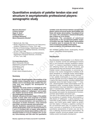
Figure 1. Representative patellar tendon scans showing the targets for the thickness measurement (upper panels), longitu-
dinal regions of interest (middle panels), and transversal regions of interest (lower panels) for the echo-intensity measure-
ments: ROI: region of interest.
©
C
IC
EdizioniInternazionali
4. dinal and in the transversal scans by using the four-
grade scale proposed by Sunding et al.16: 0: no neo-
vascularization; 1: mild neovascularization (a few
solitary blood vessels); 2: moderate neovasculariza-
tion (moderate quantity, mostly transversal blood ves-
sels); 3: severe neovascularization (several, mostly
horizontal blood vessels spread in the whole depth of
the tendon).
We arbitrarily defined “subclinical tendinopathy” as
the presence of either light structural changes in as-
sociation with at least mild neovascularization or
moderate/severe structural changes with/without neo-
vascularization.
Measurement of tendon thickness
Tendon thickness was measured in the longitudinal
scans acquired with 0-degree knee angle: as shown
in the representative images in Figure 1 (upper pan-
els), the target for the thickness measurement was
the thickest part of the tendon both for the proximal
probe position (i.e., just below the apex of the patella:
left panel) and for the central probe position (i.e.,
midportion of the tendon: right panel).
Measurement of tendon cross sectional area
Tendon CSA was measured in the transversal scans
acquired with 0-degree knee angle: as shown in the
representative images in Figure 1 (lower panels), a
region of interest was chosen in each scan to include
as much of the tendon as possible. The area was de-
termined for the selected region of interest by a cus-
tom software developed in MATLAB (The Math-
Works, Inc., Natick, MA, USA).
Measurement of tendon echo-intensity
Tendon echo-intensity (for images acquired with 0-
degree knee angle) was measured both in the longi-
tudinal (Fig. 1: middle panels) and in the transversal
scan (Fig. 1: lower panels) regions of interest: the
mean echo intensity (8-bit resolution, resulting in a
number between 0 and 255, where black=0,
white=255) was determined by a custom software de-
veloped in MATLAB (The MathWorks, Inc., Natick,
MA, USA).
Statistical analysis
Since the Shapiro-Wilk test for the normal distribution
of the data failed, non-parametric tests were used.
The Friedman’s ANOVA followed by Dunn’s post-hoc
test and the Wilcoxon test were adopted for compar-
isons of size parameters (i.e., thickness and cross
sectional area) and echo-intensity both between the
two tendon portions investigated (proximal vs central)
and between the two sides (left vs right).
K-means cluster analysis followed by F-ratio calcula-
tion were applied to the side-to-side differences in
size and echo-intensity of the tendon proximal portion
in order to discern between different groups of play-
ers.
Data are expressed as median (and range) and are
represented as box-and-whisker plots. Threshold for
statistical significance was set at P<0.05. Statistical
tests were performed with the IBM SPSS Statistics
(version 20 - IBM Corporation, Armonk, NY, USA)
software package.
Results
Qualitative assessments of tendon structure and
neovascularization
Figure 2 reports a representative example of one
thrower player showing in both sides a normal struc-
ture of the patellar tendon without neovascularization,
while Figure 3 shows for another representative
thrower player moderate structural changes and mild
neovascularization in the proximal portion of the left
patellar tendon and normal structure in the homolo-
gous portion of the contralateral tendon.
Similar to these representative examples, the qualita-
tive assessment of the longitudinal and transversal
scans of all players showed normal structure without
neovascularization in 7 out of 14 throwers and struc-
tural changes with or without neovasculation in the
proximal portion of the non-dominant tendon of the
other 7 throwers. The structure and neovasculariza-
tion scores of the non-dominant tendon of all players
are listed in Table II: in total, 5 out of 14 throwers
(35% of cases: # 4-8-9-11-13 in Tab. II) were consid-
ered affected by “subclinical tendinopathy” of the
non-dominant tendon because of the presence of
light structural changes in association with at least
mild neovascularization (two out of five players) or
moderate/severe structural changes with or without
neovascularization (three out of five players).
Quantitative assessments of tendon size and
echo-intensity
Figure 4 reports the box-and-whisker plots of the ten-
don size parameters (thickness and CSA) and of the
eco-intensity for both sides (left and right) and for
both portions (proximal and central) investigated.
As expected, the thickness of the proximal portion of
both sides of the tendon was higher compared to that
of the central portion (left side: P<0.001; right side:
P<0.01), while no differences were observed in ten-
don CSA and echo-intensity between the proximal
and central portion of both sides (P values > 0.05).
Further, no differences in thickness, CSA, and echo-
intensity of the tendon proximal portion were ob-
served between the two sides (P values > 0.05).
Analysis of individual data and K-means cluster
analysis unraveled a remarkable interindividual vari-
ability in the side-to-side differences in size and echo-
intensity of the tendon proximal portion. In fact, 6 out
of 14 players (43% of cases) presenting comparable
values of thickness, CSA, and echo-intensity between
the proximal portion of the two sides were classified
in cluster 1 (Tab. III: P≥0.05 for all side-to-side com-
parisons), while 8 out of 14 players (57% of cases)
Muscles, Ligaments and Tendons Journal 2017;7 (3):449-458452
M. Giacchino et al.
©
C
IC
EdizioniInternazionali
5. presenting side-to-side differences in CSA (P=0.01)
and echo-intensity (P=0.01) were included in cluster
2 (Tab. III: P<0.05 for side-to-side comparisons in
CSA and echo-intensity). In this subgroup of 8 play-
ers, CSA was significantly higher and echo-intensity
was significantly lower in the non-dominant tendon
compared to the dominant one. The following signifi-
cances (relative to the F-ratio for each variable) were
obtained for the differences between the two clusters:
thickness: P=0.165; CSA: P=0.005; longitudinal echo-
Muscles, Ligaments and Tendons Journal 2017;7 (3):449-458 453
Quantitative analysis of patellar tendon size and structure in asymptomatic professional players: sonographic study
Figure 2. Representative patellar tendon scans of one thrower player showing in both sides a normal structure of the tendon
without neovascularization.
©
C
IC
EdizioniInternazionali
6. intensity: P=0.001; transversal echo-intensity:
P=0.003.
Briefly, CSA and echo-intensity were the quantitative
variables most discriminant between the two clusters
and resulted significantly different between the domi-
nant and the non-dominant tendon in a large sub-
group (57% of cases) of asymptomatic thrower play-
ers.
Muscles, Ligaments and Tendons Journal 2017;7 (3):449-458454
M. Giacchino et al.
Figure 3. Representative patellar tendon scans of one thrower player showing moderate structural changes and mild neo-
vascularization in the proximal portion of the left patellar tendon and normal structure in the homologous portion of the right
tendon.
©
C
IC
EdizioniInternazionali
7. Correspondence between qualitative and quanti-
tative assessments
The 5 players identified by the qualitative analysis as
affected by subclinical tendinopathy (# 4-8-9-11-13 in
Tab. II) were a subgroup of the 8 players classified in
cluster 2.
Briefly, the qualitative assessments of tendon struc-
ture and neovascularization were in good agreement
with the results of the cluster analysis applied to
quantitative data of CSA and echo-intensity.
Do patellar tendon abnormalities predict tendi-
nopathy?
Two players (# 4 and 9 in Tab. II) identified by quali-
tative analysis as affected by subclinical tendinopathy
and classified in cluster 2 (2 out of 8 cases: 25%) de-
veloped a clinical tendinopathy (that was confirmed
by ultrasonography through qualitative assessments
of tendon structure and neovascularization) after a
follow-up of six months, while no players classified in
cluster 1 (0 out of 6 cases: 0%) developed a
tendinopathy (Tab. II, last column). Therefore, patel-
lar tendon abnormalities at baseline seems to in-
crease (although not significantly) the risk of develop-
ment of subsequent patellar tendinopathy (relative
risk=3.89, 95% CI 0.22 - 68.67, P=0.35).
Muscles, Ligaments and Tendons Journal 2017;7 (3):449-458 455
Quantitative analysis of patellar tendon size and structure in asymptomatic professional players: sonographic study
Table II. Results of the qualitative assessments of ten-
don structure and neovascularization (second and
third column: 5 players affected by “subclinical
tendinopathy” of the non-dominant patellar tendon are
highlighted in bold) and of the cluster analysis applied
to quantitative data of cross-sectional area and echo-
intensity (fourth column). The last column reports that
a clinical tendinopathy of the non-dominant patellar
tendon was found in two players (# 4 and 9) after a fol-
low-up of six months.
Player Structure Neovascu- Cluster Follow-up
score larisation
score
1 0 0 1 -
2 1 0 2 -
3 0 0 2 -
4 1 1 2 +
5 0 0 1 -
6 0 0 1 -
7 0 0 1 -
8 2 1 2 -
9 2 1 2 +
10 0 0 2 -
11 1 1 2 -
12 1 0 1 -
13 2 0 2 -
14 0 0 1 -
Figure 4. Patellar tendon
thickness, cross sectional
area, longitudinal echo-in-
tensity, and transversal
echo-intensity for both
sides (left and right) and for
both portions (proximal and
central) investigated in the
whole group of 14 thrower
players.
©
C
IC
EdizioniInternazionali
8. Discussion
In the present study, patellar tendon ultrasound im-
ages were acquired from both sides of 14 asympto-
matic thrower players and qualitative assessments (of
tendon structure and neovascularization) and quanti-
tative measurements (of tendon thickness, CSA, and
echo-intensity) were performed.
Referring to the aims listed in the Introduction, the
main results can be summarized as follows: I) qualita-
tive assessments showed a subclinical tendinopathy
of the non-dominant tendon in 5 out of 14 players
(35% of cases); II) quantitative assessments showed
abnormalities of the non-dominant tendon in 8 out of
14 players (57% of cases); III) qualitative assess-
ments of tendon structure and neovascularization
were in good agreement with the results of the cluster
analysis applied to quantitative data of CSA and
echo-intensity; IV) CSA and echo-intensity were the
quantitative variables most discriminant between
asymptomatic players without structural tendon ab-
normalities and those with tendon abnormalities; V)
patellar tendon abnormalities at baseline seem to in-
crease the risk of development of subsequent patellar
tendinopathy.
The high prevalence of subclinical tendinopathy and
structural tendon abnormalities in the non-dominant
side of the investigated group of thrower players can
be related to the repetitive overload of the non-domi-
nant lower limb: in fact, the throwing performance of
these players is similar to that of javelin throwers who
commonly present both subclinical and clinical patel-
lar tendinopathy.
Patellar tendinopathy is commonly evaluated through
the ultrasonographic assessment of localized tendon
thickening, presence of hypoechoic areas, and altered
vascularity8,13,17. The two approaches (qualitative and
quantitative) we adopted to evaluate the patellar ten-
dons of throwers enabled to assess all these ultra-
sonographic features: tendon size, tendon echogenici-
ty, and tendon vascularity. The main findings observed
in players classified as affected by subclinical
tendinopathy (and included in cluster 2) were presence
of hypo-echogenic areas and decreased echo-intensity
of the non-dominant tendon compared to the dominant
one, presence of vessels on Power Doppler analysis,
increased thickness and CSA of the non-dominant ten-
don compared to the dominant one. All these findings
represent valid signs of some of the structural changes
that occur during the tendinopathic process: increase
in tendon thickness, increase in the number of vessels,
disorganization of collagen fibers, increase in the hy-
drated components of the extracellular matrix, and
breakdown of tissue organization18-21. Other histopato-
logic findings (that cannot be assessed through ultra-
sonography) include increase in the number of sensory
nerves, proliferation of type III collagen fibers, hypocel-
lularity, increased number of inflammatory cells18-20. All
these features underlie tendon pathomechanics (i.e.,
reduced load-bearing capacity) and ultimately result in
tendon pain (and increased risk of tendon rupture)18,21.
Muscles, Ligaments and Tendons Journal 2017;7 (3):449-458456
M. Giacchino et al.
Table III. Median and min-max values of the size parameters and echo-intensity for the tendon proximal portion of the right
and left side. Statistically significant differences are highlighted in bold. CSA: cross sectional area, a.u.: arbitrary units.
Variable Left side Right side P value
Cluster 1 (n= 6 players)
Thickness (mm)
5.3
(4.2 - 6.4)
5.4
(4.8 - 6.3)
0.75
CSA (mm
2
)
98.8
(81.3 - 121.7)
95.9
(83.0 - 112.1)
0.92
Longitudinal echo-intensity (a.u.)
96.4
(70.6 - 109.2)
91.4
(65.6 - 107.6)
0.05
Transversal echo-intensity (a.u.)
73.2
(55.3 - 81.4)
64.3
(61.2 - 68.1)
0.46
Cluster 2 (n= 8 players)
Thickness (mm)
7.7
(5.6 - 9.3)
6.8
(5.3 - 7.8)
0.12
CSA (mm
2
)
120.3
(107.2 - 133.8)
93.4
(81.2 - 109.1)
0.01
Longitudinal echo-intensity (a.u.)
84.0
(74.9 - 112.5)
94.6
(85.1 - 115.5)
0.01
Transversal echo-intensity (a.u.)
63.1
(40.3 - 70.6)
80.1
(77.0 - 91.9)
0.01
Table III. Median (min-max values) of the size parameters and echo-intensity for the tendon proximal portion of the
right and left side. Statistically significant differences are highlighted in bold. CSA: cross sectional area, a.u.: arbi-
trary units.
Variable Left side Right side P value
Cluster 1 (n= 6 players)
Thickness (mm) 5.3 5.4 0.75
(4.2 - 6.4) (4.8 - 6.3)
CSA (mm2) 98.8 95.9 0.92
(81.3 - 121.7) (83.0 - 112.1)
Longitudinal echo-intensity (a.u.) 96.4) 91.4 0.05
(70.6 - 109.2 (65.6 - 107.6)
Transversal echo-intensity (a.u.) 73.2 64.3 0.46
(55.3 - 81.4) (61.2 - 68.1)
Cluster 2 (n= 8 players)
Thickness (mm) 7.7 6.8 0.12
(5.6 - 9.3) (5.3 - 7.8)
CSA (mm2) 120.3 93.4 0.01
(107.2 - 133.8) (81.2 - 109.1)
Longitudinal echo-intensity (a.u.) 84.0 94.6 0.01
(74.9 - 112.5) (85.1 - 115.5)
Transversal echo-intensity (a.u.) 63.1 80.1 0.01
(40.3 - 70.6) (77.0 - 91.9)
©
C
IC
EdizioniInternazionali
9. In the present study, the percentage of structurally
abnormal asymptomatic patellar tendons that became
symptomatic was 25%. This percentage and the rela-
tive risk of 3.89 are in agreement with the results of
previous studies22 indicating that the ultrasonograph-
ic structural changes in asymptomatic subjects may
represent markers of an early presymptomatic pathol-
ogy. However, a frequently cited criticism of ultra-
sound findings is their poor reliability. For instance,
ultrasonography is perceived to have a high risk of
error when evaluating tendon thickness and CSA22,23.
Moreover, the qualitative assessment of Achilles and
patellar tendon echogenicity showed poor to moder-
ate inter-observer agreement16. On the contrary,
quantitative analyses of tendon thickness and CSA
showed excellent inter-rater and intra-rater reliability5.
To our knowledge, this is the first study investigating
the longitudinal and transversal echo-intensity in
patellar tendons of asymptomatic humans. Previous
experimental works assessing the echo-intensity of
Achilles tendons in rats24 and of infraspinatus ten-
dons in sheeps25 found that the echo intensity of the
injured tendons was significantly lower compared to
intact tendons. Consistently, we observed in the 8
players classified in cluster 2 that both the longitudi-
nal and the transversal echo-intensity were signifi-
cantly lower in the proximal portion of the left (non-
dominant) tendon compared to the right (dominant)
tendon. Further, we showed that the quantitative vari-
ables most discriminant between structurally abnor-
mal and normal patellar tendons were echo-intensity
and CSA. Therefore, we recommend the systematic
use of these quantitative ultrasound features in cross-
sectional studies aimed to compare the structural ten-
don adaptations between different populations26 as
well as in longitudinal studies aimed to establish the
time course of the tendinopathic process8,13. It may
be hypothesized that the decrease in tendon echo-in-
tensity (that is possibly related to the increase in the
hydrated components of the extracellular matrix and
to the local inflammatory response) precede the in-
crease in tendon CSA (that is possibly related to dis-
organization of collagen fibers and proliferation of
type III collagen fibers). However, further studies are
required to confirm this hypothesis.
There are several limitations to this study. First, the
small sample size and the peculiarity of the investi-
gated population of elite throwers make generaliza-
tion to other (tendons of other) populations of athletes
difficult. Second, the short duration of the follow-up
(six months) could have implied an underestimation
of the risk of development of patellar tendinopathy.
Third, the causal association between (ultrasound
markers of) tendon histopathology and development
of tendinopathy was not demonstrated.
In conclusion, this study showed a high prevalence of
abnormalities of the non-dominant patellar tendon in
the recruited group of elite players (35% and 57% of
cases according to the qualitative and quantitative
assessments, respectively). Qualitative assessments
of tendon structure and neovascularization were in
good agreement with the results of the cluster analy-
sis applied to quantitative data of CSA and echo-in-
tensity. Finally, we found that patellar tendon abnor-
malities at baseline seem to increase the risk of de-
velopment of subsequent patellar tendinopathy.
Conflict of interest
None.
References
1. Martinoli C, Derchi LE, Pastorino C, Bertolotto M, Silvestri E.
Analysis of echotexture of tendons with US. Radiology.
1993;186:839-843.
2. Rasmussen OS. Sonography of tendons. Scand J Med Sci
Sports. 2000;10:360-364.
3. Fredberg U, Bolvig L, Andersen NT, Stengaard-Pedersen K.
Ultrasonography in evaluation of Achilles and patella tendon
thickness. Ultraschall Med. 2008;29:60-65.
4. Gellhorn AC, Morgenroth DC, Goldstein B. A novel sono-
graphic method of measuring patellar tendon length. Ultra-
sound Med Biol. 2012;38:719-726.
5. Gellhorn AC, Carlson MJ. Inter-rater, intra-rater, and inter-ma-
chine reliability of quantitative ultrasound measurements of the
patellar tendon. Ultrasound Med Biol. 2013;39:791-796.
6. Moustafa AM, Hassanein E, Foti C. Objective assessment of
corticosteroid effect in plantar fasciitis: additional utility of ultra-
sound. Muscles Ligaments Tendons J. 2016;5:289-296.
7. Kulig K, Landel R, Chang YJ, Hannanvash N, Reischl SF,
Song P, Bashford GR. Patellar tendon morphology in volley-
ball athletes with and without patellar tendinopathy. Scand J
Med Sci Sports. 2013;23:e81-88.
8. Giombini A, Dragoni S, Di Cesare A, Di Cesare M, Del Buono
A, Maffulli N. Asymptomatic Achilles, patellar, and quadriceps
tendinopathy: a longitudinal clinical and ultrasonographic
study in elite fencers. Scand J Med Sci Sports. 2013;23:311-
316.
9. Comin J, Cook JL, Malliaras P, McCormack M, Calleja M,
Clarke A, Connell D. The prevalence and clinical significance
of sonographic tendon abnormalities in asymptomatic ballet
dancers: a 24-month longitudinal study. Br J Sports Med.
2013;47:89-92.
10. Visnes H, Tegnander A, Bahr R. Ultrasound characteristics of
the patellar and quadriceps tendons among young elite ath-
letes. Scand J Med Sci Sports. 2015;25:205-215.
11. Van der Worp H, de Poel HJ, Diercks RL, van den Akker-
Scheek I, Zwerver J. Jumper’s knee or lander’s knee? A sys-
tematic review of the relation between jump biomechanics and
patellar tendinopathy. Int J Sports Med. 2014;35:714-722.
12. Reinking MF. Current concepts in the treatment of patellar
tendinopathy. Int J Sports Phys Ther. 2016;11:854-866.
13. Cook JL, Khan KM, Kiss ZS, Coleman BD, Griffiths L. Asymp-
tomatichypoechoicregionsonpatellartendonultrasound:A4-
year clinical and ultrasound follow-up of 46 tendons. Scand J
Med Sci Sports. 2001;11:321-327.
14. Elias LJ, Bryden MP, Bulman-Fleming MB. Footedness is a
better predictor than is handedness of emotional lateralization.
Neuropsychologia. 1998;36:37-43.
15. Padulo J, Oliva F, Frizziero A, Maffulli N. Muscles, Ligaments
and Tendons Journal - Basic principles and recommendations
in clinical and field science research: 2016 update. MLTJ.
2016;6(1):1-5.
16. Sunding K, Fahlström M, Werner S, Forssblad M, Willberg L.
Evaluation of Achilles and patellar tendinopathy with greyscale
ultrasound and colour Doppler: using a four-grade scale. Knee
Muscles, Ligaments and Tendons Journal 2017;7 (3):449-458 457
Quantitative analysis of patellar tendon size and structure in asymptomatic professional players: sonographic study
©
C
IC
EdizioniInternazionali
10. Surg Sports Traumatol Arthrosc. 2016;24:1988-1996.
17. Ooi CC, Richards PJ, Maffulli N, Ede D, Schneider ME, Con-
nell D, Morrissey D, Malliaras P. A soft patellar tendon on ul-
trasound elastography is associated with pain and functional
deficit in volleyball players. J Sci Med Sport. 2016;19:373-378.
18. Sharma P, Maffulli N. Biology of tendon injury: healing, model-
ing and remodeling. J Musculoskelet Neuronal Interact.
2006;6:181-190.
19. Khan KM, Maffulli N, Coleman BD, Cook JL, Taunton JE.
Patellar tendinopathy: some aspects of basic science and clin-
ical management. Br J Sports Med. 1998;32:346-355.
20. Khan KM, Cook JL, Bonar F, Harcourt P, Astrom M. Histopa-
thology of common tendinopathies. Update and implications
for clinical management. Sports Med. 1999;27:393-408.
21. Scott A, Backman LJ, Speed C. Tendinopathy: Update on
Pathophysiology. J Orthop Sports Phys Ther. 2015;45:833-
841.
22. McAuliffe S, McCreesh K, Culloty F, Purtill H, O’Sullivan K.
Can ultrasound imaging predict the development of Achilles
and patellar tendinopathy? A systematic review and meta-
analysis. Br J Sports Med. 2016;50:1516-1523.
23. Ekizos A, Papatzika F, Charcharis G, Bohm S, Mersmann F,
Arampatzis A. Ultrasound does not provide reliable results for
the measurement of the patellar tendon cross sectional area. J
Electromyogr Kinesiol. 2013;23:1278-1282.
24. Chamberlain CS, Duenwald-Kuehl SE, Okotie G, Brounts SH,
Baer GS, Vanderby R. Temporal healing in rat achilles tendon:
ultrasound correlations. Ann Biomed Eng. 2013;41:477-487.
25. Frisch KE, Marcu D, Baer GS, Thelen DG, Vanderby R. Influ-
ence of tendon tears on ultrasound echo intensity in response
to loading. J Biomech. 2014;47:3813-3819.
26. Malliaras P, Kamal B, Nowell A, Farley T, Dhamu H, Simpson
V, Morrissey D, Langberg H, Maffulli N, Reeves ND. Patellar
tendon adaptation in relation to load-intensity and contraction
type. J Biomech. 2013;46:1893-1899.
Muscles, Ligaments and Tendons Journal 2017;7 (3):449-458458
M. Giacchino et al.
©
C
IC
EdizioniInternazionali