
Recomendados
Recomendados
Mais conteúdo relacionado
Mais procurados
Semelhante a 1. microscopic study of parasites
Semelhante a 1. microscopic study of parasites (20)
Último
Último (20)
1. microscopic study of parasites
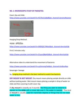
- 1. Cover slip and slide: https://www.youtube.com/watch?v=FG74x15a0y8&ab_channel=JeromeReamer Hanging Drop Method: https://www.youtube.com/watch?v=O0DQ2LTARaU&ab_channel=AmritaVlab First 2 minutes only: https://www.youtube.com/watch?v=Z1yyuQ8qHQI&ab_channel=AmritaVlab Alternative video to understand the movement of bacteria: https://www.youtube.com/watch?v=ujzSmsmg7ok&ab_channel=SridharRao Sewarage= Sewage So, hanging drop method is the best method to watch live bacteria. : Dry mount means placing sample directly on slide without putting water. Wet mount means placing a sample in drop of water on slide and then covering it with cover slip. A dry mount is exactly as it sounds: the thing you plan to observe is placed on the slide with a cover slip over it. No water is needed. This kind of mount is usually used for inanimate objects or things that do not need water to live.
- 2. Why is a wet mount better than a dry one? The hanging drop and wet mount techniques allow for observation of living organisms. The wet mount tends to dry out quickly under the heat of the microscope light; it is simpler to perform, but it is useful for short-term observation only. Which organisms are used in the method? Generally, Pseudomonas aeruginosa, Bacillus cereus, Staphylococcus aureus, Proteus. HANGING DROP PREPARATION TO EXAMINE THE MOTILITY OF MICROORGANISM / BACTERIA Hanging Drop Preparation is the useful technique employed in the laboratory for the microscopic examination of living microorganisms (viable microorganisms), especially the bacteria without staining them and to see their motility due to flagella. PRINCIPLE OF HANGING DROP PREPARATION The Hanging drop preparations is a special type of Wet mount in which a drop of broth culture of microorganism or bacterial suspension to be analyzed is placed on a glass cover slip which is encircled with a stick substance, preferably the Petroleum jelly and this cover slip containing the specimen is promptly inverted over the special type of Glass slide known as Cavity slide or depression slide containing a well (depression or cavity) in order that the drop hangs freely on the cover slip in the concavity of slide and the Petroleum jelly forms a seal that prevents the evaporation of the specimen and preserves it temporarily. The hanging drop preparation is then examined under the microscope to check the motility of the organism, preferably under reduced light to enhance the visibility and for better contrast. This method is ideally used in the laboratory to check the motility of Bacteria.
- 3. REQUIREMENTS FOR HANGING DROP PREPARATION: Soil/ Sewage sample Normal saline (0.9 %) Hanging drop slide / Cavity Slide Glass Coverslips Vaseline / Petroleum Jelly Matchsticks / cotton swab (cotton swab, cotton bar, swab stick Same) Inoculation loop Microscope Bunsen Burner PROCEDURE: ⇒ Collect soil/ sewerage (sewage) sample and mix the sample with normal saline. ⇒ Clean and flame the hanging drop slide/cavity slide and place it on the table with concavity/depression side up. ⇒ Now, Clean a coverslip and apply petroleum jelly or vaseline on each of the four corners of the coverslip, using a matchstick/ cotton swab. ⇒ Place the jelly coated coverslip on a clean paper with the petroleum jelly side up. ⇒ Transfer one loopful of broth culture or Bacterial suspension in the center of the coverslip.
- 4. ⇒ Now, Place the depression slide onto the coverslip, with the cavity facing down so that the depression covers the suspension drop. ⇒ Press the slide gently to form a seal between the coverslip and the slide to prevent the evaporation of specimen. ⇒ Lift the preparation and quickly turn the hanging drop preparation coverslip up so that the culture drop is suspended in the concavity of Depression slide. ⇒ Examine the preparation under low power objective lens, with reduced light and close the diaphragm of the microscope. Focus the edge (appeared as irregular lines crossing the field) of the slide using the coarse adjustment knob. ⇒ Without moving the microscope tube, switch to high power objective lens and examine the preparation again.
- 5. RESULT: Motile bacteria are observed on the edge of the drop. The motile bacteria were differentiated from bacteria establishing Brownian motion. OBSERVATIONS OF HANGING DROP PREPARATION Observe under the microscope by focusing the edge of the drop and carefully find the tiny objects which are bacteria. The bacterial cells will appear as either a dark or slightly greenish tiny bodies, very small rods (bacilli) or spheres (round or cocci). Movements are seen under microscope. PRECAUTIONS TO BE TAKEN WHILE PERFORMING HANGING DROP PREPARATION ⇒ The use of PPE (Gloves, Mask, Lab coat / Gown, Safety goggles etc.) is mandatory as you are going to deal with highly infections viable microorganism. ⇒ Use the young culture of the organism as in the old cultures, most probably the organisms are dead. ⇒ Put the appropriate size of the drop onto the cover glass which should not be too large or too small and hang freely in the concavity of depression slide. ⇒ First, observe under the low power objectives & then switch to high power & oil immersion objective for easy findings. ⇒ Adjust the diaphragm accordingly for better contrast which minimizes the errors in observations. ⇒ Observe carefully before reporting as Motile or Non- motile organisms. Discussion:
- 6. Hanging drop method is an aseptic method for examining the specimens from liquid culture instead of a solid culture medium. It is extensively used to study bacterial shape and arrangement and presence of flagella. Specimens in the hanging drop method will show Brownian movement, due to which the microscopic objects in the fluid will swim erratically through the kinetic energy possessed by the molecules in the surrounding fluid. The true mobility is observed through the multi-directional movement of bacterial cells to longer distances, instead of the cells moving back and forth. The bacterial motility can be observed under 10X and 40X objective, which is shown in a diagram. In a hanging drop method, 10X objective is initially used to focus the microscopic image, then later the objective is raised to 40X to get a magnified view of the sample taken and to distinguish between the motile and immotile cells. The petroleum jelly used on the corner of the coverslip acts as a sealing material between the coverslip and the concave depression glass. Besides, it also reduces the evaporation and excludes the effect of air currents. Overuse of petroleum jelly can give false results, as it may squeeze towards the centre of the drop containing microorganisms or may squeeze out of the edges and can stick to the objective lens of the microscope. The morphology of spiral bacteria can be explicitly studied in hanging drop method, as its shape becomes distorted in the heat fixing method. Bacterial motility or mobility can be well studied by employing a hanging drop method, in which a bacterial cell can freely move in the liquid medium. It is a wet mount technique as the microbes were first mixed with normal saline. The cytological changes that occur during the cell division, spore formation and germination of the bacteria are the events that need to be studied in a living condition or hanging drop method. The cytoplasmic inclusions, including vacuoles, granules etc. are easily noticeable by using this method.
- 7. We should be careful because, It is risky for the study of pathogenic bacteria in a living condition. And the depression slide is cost-effective, and the coverslip is fragile to work with.