SlideShare uma empresa Scribd logo
1 de 8
Maternal and Child Health Nursing
Pregnancy


                                    MATERNAL and CHILD HEALTH NURSING

                                                   PREGNANCY

                                     Lecturer: Mark Fredderick R. Abejo RN, MAN
 __________________________________________________________________________________

                                             Fertilization to Conception


      Fertilization: the union of the ovum & sperm. The start of Mitotic cell division &fetal sex determination.

            > Primary oocyte (immature ovum) contains Diploid number of chromosomes (46).
            > One oocyte contains a haploid (23) number of chromosomes after division.
            > Gamete (mature ovum): is a cell or ovum that has undergone Maturation & will be ready for
              fertilization.
            > One gamete carries 23 chromosomes.
            > A sperm carries 2 types of sex chromosomes. X & Y.
            > 400 million sperm cells in one ejaculation.
            > Functional Life of spermatozoa is 48 hours
            > XX= female, XY= male.


                                                      Morula


Process of Fertilization:
After ovulation          ovum will be expelled from the Graafian follicles        ovum will be surrounded byZona
Pellucida(mucopolysaccharide fluid) & a circle of cells (Corona Radiata) which increases the bulk of the Ovum
expelled from the Fallopian Tube by the Fimbriae (infundibulum). Sperms move by flagella & Penetrate the &
dissolve the cell wall of the ovum by releasing a proteolytic enzyme                 (Hyaluronidase)           After
penetration Fusion will result to Zygote.         Zygote migrate for 4 days in the body of the uterus (Mitosis will
take place-Cleavage formation will begin)          After 16-50 cell formation from mitosis, a mulberry & Bumpy
appearance will follow morula after 3-4 days, the structure will be ball like in appearance which will be called
Blastocyst. Cells in the outer ring are called Trophoblast (later it forms the placenta, responsible for the dev’t of
placenta & fetal membrane; Cells in the inner ring are called Erythroblast cells (which will be the embryo).


Terms to remember:
       Ovum: From ovulation to fertilization
       Zygote: From fertilization to implantation
       Embryo: From implantation to 5-8 weeks.
       Fetus:From 5-8 weeks until term
       The ovum is said to be viable for 24-36 hours.
       Sodium Bicarbonate- the frequent medication to alter the vaginal ph, decrease the acidity of the vagina
       so as to INCREASE THE MOTILITY OF THE SPERM.


Pregnancy                                                                                                   Abejo
Maternal and Child Health Nursing
Pregnancy


                                                    Fetal Membranes

     Fetal Membranes: membranes that surround the fetus, & give the placenta the shiny appearance.

2 Layers:

1. Amnion: shiny membrane on the 2nd week of Embryonic Development & encloses the Amniotic Cavity
        2. Chorion: Outer membrane that supports the sac of the amniotic fluid.

            Chorionic Villi: finger like projections from the chorion. This is the place where gases, nutrients and
                        waste products between the maternal & fetal blood takes place.

     Amniotic Fluid: surrounds the embryo, contains fetal urine, lanugo from fetal skin & epithelial cells.
       Ph is 7. 2. Specific Gravity: 1.005 – 1.025
       Normal Amount:          500 – 1000 ml.
       Oligohydramnios-       less than 300 ml.
       Polyhydramnios-        more than 2000 ml. observe for Down syndrome & congenital defects

            Functions of Amniotic Fluid:
              a. Protects the fetus from changes in the temperature & cushion against injury.
              b. Protects the umbilical cord from pressure, the fetus drinks & breaths the fluid into the lungs.

     Amniotic Fluid Colors: Normal color: transparent, clear, with white tiny specks
       Dark amber or yellow: Ominous sign of presence of Bilirubin, hemolytic disease
       Port Wine Colored: Abruptio Placenta
       Greenish: Meconium Stained / FETAL DISTRESS: always go for Cesarian Section! Also if ph is less
          than 7.2
       If with odor: deliver within 24 hours, may indicate infection.

     Umbilical Cord: 21 inches in length & 2 cm in thickness, circulatory communication of the fetus to the
         mother. CONTAINS 2 ARTERIES & 1 VEIN. Covered by a gelatinous mucopolysaccharide called
         Whartons jelly.

Implantation occurs at the end of the 1st week after fertilization, when the blastocyst attaches to the endometrium.
During the 2nd week (14 days after implantation), implantation progresses and two germ layers, cavities, and cell
layers develop. During the 3rd week of development (21 days after implantation), the embryonic disk evolves into
three layers, and three new structures — the primitive streak, notochord, and allantois — form. Early during the
4th week (28 days after implantation), cellular differentiation and organization occur.


                                                   Fertilization Cycle

Pregnancy                                                                                                      Abejo
Maternal and Child Health Nursing
Pregnancy


  PRE-FERTILIZATION                                        CONCEPTION                               IMPLANTATION
  ACTIVITIES
                                                     Zona reaction                                Morula (after 3-4
      Ovum moves to amulla of                                                                      days implantation)
          fallopian tubes                            Zygote (fertilized ovum;                     Blastocyst
                                                      about 24-48 hrs, divides;                     (trophoblast;
      Capacitation                                   cleavage divides, travels to                  embryolast)
                                                      the uterus                                   Implants complete
      Acrosome reaction                                                                            w/n 7-10 days


THREE PREGNANCY SIGNS & SYMPTOMS

        PRESUMPTIVE                                     PROBABLE                                    POSITIVE

      Amenorrhea                    Pregnancy test (presence of HCG)                               Auscultation of fetal
       Nausea/Vomiting              Softening of the uterine isthmus (Hegar’s sign)                heart by week 8
      Breast sensitivity and        Cervical softening (Goodell’s sign)                            Ultrasound imaging
      increased size                                                                               of fetal heart motion
       Fatigue Abdominal            Braxton-Hicks contractions                                     by week 7
      enlargement                   Ballotment: bouncing of the fetus in the amniotic fluid against the
                                                                                                   Ultrasound
      Skin pigmentation             examiners hand. During the 16th-20th week.                     confirmation of
      changes                                                                                      gestational sac by
      (Melasma chloasma,                 Braxton Hicks Contractions: painless                      week 6
      linea nigra- a brown line          contractions felt for 20-30 minutes occurs on
      running from the                   the 16th week.                                      Ultrasound: 6 weeks can
      umbilicus to the                                                                       auscultate the fetal heart.
      symphysis pubis               Chadwick’s sign is a bluish coloring of the vaginal
                                    mucosal that occurs as early as 6 weeks gestation.       Fetal movements palpated
 Stretch marks will                 Rationale: due to increase vascularity & blood           by the provider by week
 eventually fade to a silvery       vessel engorgement.                                      20.
 white color, but it is highly      Increase size of the uterus
 unlikely that they will                                                                     The most objective sign of
 completely disappear.              + Pregnancy Test                                         pregnancy is fetal
                                    > Secretion of HCG in the urine (Frog Test).             movement felt by the
 Breast changes- increase in        Detectable 10 days after the missed period               examiner.
 fullness, darker areola.
                                    . The fetal heartbeat typically can be heard and fetal
      Quickening: first fetal       rebound is possible between 18 and 22 weeks. The
           mov’t.                   fetal outline becomes palpable and the fetus is highly
                                    mobile between 28 and 31 weeks. Braxton Hicks
      Urinary Frequency
                                    contractions increase in frequency and intensity
      Melasma .
                                    between 32 and 35 weeks.


FETAL DEVELOPMENT


                                                 ORIGIN OF BODY TISSUE

       Tissue Layer                                                     Body Portion Formed
       Ectoderm                                         Nervous system, mucus membranes, anus & mouth
       Mesoderm                                         Connective Tissue, Reproductive, circulatory & upper Urinary
                                                        system, bones, cartillage
       Endoderm                                          Lining of the GI tract, Respiratory Tract, bladder & urethra


Pregnancy                                                                                                                  Abejo
Maternal and Child Health Nursing
Pregnancy


                                    Embryo is 4-5 mm length
                                    Trophoblasts embedded in deciduas
1 mo/ 4 weeks                       Foundations for nervous system, genitourinary system, skin, bones, and
                                    lungs are formed
                                    Rudiments of eyes, ears, nose appear
                                   Cardiovascular system functioning, heart beginning to beat, beginning of heart circulation.
                                    Placenta dev’t.
                                    Placental transport of substances ( 5 weeks)
                                    The fetus is 27-31 mm and weighs 2-4 grams
                                    Fetus s markedly bent
2 mo/ 5-8 weeks                     Head is disproportionately large due to brain development
                                    Centers of bone begin to ossify
                                    Ganglionic cells (5th to 12th weeks)
                                    Placenta and meconium are present, with facial features
3 mos./9-12 wks                  CVS done (8 12 weeks) every organ present, Head greatly enlarged
                                  Average length is 50-55 mm and weighs 45 gms.
                                  Fingers and toes are distinct.
                                  Rudimentary kidneys secrete urine.
                                  Fetal circulation is complete.
                                  External genitalia show definite characteristics.
                                  Ganglionic cells
                               SEX IS VISUALLY RECOGNIZABLE. Heart is audible in a Doppler ( 11th week)
                                  Fetus swallows. With nails. Kidneys able to secrete.
4 mos. /13-16 weeks               94-140 mm length and weighs 97-200 gms.
                                  Head is erected, lower limbs are well developed.
                                  Heartbeat is present
                                  Nasal septum and palate close
                                  Fingerprints are set
                               LANUGO APPEARS IN THE BODY
5 mos. /17-20 weeks               Fetus is 150-190 mm. In length and weighs approximately 260-460 gms.
                                  Lanugo covers entire body.
                                  Eyebrows and scalp hair is present.
                                  Heart sounds are perceptible by auscultation.
                                  Vernix caseosa covers skin.
                               Heartbeat can be heard in the fetoscope ( 18 weeks—20 weeks). Liver is already    pancreas
                                  functioning.
                               Quickening felt by a mother. Skeleton begins to develop.
                                  Brown Fats begin to form. Heart sounds in the stethoscope
                                  Can be heard ( 17- 20 weeks)
                              NOTE: There is a placental barrier to syphilis until the 18th week of pregnancy. If
                              the mother is treated before 18th week, the baby will most likely not be affected.
6 mos. /21-25 weeks               21-25 WEEKS… OLD MAN’s FACE
                                  Length 200-240 mm. Wt. 495-910 gms.
                                  Skin appears wrinkled and pink to red.
                                  REM begins
                                  Eyebrows and fingernails develop.
                               VERNIX COVERS THE ENTIRE BODY. Has the ability to hear. Production of lung surfactants.
                                  Passive Antibody transfer ( placental immunoglobulin G)
                                  Sustained weight gain occurs.
7 mos. /26-29 weeks               Length 250-275; weight 910-1500 gms.
                                  Skin red
                                  Rhythmic breathing occurs
                                  Pupillary membrane disappears from eyes.
                                  Fetus often survives if born prematurely
                                  Brain develops rapidly. Lecithin- Sphingomyelin (L/S ratio is already 2:1)
                               Brains fully developed. If born, neonate may survive.
8 mos. /30-34 weeks               Length 280-320 mm. weight 1700-2500 gms.
                                  Toenails become visible
                                  Steady weight gain occurs
                                  Vigorous fetal movement occurs.
                               LANUGO DISAPPEARS. Bones are fully developed.
                                  Aware of sounds outside the body. Assumes the delivery position. Increased
                              chance of survival.
9 mos. /35-37 weeks               Length 330-360 mm. weight 2700-3400 gms.
                                  Face and body has a loose wrinkled appearance because of subcutaneous fat
                                  deposit.
                                  Body is usually lump and lanugo disappears
                                  Nails reach fingertip edge
                                  Amniotic fluid decreases.
                               Increase Development. Sole of the foot have already
                               creases. Good chance of survival.

Pregnancy                                                                                                         Abejo
Maternal and Child Health Nursing
Pregnancy


10 mos. / 38-40 weeks               Length 360 mm.; Weight 3400-3600 gms.
                                    Skin is smooth, chest is prominent
                                    Eyes are uniformly slate colored
                                    Bones of skull are ossified and are nearly together at sutures.
                                   Testes are in scrotum.


                                                           Fetal Circulation

       As early as 3rd week of intra-uterine life, fetal blood is already is circulating, specifically there is already exchange of
      nutrients with the maternal circulation in the chorionic villi.
      > Arteries carry UNOXYGENATED BLOOD. VEINS carry OXYGENATED BLOOD.
      > Fetal Circulation Bypass: Why:

      DUE TO NON-FUNCTIONING LUNGS:
      ----- Ductus arteriousus (between pulmonary artery & Aorta, OPENS AT BIRTH & CLOSES 24 –48 hours after
            delivery.) It CONTAINS a mixture of arterial & venous blood.
      ----- Foramen Ovale : between right & left atrium

      DUE TO NON-FUNCTIONING LIVER:
      ----- Ductus Venosus (by pass the liver, closes at birth; an umbilical vein that carries High oxygen from the placenta.


Maternal & Fetal Diagnostic Test


      CHORIONIC VILLI SAMPLING                               Earliest test possible on fetal cells; sample obtained
                                                             by slender catheter passed through cervix to
                                                             implantation site.
                                                             a.     Chorionic Villi Sampling: removal of a
                                                                   small piece of Chorionic villi sampling to
                                                                   detect the ff: fetal chromosome, enzyme, DNA
                                                                   & biochemical abnormalities. Performed
                                                                   between the 8th – 11th weeks of gestation. Can
                                                                   detect the ff; Genetic Defects:
                                                                    Cystic fibrosis, trisomy 21, Tay Sachs, sickle
                                                                   cell     anemia,      thallasemia,    Duchenne
                                                                   muscular dystrophy & hemophilia.
                                                                 Most common indication: advance maternal
                                                                   age: increases risk of chromosomal damage
                                                                   from aging of oocyte.
                                                                 Greatest Advantage over Amniocentesis:
                                                                   PERFORMED DURING THE FIRST
                                                                   TRIMESTER. (16th- 20th week of gestation). .
                                                                   Laboratory results are obtained in 1 - 7 days
                                                                   compared to 20-28 days for an amniocentesis.
                                                                   Disadvantages:
                                                                        1. Risk of Abortion
                                                                        2. Infection
                                                                        3. Embryo-fetal/placental damage
                                                                        4. Spontaneous abortion
                                                                        5. Premature rupture of the membranes
                                                                 After an Rh-negative patient undergoes
                                                                   amniocentesis or CVS, the nurse should
                                                                   administer Rh (D) immune globulin
                                                                   (RhoGAM), to prevent Rh sesnsitization, an
                                                                   antigen antibody immunologic reaction that
                                                                   sometimes occurs when an Rh negative mother
                                                                   carries an Rh + fetus.
                                                                 The patient does not require complete bed
                                                                   rest after CVS---SHE SHOULD REFRAIN
                                                                   FROM SEXUAL INTERCOURSE AND
                                                                   PHYSICAL ACTIVITY FOR 48 hours. A
                                                                   small amount of spotting is normal for the 1st
                                                                   24-48 hours.

                   ULTRASOUND                                Use of sound and returning echo patterns to identify
                                                             intrabody structures; useful early in pregnancy to
                                                             identify gestational sacs; later uses include
                                                             assessment of fetal viability, growth patterns,
                                                             anomalies, fluid volume, uterine anomalies and
                                                             adnexal masses. Use adjunct to amniocentesis; safe
                                                             for fetus (no ionizing radiation)

Pregnancy                                                                                                                Abejo
Maternal and Child Health Nursing
Pregnancy


                                     Ultrasound:        done 18-40 weeks for fetal
                                     abnormalities,

                                     THE   BEST           TEST      FOR       ECTOPIC
                                     PREGNANCIES

                                     - Non-invasive procedure with high frequency sound
                                         waves to obtain outline of the fetus, placenta &
                                         uterine cavities and to confirm gestational age &
                                         EDD.
                                     -  NEEDS A FULL BLADDER TO OBTAIN A
                                         BETTER IMAGE (drink a full glass every 15
                                         minutes beginning an hour & half the procedure)
                                     -  COMMON METHOD IN LOCATING THE
                                         PRECISE POSITION OF THE FETUS &
                                         PLACENTA BEFORE AMNIOCENTESIS.

                 AMNIOCENTESIS       Location and aspiration of amniotic fluid for
                                     examination; possible after the 14th week when
                                     sufficient amounts are present; used to identify
                                     chromosomal aberration, sex of fetus, levels of
                                     alpha-fetoprotein and other chemicals indicative of
                                     neural tube defects and inborn error of metabolism,
                                     gestational age, RH factor.
                                     I.V. anesthesia isn't given for amniocentesis. The
                                          client should be supine during the procedure;
                                          afterward, she should be placed on her left side
                                          to avoid supine hypotension, promote venous
                                          return, and ensure adequate cardiac output.

                                     Amniocentesis: invasive procedure for amniotic
                                     fluid analysis, & fetal lung maturity.
                                     Procedure: Ultrasound 1st: the rationale: to locate
                                         the Placenta. The patient MUST EMPTY THE
                                         BLADDER TO REDUCE THE SIZE OF THE
                                         BLADDER. Vital signs are assessed every 15
                                         minutes.

                                      Typically performed on the 3rd trimester to assess
                                        LECITHIN-SPHINGOMYELIN RATIO IN THE
                                        AMNIOTIC FLUID (this ratio indicates fetal
                                        lung maturity), which is commonly delayed in a
                                         diabetic client, Cesarean Delivery  should
                                        not be done, unless the fetal lungs are matured.

                                    Position: Supine.
                                      PLACE A FOLDED TOWEL ON HER RIGHT
                                         BUTTOCKS TO TIP HER SLIGHTLY TO THE
                                         LEFT & MOVE THE UTERUS OFF THE VENA
                                         CAVA TO PREVENT SUPINE HYPOTENSION
                                         SYNDROME.
                                      ABDOMINAL PREP IS DONE, then, needle
                                          insertion in a 20-22 gauge spinal needle,
                                          withdrawing amniotic fluid.
                                      NORMAL L/S RATIO (lecithin/sphingomyelin):
                                          2:1 = normal fetal lung maturity ratio

                                      Most important factor affecting Amniocentesis:
                                         NEEDLE INSERTION-because of the risk of
                                         puncture or damage to the placenta, fetus,
                                         umbilical cord, bladder & uterine arteries.
                                      Disadvantages:
                                     Risk for:
                                        1. Maternal hemorrhage
                                        2. Infection
                                        3. Rh immunization
                                        4. abruptio placenta
                                        5. Amniotic fluid embolism

                                     CALL THE PHYSICIAN FOR THE FF: Chills,
                                        fever, leakage of fluid, decrease fetal movement
                                        or uterine contractions.


Pregnancy                                                                                    Abejo
Maternal and Child Health Nursing
Pregnancy


                                                                  After amniocentesis, the patient is
                                                              monitored for uterine contractions, fetal heart
                                                              rate changes and leakage of amniotic fluid
                                                              from the puncture site. During this period, the
                                                              patient isn’t ambulated.


                          X-RAY                          Can be used late in pregnancy (after ossification of
                                                         fetal bones) to confirm position and presentation; not
                                                         used in early pregnancy to avoid possibility of
                                                         causing damage to fetus and mother.

             ALPHA-FETOPROTEIN                           Maternal serum screens for open neural tube defects.
                 SCREENING                               It is a glucoprote in produced by fetal yolk sac, GI
                                                         tract and liver. Test done between 16 and 18 weeks
                                                         gestation.
                                                             Alpha Fetoprotein:
                                                                    PRINCIPAL SCREENING TEST DOR
                                                                   THE DETECTION OF NEURAL TUBE
                                                                   DEFECTS (spina bifida, hydrocephalus-
                                                                   can be reduced through increase folic acid-
                                                                   0.4 mg/day in the 1st trimester)
                                                                   > Maternal blood sampling between 16-20
                                                             weeks.
                                                                   LOW: chromosomal defects (Downs
                                                             syndrome)
                                                         HIGH: (greater than 10 mg/dl) Neural tube defects,
                                                         anencephaly & the absence of ventral abdominal
                                                         wall, premature delivery, toxemia & fetal distress &
                                                         Rh immunization.
                       L/S RATIO                         Uses amniotic fluid to ascertain fetal lung maturity
                                                         through measurement of presence and amounts of the
                                                         lung surfactants lecithin and sphingomyelin. At 35-
                                                         36 weeks; ratio is 2:1 indicative of mature levels.
         PHOSPHATIDYL GLCEROL                            Found in amniotic fluid after 35 weeks. In
                                                         conjunction with the L/S ratio; it contributes to
                                                         increased reliability of fetal lung maturity testing.
                                                         Maybe done in laboratory.

                                                         Phosphatidyl Glycerol (PG): when present in the
                                                         amniotic fluid, it can be predicted that respiratory
                                                         distresss will not occur, or RDS will not occur.
               CREATININE LEVEL                          Estimates fetal renal maturity and function, uses
                                                         amniotic fluid.
                      BILIRUBIN                          Level-high early in pregnancy; drops after 36 weeks
                                                         gestation; uses amniotic fluid.
                                                         The yellow color is the result of fetal anemia and
                                                         bilirubin.
        FETAL MOVEMENT COUNT                             Teach mother to count 2-3 times daily, 30-60
                                                         minutes each time, should feel 5-6 movements per
                                                         counting time; mother should notify care giver
                                                         immediately of abrupt change or no movement.
       PERCUTANEOUS UMBILICAL                            Uses ultrasound to locate umbilical cord. Cord blood
           BLOOD SAMPLING                                aspirated and tested. Used in second and third
                                                         trimesters.
             BIOPHYSICAL PROFILE                         A collection of data on fetal breathing movements,
                                                         body movements, muscle tone, reactive heart rate
                                                         and amniotic fluid volume.


                                            ELECTRONIC MONITORING

     A. Non-Stress Test – accelerations in heart rate accompany normal fetal movement; non-invasive
        Tocodynamometer records fetal movements and Doppler ultrasound measures
       - Observation of fetal heart rate related to fetal movement. Fetal well-being.
         Indicated for: assess placental function & oxygenation, fetal well being, evaluates fetal heart rate in response to
         fetal movement especially for: Maternal Problems such as chronic hypertension, diabetes and Pre-eclampsia, given
         after the 32nd week.

            PREPARATION:
            Patient should eat snacks.
            Position: Semi-Fowlers or left lateral positions the mother may ask tom press the button every time she feels fetal
                       movements; the monitor records a mark at each point of fetal movement.


Pregnancy                                                                                                             Abejo
Maternal and Child Health Nursing
Pregnancy


            RESULTS:
              1. Reactive (normal): indicates a fetal fetus
                       Greater than 15 beats per minute- occur with fetal movement in a 10 or 20 minute period.
                        FAVORABLE RESULTS:
                - 2 or more FHR accelerations of 15 seconds over a 20 minutes interval and return of FHR to normal baseline.
              2. Non-Reactive (Abnormal): No fetal movement occurs or there is short-term fetal heart rate variability (less
                   than 6 beats per minute). The doctor will order an Oxytocin Test AFTER the patient has non-reactive test.

        NOTE: COMMONLY PERFORMED ON DIABETIC PATIENTS BECAUSE OF THE INCREASE RISK FOR STILL
                                               BIRTH.

     B. Contraction Stress Test (CST) – based on the principle that healthy fetus can withstand decreased oxygen during
        contraction but compromised fetus cannot. Response of the fetus to induced uterine contractions as an INDICATOR
        OF UTEROPLACENTAL & FETAL PHYSIOLOGICAL INTEGRITY.
        PREPARATION:
                 Woman in semi-Fowler’s or side-lying position.
                 Monitor for post-test labor onset.
        TYPES:
                  a. Mammary stimulation Test or Breast Stimulation Exam or
                      Nipple Stimulated CST – non-invasive
                  b. Oxytocin Challenge test

            Indications: ALL PREGNANCIES AFTER 28 WEEKS WITH HIGH RISK CLIENTS.
            Contraindicated for history of PRE-TERM LABOR.

Interpretations:
POSITIVE RESULT: Late decelerations with at least 50% of contractions. Potential risks to the fetus, which may
         necessitate to C-section.
        Abnormal and known as “Positive window”. Abnormal: ―Positive Window‖: (+) LATE DECELERATIONS OF
        FHR with three contractions a 10 minute interval. Indicates Uteroplacental Insufficiency.

NEGATIVE RESULTS: No late decelerations with a minimum of 3 contractions lasting 40-60 seconds in 10 minutes
     period. Normal: ―Negative Window‖: (-) LATE DECELERATIONS OF FHR with three contractions a 10m minute
     interval
     Normal and known as “Negative window

Laboratory Studies

            1. Estriol excretion: measures placental functioning through urine test.
                            Collect a 24-hour urine specimen or serum blood levels.
                            High Estriol: Good placental function
                            Low Estriol: Fetal hypoxia
             Estriol: estrogenic hormone, synthesized by the placenta & adrenal gland of the fetus which secreted by the
                             ovaries

Rh Incompatibility Test:
Purpose: to discover presence of antibodies present in Rh-negative mother’s blood
       > Test will confirm the diagnosis for Hemolytic Disease in the Newborn.

            Types:
                       1. Indirect Coomb’s Test: women who have Rh negative have this test done to determine if they have
                                   antibodies to the factor present. Repeated 28 weeks pregnancy. Mothers reveal antibodies as
                                   a result of previous transfusion or pregnancy.
                       2. Direct Coomb’s test: tests for newborns cord blood- determines presence of maternal antibodies
                                   attached to the baby’s cell.

                                       Rh (D) & D negative who hasn’t formed antibodies should receive Rhogam at 28
                                        weeks gestation or after 72 hours after delivery.

 Nitrazine Test: use of nitrazin strip to detect the presence of amniotic fluid.
        Vaginal Secretions: PH: 4.5- 5.5
        Amniotic fluid: PH: 7.2 – 7.5 (turns the yellow Nitrazine blue gray, blue green – Ruptured Membranes)
 Kicks count: fetal movement counting mother sits quietly on the LEFT SIDE for 1 hour after meals & count fetal kicks for
        30 minutes. Notify the physician or health care provider if FEWER THAN 3 KICKS.

 Biophysical Profile : surveillance of fetal well being base on 5 categories:
       1. Fetal breath mov’t
       2. Fetal tone
       3. Amniotic fluid
       4. Fetal heart reactivity
       5. Placental Grade

Interpretation:
        Fetal score of 8 – 10: normal fetal well-being
        Fetal score of 4 – 6: fetal distress

Pregnancy                                                                                                            Abejo

Mais conteúdo relacionado

Mais procurados

Prenatal ( Health Center) Handouts
Prenatal ( Health Center)  HandoutsPrenatal ( Health Center)  Handouts
Prenatal ( Health Center) HandoutsMarkFredderickAbejo
 
Complication on Labor and Delivery
Complication on Labor and DeliveryComplication on Labor and Delivery
Complication on Labor and DeliveryMarkFredderickAbejo
 
Intrapartum Care: Introduction
Intrapartum Care: IntroductionIntrapartum Care: Introduction
Intrapartum Care: IntroductionSaide OER Africa
 
Ncp for cae press
Ncp for cae pressNcp for cae press
Ncp for cae pressKath Gindap
 
PNLE Medical Surgical Nursing Exam 1.docx
PNLE Medical Surgical Nursing Exam 1.docxPNLE Medical Surgical Nursing Exam 1.docx
PNLE Medical Surgical Nursing Exam 1.docxAbdhaniePanontongan
 
Nursing care for abortion
Nursing care for abortionNursing care for abortion
Nursing care for abortionIda Hui-Ming
 
Pathophysiology of Normal Labour by Sunil Kumar Daha
Pathophysiology  of Normal Labour by Sunil Kumar DahaPathophysiology  of Normal Labour by Sunil Kumar Daha
Pathophysiology of Normal Labour by Sunil Kumar Dahasunil kumar daha
 
Antihypertensive and Anticonvulsant drugs in OBG
Antihypertensive and Anticonvulsant drugs in OBGAntihypertensive and Anticonvulsant drugs in OBG
Antihypertensive and Anticonvulsant drugs in OBGSantosh Kumari
 

Mais procurados (20)

July 2012 nle tips mchn
July 2012 nle tips mchnJuly 2012 nle tips mchn
July 2012 nle tips mchn
 
Prenatal ( Health Center) Handouts
Prenatal ( Health Center)  HandoutsPrenatal ( Health Center)  Handouts
Prenatal ( Health Center) Handouts
 
Dec 2012 NLE TIPS CHD and CD
Dec 2012  NLE TIPS CHD and CDDec 2012  NLE TIPS CHD and CD
Dec 2012 NLE TIPS CHD and CD
 
Complication on Labor and Delivery
Complication on Labor and DeliveryComplication on Labor and Delivery
Complication on Labor and Delivery
 
Intrapartum Care: Introduction
Intrapartum Care: IntroductionIntrapartum Care: Introduction
Intrapartum Care: Introduction
 
July 2012 nle tips ms
July 2012 nle tips msJuly 2012 nle tips ms
July 2012 nle tips ms
 
July 2012 nle tips chn and cd
July 2012 nle tips chn and cdJuly 2012 nle tips chn and cd
July 2012 nle tips chn and cd
 
Female Reproductive System
Female Reproductive SystemFemale Reproductive System
Female Reproductive System
 
Ncm 102 (new) lea 1 midterm
Ncm 102 (new)  lea 1 midtermNcm 102 (new)  lea 1 midterm
Ncm 102 (new) lea 1 midterm
 
July 2012 nle tips funda
July 2012 nle tips fundaJuly 2012 nle tips funda
July 2012 nle tips funda
 
Ncp for cae press
Ncp for cae pressNcp for cae press
Ncp for cae press
 
EPI DISEASES Handouts
EPI DISEASES HandoutsEPI DISEASES Handouts
EPI DISEASES Handouts
 
PNLE Medical Surgical Nursing Exam 1.docx
PNLE Medical Surgical Nursing Exam 1.docxPNLE Medical Surgical Nursing Exam 1.docx
PNLE Medical Surgical Nursing Exam 1.docx
 
Gastrointestinal Nursing
Gastrointestinal NursingGastrointestinal Nursing
Gastrointestinal Nursing
 
Newborn Assessment
Newborn AssessmentNewborn Assessment
Newborn Assessment
 
Care of the Newborn Handouts
Care of the Newborn  HandoutsCare of the Newborn  Handouts
Care of the Newborn Handouts
 
Nursing care for abortion
Nursing care for abortionNursing care for abortion
Nursing care for abortion
 
Pathophysiology of Normal Labour by Sunil Kumar Daha
Pathophysiology  of Normal Labour by Sunil Kumar DahaPathophysiology  of Normal Labour by Sunil Kumar Daha
Pathophysiology of Normal Labour by Sunil Kumar Daha
 
Antihypertensive and Anticonvulsant drugs in OBG
Antihypertensive and Anticonvulsant drugs in OBGAntihypertensive and Anticonvulsant drugs in OBG
Antihypertensive and Anticonvulsant drugs in OBG
 
July 2012 nle tips psych
July 2012 nle tips psychJuly 2012 nle tips psych
July 2012 nle tips psych
 

Semelhante a Pregnancy Handouts

The physiological changes of pregnancy promoting maternal health
The physiological changes of pregnancy   promoting maternal healthThe physiological changes of pregnancy   promoting maternal health
The physiological changes of pregnancy promoting maternal healthReynel Dan
 
RbGrp3 - Fertilization and Development
RbGrp3 - Fertilization and Development RbGrp3 - Fertilization and Development
RbGrp3 - Fertilization and Development Jessica Kabigting
 
ASSESSMENT AND MANAGEMENT OF WOMEN DURING POSTNATAL PERIOD.pptx
ASSESSMENT AND MANAGEMENT OF WOMEN DURING POSTNATAL PERIOD.pptxASSESSMENT AND MANAGEMENT OF WOMEN DURING POSTNATAL PERIOD.pptx
ASSESSMENT AND MANAGEMENT OF WOMEN DURING POSTNATAL PERIOD.pptxRameeThj
 
fertilization, pregnancy, and lactation.ppt
fertilization, pregnancy, and lactation.pptfertilization, pregnancy, and lactation.ppt
fertilization, pregnancy, and lactation.pptHumaMuth
 
fertilization, pregnancy, and lactation.ppt
fertilization, pregnancy, and lactation.pptfertilization, pregnancy, and lactation.ppt
fertilization, pregnancy, and lactation.pptHumaMuth
 
Diagnosis of pregnancy
Diagnosis of pregnancy Diagnosis of pregnancy
Diagnosis of pregnancy BRITO MARY
 
2 gy embryology for anesthesia
2 gy embryology for anesthesia2 gy embryology for anesthesia
2 gy embryology for anesthesiagishabay
 
Embryology for anesthesia
Embryology for anesthesiaEmbryology for anesthesia
Embryology for anesthesiaEngidaw Ambelu
 
embryologymo-150405023631-conversion-gate01.pdf
embryologymo-150405023631-conversion-gate01.pdfembryologymo-150405023631-conversion-gate01.pdf
embryologymo-150405023631-conversion-gate01.pdfUllas Chandra Sahoo
 
5_6282657037939640532.pptx
5_6282657037939640532.pptx5_6282657037939640532.pptx
5_6282657037939640532.pptxSrinath Chowdary
 
fertilization.pptx
fertilization.pptxfertilization.pptx
fertilization.pptxSujatha A
 
Reproductive system
Reproductive systemReproductive system
Reproductive systemkrisgonewild
 
Human Reproduction
Human ReproductionHuman Reproduction
Human Reproductiongreinejl
 
Development
DevelopmentDevelopment
Developmentktrainor
 
Chapter24 reprofemalemarieb
Chapter24 reprofemalemariebChapter24 reprofemalemarieb
Chapter24 reprofemalemariebLawrence James
 

Semelhante a Pregnancy Handouts (20)

The physiological changes of pregnancy promoting maternal health
The physiological changes of pregnancy   promoting maternal healthThe physiological changes of pregnancy   promoting maternal health
The physiological changes of pregnancy promoting maternal health
 
Beyond fertilization
Beyond fertilizationBeyond fertilization
Beyond fertilization
 
Conception/Obstetrics Nursing
Conception/Obstetrics NursingConception/Obstetrics Nursing
Conception/Obstetrics Nursing
 
RbGrp3 - Fertilization and Development
RbGrp3 - Fertilization and Development RbGrp3 - Fertilization and Development
RbGrp3 - Fertilization and Development
 
ASSESSMENT AND MANAGEMENT OF WOMEN DURING POSTNATAL PERIOD.pptx
ASSESSMENT AND MANAGEMENT OF WOMEN DURING POSTNATAL PERIOD.pptxASSESSMENT AND MANAGEMENT OF WOMEN DURING POSTNATAL PERIOD.pptx
ASSESSMENT AND MANAGEMENT OF WOMEN DURING POSTNATAL PERIOD.pptx
 
fertilization, pregnancy, and lactation.ppt
fertilization, pregnancy, and lactation.pptfertilization, pregnancy, and lactation.ppt
fertilization, pregnancy, and lactation.ppt
 
fertilization, pregnancy, and lactation.ppt
fertilization, pregnancy, and lactation.pptfertilization, pregnancy, and lactation.ppt
fertilization, pregnancy, and lactation.ppt
 
Diagnosis of pregnancy
Diagnosis of pregnancy Diagnosis of pregnancy
Diagnosis of pregnancy
 
Chapter25 reprofemalemarieb
Chapter25 reprofemalemariebChapter25 reprofemalemarieb
Chapter25 reprofemalemarieb
 
2 gy embryology for anesthesia
2 gy embryology for anesthesia2 gy embryology for anesthesia
2 gy embryology for anesthesia
 
Embryology for anesthesia
Embryology for anesthesiaEmbryology for anesthesia
Embryology for anesthesia
 
embryologymo-150405023631-conversion-gate01.pdf
embryologymo-150405023631-conversion-gate01.pdfembryologymo-150405023631-conversion-gate01.pdf
embryologymo-150405023631-conversion-gate01.pdf
 
Embryology
Embryology Embryology
Embryology
 
5_6282657037939640532.pptx
5_6282657037939640532.pptx5_6282657037939640532.pptx
5_6282657037939640532.pptx
 
1 physiology of pregnancy
1 physiology of pregnancy1 physiology of pregnancy
1 physiology of pregnancy
 
fertilization.pptx
fertilization.pptxfertilization.pptx
fertilization.pptx
 
Reproductive system
Reproductive systemReproductive system
Reproductive system
 
Human Reproduction
Human ReproductionHuman Reproduction
Human Reproduction
 
Development
DevelopmentDevelopment
Development
 
Chapter24 reprofemalemarieb
Chapter24 reprofemalemariebChapter24 reprofemalemarieb
Chapter24 reprofemalemarieb
 

Mais de MarkFredderickAbejo

10 Good Reason to Pass RH Bill (Tag)
10 Good Reason to Pass RH Bill (Tag)10 Good Reason to Pass RH Bill (Tag)
10 Good Reason to Pass RH Bill (Tag)MarkFredderickAbejo
 
10 Good Reason to Pass RH Bill (Eng)
10 Good Reason to Pass RH Bill (Eng)10 Good Reason to Pass RH Bill (Eng)
10 Good Reason to Pass RH Bill (Eng)MarkFredderickAbejo
 
Get set for a healthy 2012 london olympic games
Get set for a healthy 2012 london olympic gamesGet set for a healthy 2012 london olympic games
Get set for a healthy 2012 london olympic gamesMarkFredderickAbejo
 
Stay healthy during london olympics 2012
Stay healthy during london olympics 2012Stay healthy during london olympics 2012
Stay healthy during london olympics 2012MarkFredderickAbejo
 
Perioperative Nursing (complete)
Perioperative Nursing (complete)Perioperative Nursing (complete)
Perioperative Nursing (complete)MarkFredderickAbejo
 
Community Health Nursing (complete)
Community Health Nursing (complete)Community Health Nursing (complete)
Community Health Nursing (complete)MarkFredderickAbejo
 
Prc bon memorandum-order-no-2 b-odc form-series-of-2009
Prc bon memorandum-order-no-2 b-odc form-series-of-2009Prc bon memorandum-order-no-2 b-odc form-series-of-2009
Prc bon memorandum-order-no-2 b-odc form-series-of-2009MarkFredderickAbejo
 
Prc bon memorandum-order-no-2-series-of-2009
Prc bon memorandum-order-no-2-series-of-2009Prc bon memorandum-order-no-2-series-of-2009
Prc bon memorandum-order-no-2-series-of-2009MarkFredderickAbejo
 
Medical and Surgical Nursing Review Bullets
Medical and Surgical Nursing Review BulletsMedical and Surgical Nursing Review Bullets
Medical and Surgical Nursing Review BulletsMarkFredderickAbejo
 

Mais de MarkFredderickAbejo (19)

December 2012 NLE Tips Funda
December 2012 NLE Tips FundaDecember 2012 NLE Tips Funda
December 2012 NLE Tips Funda
 
Cybercrime Prevention Act
Cybercrime Prevention ActCybercrime Prevention Act
Cybercrime Prevention Act
 
10 Good Reason to Pass RH Bill (Tag)
10 Good Reason to Pass RH Bill (Tag)10 Good Reason to Pass RH Bill (Tag)
10 Good Reason to Pass RH Bill (Tag)
 
10 Good Reason to Pass RH Bill (Eng)
10 Good Reason to Pass RH Bill (Eng)10 Good Reason to Pass RH Bill (Eng)
10 Good Reason to Pass RH Bill (Eng)
 
Get set for a healthy 2012 london olympic games
Get set for a healthy 2012 london olympic gamesGet set for a healthy 2012 london olympic games
Get set for a healthy 2012 london olympic games
 
Stay healthy during london olympics 2012
Stay healthy during london olympics 2012Stay healthy during london olympics 2012
Stay healthy during london olympics 2012
 
IMCI 2008 Edition by WHO
IMCI 2008 Edition by WHOIMCI 2008 Edition by WHO
IMCI 2008 Edition by WHO
 
Project entrepre nurse
Project entrepre nurseProject entrepre nurse
Project entrepre nurse
 
July 2012 nle tips palmer
July 2012 nle tips palmerJuly 2012 nle tips palmer
July 2012 nle tips palmer
 
Perioperative Nursing (complete)
Perioperative Nursing (complete)Perioperative Nursing (complete)
Perioperative Nursing (complete)
 
Community Health Nursing (complete)
Community Health Nursing (complete)Community Health Nursing (complete)
Community Health Nursing (complete)
 
Prc bon memorandum-order-no-2 b-odc form-series-of-2009
Prc bon memorandum-order-no-2 b-odc form-series-of-2009Prc bon memorandum-order-no-2 b-odc form-series-of-2009
Prc bon memorandum-order-no-2 b-odc form-series-of-2009
 
Prc bon memorandum-order-no-2-series-of-2009
Prc bon memorandum-order-no-2-series-of-2009Prc bon memorandum-order-no-2-series-of-2009
Prc bon memorandum-order-no-2-series-of-2009
 
Prc office order no. 2012 142
Prc office order no. 2012 142Prc office order no. 2012 142
Prc office order no. 2012 142
 
Genito Urinary Nursing
Genito Urinary NursingGenito Urinary Nursing
Genito Urinary Nursing
 
General Psychology: Chapter 4
General Psychology: Chapter 4General Psychology: Chapter 4
General Psychology: Chapter 4
 
General Psychology: Chapter 1
General Psychology: Chapter 1General Psychology: Chapter 1
General Psychology: Chapter 1
 
General Psychology: Chapter 1
General Psychology: Chapter 1General Psychology: Chapter 1
General Psychology: Chapter 1
 
Medical and Surgical Nursing Review Bullets
Medical and Surgical Nursing Review BulletsMedical and Surgical Nursing Review Bullets
Medical and Surgical Nursing Review Bullets
 

Pregnancy Handouts

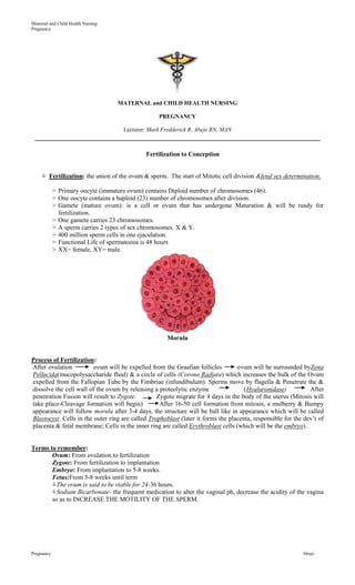
  • 1. Maternal and Child Health Nursing Pregnancy MATERNAL and CHILD HEALTH NURSING PREGNANCY Lecturer: Mark Fredderick R. Abejo RN, MAN __________________________________________________________________________________ Fertilization to Conception  Fertilization: the union of the ovum & sperm. The start of Mitotic cell division &fetal sex determination. > Primary oocyte (immature ovum) contains Diploid number of chromosomes (46). > One oocyte contains a haploid (23) number of chromosomes after division. > Gamete (mature ovum): is a cell or ovum that has undergone Maturation & will be ready for fertilization. > One gamete carries 23 chromosomes. > A sperm carries 2 types of sex chromosomes. X & Y. > 400 million sperm cells in one ejaculation. > Functional Life of spermatozoa is 48 hours > XX= female, XY= male. Morula Process of Fertilization: After ovulation ovum will be expelled from the Graafian follicles ovum will be surrounded byZona Pellucida(mucopolysaccharide fluid) & a circle of cells (Corona Radiata) which increases the bulk of the Ovum expelled from the Fallopian Tube by the Fimbriae (infundibulum). Sperms move by flagella & Penetrate the & dissolve the cell wall of the ovum by releasing a proteolytic enzyme (Hyaluronidase) After penetration Fusion will result to Zygote. Zygote migrate for 4 days in the body of the uterus (Mitosis will take place-Cleavage formation will begin) After 16-50 cell formation from mitosis, a mulberry & Bumpy appearance will follow morula after 3-4 days, the structure will be ball like in appearance which will be called Blastocyst. Cells in the outer ring are called Trophoblast (later it forms the placenta, responsible for the dev’t of placenta & fetal membrane; Cells in the inner ring are called Erythroblast cells (which will be the embryo). Terms to remember: Ovum: From ovulation to fertilization Zygote: From fertilization to implantation Embryo: From implantation to 5-8 weeks. Fetus:From 5-8 weeks until term The ovum is said to be viable for 24-36 hours. Sodium Bicarbonate- the frequent medication to alter the vaginal ph, decrease the acidity of the vagina so as to INCREASE THE MOTILITY OF THE SPERM. Pregnancy Abejo
  • 2. Maternal and Child Health Nursing Pregnancy Fetal Membranes Fetal Membranes: membranes that surround the fetus, & give the placenta the shiny appearance. 2 Layers: 1. Amnion: shiny membrane on the 2nd week of Embryonic Development & encloses the Amniotic Cavity 2. Chorion: Outer membrane that supports the sac of the amniotic fluid. Chorionic Villi: finger like projections from the chorion. This is the place where gases, nutrients and waste products between the maternal & fetal blood takes place. Amniotic Fluid: surrounds the embryo, contains fetal urine, lanugo from fetal skin & epithelial cells. Ph is 7. 2. Specific Gravity: 1.005 – 1.025 Normal Amount: 500 – 1000 ml. Oligohydramnios- less than 300 ml. Polyhydramnios- more than 2000 ml. observe for Down syndrome & congenital defects Functions of Amniotic Fluid: a. Protects the fetus from changes in the temperature & cushion against injury. b. Protects the umbilical cord from pressure, the fetus drinks & breaths the fluid into the lungs. Amniotic Fluid Colors: Normal color: transparent, clear, with white tiny specks Dark amber or yellow: Ominous sign of presence of Bilirubin, hemolytic disease Port Wine Colored: Abruptio Placenta Greenish: Meconium Stained / FETAL DISTRESS: always go for Cesarian Section! Also if ph is less than 7.2 If with odor: deliver within 24 hours, may indicate infection. Umbilical Cord: 21 inches in length & 2 cm in thickness, circulatory communication of the fetus to the mother. CONTAINS 2 ARTERIES & 1 VEIN. Covered by a gelatinous mucopolysaccharide called Whartons jelly. Implantation occurs at the end of the 1st week after fertilization, when the blastocyst attaches to the endometrium. During the 2nd week (14 days after implantation), implantation progresses and two germ layers, cavities, and cell layers develop. During the 3rd week of development (21 days after implantation), the embryonic disk evolves into three layers, and three new structures — the primitive streak, notochord, and allantois — form. Early during the 4th week (28 days after implantation), cellular differentiation and organization occur. Fertilization Cycle Pregnancy Abejo
  • 3. Maternal and Child Health Nursing Pregnancy PRE-FERTILIZATION CONCEPTION IMPLANTATION ACTIVITIES  Zona reaction  Morula (after 3-4  Ovum moves to amulla of days implantation) fallopian tubes  Zygote (fertilized ovum;  Blastocyst about 24-48 hrs, divides; (trophoblast;  Capacitation cleavage divides, travels to embryolast) the uterus  Implants complete  Acrosome reaction w/n 7-10 days THREE PREGNANCY SIGNS & SYMPTOMS PRESUMPTIVE PROBABLE POSITIVE Amenorrhea Pregnancy test (presence of HCG) Auscultation of fetal Nausea/Vomiting Softening of the uterine isthmus (Hegar’s sign) heart by week 8 Breast sensitivity and Cervical softening (Goodell’s sign) Ultrasound imaging increased size of fetal heart motion Fatigue Abdominal Braxton-Hicks contractions by week 7 enlargement Ballotment: bouncing of the fetus in the amniotic fluid against the Ultrasound Skin pigmentation examiners hand. During the 16th-20th week. confirmation of changes gestational sac by (Melasma chloasma, Braxton Hicks Contractions: painless week 6 linea nigra- a brown line contractions felt for 20-30 minutes occurs on running from the the 16th week. Ultrasound: 6 weeks can umbilicus to the auscultate the fetal heart. symphysis pubis Chadwick’s sign is a bluish coloring of the vaginal mucosal that occurs as early as 6 weeks gestation. Fetal movements palpated Stretch marks will Rationale: due to increase vascularity & blood by the provider by week eventually fade to a silvery vessel engorgement. 20. white color, but it is highly Increase size of the uterus unlikely that they will The most objective sign of completely disappear. + Pregnancy Test pregnancy is fetal > Secretion of HCG in the urine (Frog Test). movement felt by the Breast changes- increase in Detectable 10 days after the missed period examiner. fullness, darker areola. . The fetal heartbeat typically can be heard and fetal Quickening: first fetal rebound is possible between 18 and 22 weeks. The mov’t. fetal outline becomes palpable and the fetus is highly mobile between 28 and 31 weeks. Braxton Hicks Urinary Frequency contractions increase in frequency and intensity Melasma . between 32 and 35 weeks. FETAL DEVELOPMENT ORIGIN OF BODY TISSUE Tissue Layer Body Portion Formed Ectoderm Nervous system, mucus membranes, anus & mouth Mesoderm Connective Tissue, Reproductive, circulatory & upper Urinary system, bones, cartillage Endoderm Lining of the GI tract, Respiratory Tract, bladder & urethra Pregnancy Abejo
  • 4. Maternal and Child Health Nursing Pregnancy Embryo is 4-5 mm length Trophoblasts embedded in deciduas 1 mo/ 4 weeks Foundations for nervous system, genitourinary system, skin, bones, and lungs are formed Rudiments of eyes, ears, nose appear  Cardiovascular system functioning, heart beginning to beat, beginning of heart circulation. Placenta dev’t. Placental transport of substances ( 5 weeks) The fetus is 27-31 mm and weighs 2-4 grams Fetus s markedly bent 2 mo/ 5-8 weeks Head is disproportionately large due to brain development Centers of bone begin to ossify Ganglionic cells (5th to 12th weeks) Placenta and meconium are present, with facial features 3 mos./9-12 wks  CVS done (8 12 weeks) every organ present, Head greatly enlarged Average length is 50-55 mm and weighs 45 gms. Fingers and toes are distinct. Rudimentary kidneys secrete urine. Fetal circulation is complete. External genitalia show definite characteristics. Ganglionic cells  SEX IS VISUALLY RECOGNIZABLE. Heart is audible in a Doppler ( 11th week) Fetus swallows. With nails. Kidneys able to secrete. 4 mos. /13-16 weeks 94-140 mm length and weighs 97-200 gms. Head is erected, lower limbs are well developed. Heartbeat is present Nasal septum and palate close Fingerprints are set  LANUGO APPEARS IN THE BODY 5 mos. /17-20 weeks Fetus is 150-190 mm. In length and weighs approximately 260-460 gms. Lanugo covers entire body. Eyebrows and scalp hair is present. Heart sounds are perceptible by auscultation. Vernix caseosa covers skin.  Heartbeat can be heard in the fetoscope ( 18 weeks—20 weeks). Liver is already pancreas functioning.  Quickening felt by a mother. Skeleton begins to develop. Brown Fats begin to form. Heart sounds in the stethoscope Can be heard ( 17- 20 weeks) NOTE: There is a placental barrier to syphilis until the 18th week of pregnancy. If the mother is treated before 18th week, the baby will most likely not be affected. 6 mos. /21-25 weeks 21-25 WEEKS… OLD MAN’s FACE Length 200-240 mm. Wt. 495-910 gms. Skin appears wrinkled and pink to red. REM begins Eyebrows and fingernails develop.  VERNIX COVERS THE ENTIRE BODY. Has the ability to hear. Production of lung surfactants. Passive Antibody transfer ( placental immunoglobulin G) Sustained weight gain occurs. 7 mos. /26-29 weeks Length 250-275; weight 910-1500 gms. Skin red Rhythmic breathing occurs Pupillary membrane disappears from eyes. Fetus often survives if born prematurely Brain develops rapidly. Lecithin- Sphingomyelin (L/S ratio is already 2:1)  Brains fully developed. If born, neonate may survive. 8 mos. /30-34 weeks Length 280-320 mm. weight 1700-2500 gms. Toenails become visible Steady weight gain occurs Vigorous fetal movement occurs.  LANUGO DISAPPEARS. Bones are fully developed.  Aware of sounds outside the body. Assumes the delivery position. Increased chance of survival. 9 mos. /35-37 weeks Length 330-360 mm. weight 2700-3400 gms. Face and body has a loose wrinkled appearance because of subcutaneous fat deposit. Body is usually lump and lanugo disappears Nails reach fingertip edge Amniotic fluid decreases.  Increase Development. Sole of the foot have already  creases. Good chance of survival. Pregnancy Abejo
  • 5. Maternal and Child Health Nursing Pregnancy 10 mos. / 38-40 weeks Length 360 mm.; Weight 3400-3600 gms. Skin is smooth, chest is prominent Eyes are uniformly slate colored Bones of skull are ossified and are nearly together at sutures.  Testes are in scrotum. Fetal Circulation As early as 3rd week of intra-uterine life, fetal blood is already is circulating, specifically there is already exchange of nutrients with the maternal circulation in the chorionic villi. > Arteries carry UNOXYGENATED BLOOD. VEINS carry OXYGENATED BLOOD. > Fetal Circulation Bypass: Why: DUE TO NON-FUNCTIONING LUNGS: ----- Ductus arteriousus (between pulmonary artery & Aorta, OPENS AT BIRTH & CLOSES 24 –48 hours after delivery.) It CONTAINS a mixture of arterial & venous blood. ----- Foramen Ovale : between right & left atrium DUE TO NON-FUNCTIONING LIVER: ----- Ductus Venosus (by pass the liver, closes at birth; an umbilical vein that carries High oxygen from the placenta. Maternal & Fetal Diagnostic Test CHORIONIC VILLI SAMPLING Earliest test possible on fetal cells; sample obtained by slender catheter passed through cervix to implantation site. a.  Chorionic Villi Sampling: removal of a small piece of Chorionic villi sampling to detect the ff: fetal chromosome, enzyme, DNA & biochemical abnormalities. Performed between the 8th – 11th weeks of gestation. Can detect the ff; Genetic Defects: Cystic fibrosis, trisomy 21, Tay Sachs, sickle cell anemia, thallasemia, Duchenne muscular dystrophy & hemophilia.  Most common indication: advance maternal age: increases risk of chromosomal damage from aging of oocyte.  Greatest Advantage over Amniocentesis: PERFORMED DURING THE FIRST TRIMESTER. (16th- 20th week of gestation). . Laboratory results are obtained in 1 - 7 days compared to 20-28 days for an amniocentesis. Disadvantages: 1. Risk of Abortion 2. Infection 3. Embryo-fetal/placental damage 4. Spontaneous abortion 5. Premature rupture of the membranes  After an Rh-negative patient undergoes amniocentesis or CVS, the nurse should administer Rh (D) immune globulin (RhoGAM), to prevent Rh sesnsitization, an antigen antibody immunologic reaction that sometimes occurs when an Rh negative mother carries an Rh + fetus.  The patient does not require complete bed rest after CVS---SHE SHOULD REFRAIN FROM SEXUAL INTERCOURSE AND PHYSICAL ACTIVITY FOR 48 hours. A small amount of spotting is normal for the 1st 24-48 hours. ULTRASOUND Use of sound and returning echo patterns to identify intrabody structures; useful early in pregnancy to identify gestational sacs; later uses include assessment of fetal viability, growth patterns, anomalies, fluid volume, uterine anomalies and adnexal masses. Use adjunct to amniocentesis; safe for fetus (no ionizing radiation) Pregnancy Abejo
  • 6. Maternal and Child Health Nursing Pregnancy Ultrasound: done 18-40 weeks for fetal abnormalities, THE BEST TEST FOR ECTOPIC PREGNANCIES - Non-invasive procedure with high frequency sound waves to obtain outline of the fetus, placenta & uterine cavities and to confirm gestational age & EDD. -  NEEDS A FULL BLADDER TO OBTAIN A BETTER IMAGE (drink a full glass every 15 minutes beginning an hour & half the procedure) -  COMMON METHOD IN LOCATING THE PRECISE POSITION OF THE FETUS & PLACENTA BEFORE AMNIOCENTESIS. AMNIOCENTESIS Location and aspiration of amniotic fluid for examination; possible after the 14th week when sufficient amounts are present; used to identify chromosomal aberration, sex of fetus, levels of alpha-fetoprotein and other chemicals indicative of neural tube defects and inborn error of metabolism, gestational age, RH factor. I.V. anesthesia isn't given for amniocentesis. The client should be supine during the procedure; afterward, she should be placed on her left side to avoid supine hypotension, promote venous return, and ensure adequate cardiac output. Amniocentesis: invasive procedure for amniotic fluid analysis, & fetal lung maturity. Procedure: Ultrasound 1st: the rationale: to locate the Placenta. The patient MUST EMPTY THE BLADDER TO REDUCE THE SIZE OF THE BLADDER. Vital signs are assessed every 15 minutes.  Typically performed on the 3rd trimester to assess LECITHIN-SPHINGOMYELIN RATIO IN THE AMNIOTIC FLUID (this ratio indicates fetal lung maturity), which is commonly delayed in a  diabetic client, Cesarean Delivery  should not be done, unless the fetal lungs are matured. Position: Supine.  PLACE A FOLDED TOWEL ON HER RIGHT BUTTOCKS TO TIP HER SLIGHTLY TO THE LEFT & MOVE THE UTERUS OFF THE VENA CAVA TO PREVENT SUPINE HYPOTENSION SYNDROME.  ABDOMINAL PREP IS DONE, then, needle insertion in a 20-22 gauge spinal needle, withdrawing amniotic fluid.  NORMAL L/S RATIO (lecithin/sphingomyelin): 2:1 = normal fetal lung maturity ratio  Most important factor affecting Amniocentesis: NEEDLE INSERTION-because of the risk of puncture or damage to the placenta, fetus, umbilical cord, bladder & uterine arteries.  Disadvantages: Risk for: 1. Maternal hemorrhage 2. Infection 3. Rh immunization 4. abruptio placenta 5. Amniotic fluid embolism  CALL THE PHYSICIAN FOR THE FF: Chills, fever, leakage of fluid, decrease fetal movement or uterine contractions. Pregnancy Abejo
  • 7. Maternal and Child Health Nursing Pregnancy After amniocentesis, the patient is monitored for uterine contractions, fetal heart rate changes and leakage of amniotic fluid from the puncture site. During this period, the patient isn’t ambulated. X-RAY Can be used late in pregnancy (after ossification of fetal bones) to confirm position and presentation; not used in early pregnancy to avoid possibility of causing damage to fetus and mother. ALPHA-FETOPROTEIN Maternal serum screens for open neural tube defects. SCREENING It is a glucoprote in produced by fetal yolk sac, GI tract and liver. Test done between 16 and 18 weeks gestation. Alpha Fetoprotein:  PRINCIPAL SCREENING TEST DOR THE DETECTION OF NEURAL TUBE DEFECTS (spina bifida, hydrocephalus- can be reduced through increase folic acid- 0.4 mg/day in the 1st trimester) > Maternal blood sampling between 16-20 weeks. LOW: chromosomal defects (Downs syndrome) HIGH: (greater than 10 mg/dl) Neural tube defects, anencephaly & the absence of ventral abdominal wall, premature delivery, toxemia & fetal distress & Rh immunization. L/S RATIO Uses amniotic fluid to ascertain fetal lung maturity through measurement of presence and amounts of the lung surfactants lecithin and sphingomyelin. At 35- 36 weeks; ratio is 2:1 indicative of mature levels. PHOSPHATIDYL GLCEROL Found in amniotic fluid after 35 weeks. In conjunction with the L/S ratio; it contributes to increased reliability of fetal lung maturity testing. Maybe done in laboratory. Phosphatidyl Glycerol (PG): when present in the amniotic fluid, it can be predicted that respiratory distresss will not occur, or RDS will not occur. CREATININE LEVEL Estimates fetal renal maturity and function, uses amniotic fluid. BILIRUBIN Level-high early in pregnancy; drops after 36 weeks gestation; uses amniotic fluid. The yellow color is the result of fetal anemia and bilirubin. FETAL MOVEMENT COUNT Teach mother to count 2-3 times daily, 30-60 minutes each time, should feel 5-6 movements per counting time; mother should notify care giver immediately of abrupt change or no movement. PERCUTANEOUS UMBILICAL Uses ultrasound to locate umbilical cord. Cord blood BLOOD SAMPLING aspirated and tested. Used in second and third trimesters. BIOPHYSICAL PROFILE A collection of data on fetal breathing movements, body movements, muscle tone, reactive heart rate and amniotic fluid volume. ELECTRONIC MONITORING A. Non-Stress Test – accelerations in heart rate accompany normal fetal movement; non-invasive Tocodynamometer records fetal movements and Doppler ultrasound measures - Observation of fetal heart rate related to fetal movement. Fetal well-being. Indicated for: assess placental function & oxygenation, fetal well being, evaluates fetal heart rate in response to fetal movement especially for: Maternal Problems such as chronic hypertension, diabetes and Pre-eclampsia, given after the 32nd week. PREPARATION: Patient should eat snacks. Position: Semi-Fowlers or left lateral positions the mother may ask tom press the button every time she feels fetal movements; the monitor records a mark at each point of fetal movement. Pregnancy Abejo
  • 8. Maternal and Child Health Nursing Pregnancy RESULTS: 1. Reactive (normal): indicates a fetal fetus  Greater than 15 beats per minute- occur with fetal movement in a 10 or 20 minute period. FAVORABLE RESULTS: - 2 or more FHR accelerations of 15 seconds over a 20 minutes interval and return of FHR to normal baseline. 2. Non-Reactive (Abnormal): No fetal movement occurs or there is short-term fetal heart rate variability (less than 6 beats per minute). The doctor will order an Oxytocin Test AFTER the patient has non-reactive test. NOTE: COMMONLY PERFORMED ON DIABETIC PATIENTS BECAUSE OF THE INCREASE RISK FOR STILL BIRTH. B. Contraction Stress Test (CST) – based on the principle that healthy fetus can withstand decreased oxygen during contraction but compromised fetus cannot. Response of the fetus to induced uterine contractions as an INDICATOR OF UTEROPLACENTAL & FETAL PHYSIOLOGICAL INTEGRITY. PREPARATION: Woman in semi-Fowler’s or side-lying position. Monitor for post-test labor onset. TYPES: a. Mammary stimulation Test or Breast Stimulation Exam or Nipple Stimulated CST – non-invasive b. Oxytocin Challenge test Indications: ALL PREGNANCIES AFTER 28 WEEKS WITH HIGH RISK CLIENTS. Contraindicated for history of PRE-TERM LABOR. Interpretations: POSITIVE RESULT: Late decelerations with at least 50% of contractions. Potential risks to the fetus, which may necessitate to C-section. Abnormal and known as “Positive window”. Abnormal: ―Positive Window‖: (+) LATE DECELERATIONS OF FHR with three contractions a 10 minute interval. Indicates Uteroplacental Insufficiency. NEGATIVE RESULTS: No late decelerations with a minimum of 3 contractions lasting 40-60 seconds in 10 minutes period. Normal: ―Negative Window‖: (-) LATE DECELERATIONS OF FHR with three contractions a 10m minute interval Normal and known as “Negative window Laboratory Studies 1. Estriol excretion: measures placental functioning through urine test.  Collect a 24-hour urine specimen or serum blood levels.  High Estriol: Good placental function  Low Estriol: Fetal hypoxia Estriol: estrogenic hormone, synthesized by the placenta & adrenal gland of the fetus which secreted by the ovaries Rh Incompatibility Test: Purpose: to discover presence of antibodies present in Rh-negative mother’s blood > Test will confirm the diagnosis for Hemolytic Disease in the Newborn. Types: 1. Indirect Coomb’s Test: women who have Rh negative have this test done to determine if they have antibodies to the factor present. Repeated 28 weeks pregnancy. Mothers reveal antibodies as a result of previous transfusion or pregnancy. 2. Direct Coomb’s test: tests for newborns cord blood- determines presence of maternal antibodies attached to the baby’s cell.  Rh (D) & D negative who hasn’t formed antibodies should receive Rhogam at 28 weeks gestation or after 72 hours after delivery.  Nitrazine Test: use of nitrazin strip to detect the presence of amniotic fluid. Vaginal Secretions: PH: 4.5- 5.5 Amniotic fluid: PH: 7.2 – 7.5 (turns the yellow Nitrazine blue gray, blue green – Ruptured Membranes)  Kicks count: fetal movement counting mother sits quietly on the LEFT SIDE for 1 hour after meals & count fetal kicks for 30 minutes. Notify the physician or health care provider if FEWER THAN 3 KICKS.  Biophysical Profile : surveillance of fetal well being base on 5 categories: 1. Fetal breath mov’t 2. Fetal tone 3. Amniotic fluid 4. Fetal heart reactivity 5. Placental Grade Interpretation: Fetal score of 8 – 10: normal fetal well-being Fetal score of 4 – 6: fetal distress Pregnancy Abejo