SlideShare uma empresa Scribd logo
1 de 9
MARK BOTIRIUS
P a g e 1 | 9
DESCRIBE AND EXPLAIN THE STRUCTURES AND FUNCTIONS OF SEVERAL PROTEINS
Because there seemstobe an infinitenumberof differenttypesof proteins,itwasa
challenge toselectafewoutof the vastmultitude since eachproteinservesapurpose related toits
structure and function. AlthoughIcouldchoose anyprotein,Iwantedtoidentifythose thatstood
out fromthe rest forsome reasonor another. Therefore,Ichose three proteinsthatIfeltwere
especiallymeaningful. The first,andthe mostexciting,are the immunoglobulinproteinsbecause of
the unique processesthatgive rise tothem. Followingthe immunoglobulinproteinsisRubisco,
because itisby far the most abundantproteinonearththat ispart of,what couldbe consideredto
be the mostimportantbiological processonearth,photosynthesis. Formylastchoice I was goingto
selecthemoglobindue toitsrole ineveryhumanbeingasthe life sustainingcarrierof oxygen,but
that wouldhave leftme withthree globulinproteins,andfor variety,Iwantedatleastone protein
that wasrelativelystationaryaspart of a membrane. Forthis,I chose a receptortyrosine kinase.
Immunoglobulin IgG
Figure 1. The picture ontheleft is from Austin Community College, andthepictureon the right is fromtheNationalCenter for
Biotechnology Information(a nationaldatabase, similar to doing a BLASTsearch), which is partoftheNIH (National Institutes ofHealth).
On the left, thelight chains aredepictedin lightgreen, andtheheavy chains are depicted in pink. The “C”stands for the constant region,
and the “V” stands for thevariableregion. The antibody binds toantigens at the variable regions. On theright is the ribbon model. The
light chains are light brown andturquois, and theheavy chains arepink and blue.
ImmunoglobulinGiscomprisedof twopairs of proteinchainsthathave identical primary
structuresi
. The smallerchainsare calledlightchains,andthe largerchainsare calledheavychains.
Architecturally,theyare composedalmostentirelyof βpleatedsecondarystructures(ascanbe seen
inthe ribbonmodel) consistingof a diverse aminoacidcontentthatisnonethelessheldtogether
mostlybyhydrophobicinteractionsandafew covalentdisulfidebondsfromcysteine residues
(showninthe picture onthe left) thattogetherformsa stable tertiarystructure. Inthe centerof the
ribbonstructure there isan area that hasneitheranα helix orβpleatedsheet.Rather,itconsistsof
MARK BOTIRIUS
P a g e 2 | 9
a flexible hinge,whichgivesthe moleculeelasticitythatservestoaugmentitsabilitytobindtoan
antigen. Notice thatthe β pleatedsheetsof boththe heavyandlightchainsinthe ribbonmodel are
connectedbysingle strandedloops. The loopslocatedatthe top rightand topleft of the molecule
comprise the areasinthe variable regionswhere the antibodybindstoanantigen. Whenwe
considerthe variousstructural aspectsof thismolecule,itisclearthat thismolecule isstructurally
adaptedto functionasa bindingmolecule. The β pleatedsheetsgive itthe necessarystrengthand
stability,andyetthe loopingsingle strandsgive itthe neededflexibilitytobindstronglytoan
antigen. Flexibilityisalsofoundatthe centerof the molecule atthe hinge.
What isso amazingabout thisantibodymolecule (andalsoaboutall antibodymoleculesin
general) isthatif youpicture the molecule withoutitsvariable lightandheavyregionsonthe ends,it
isclear that itis an excellentplatformforamolecule whose functionisto bindavarietyof other
molecules. AsIhave just described,itisbothstructurallystable andyetflexible. Itissimilartohow
a multi-tipscrewdriverhandle isanexcellentplatformtoturnvariousscrew types. If youneedto
turn a standardscrew,you popon a standardtip. If you needtoturn a Phillip’sscrew,youpopona
Phillip’shead. Incredibly, thisissortof whathappensinour immune systems.The difference isthat
our immune systemsaren’tmakingtipstofita particularscrew,they make as manydifferentkinds
of tipsas possible,inthe eventtheyrunintoa“pathogenscrew”that fitstheirparticulartip. The
genesthatencode the variable regionsatthe endsof the “Y” onthe antibodymolecule are
composedof manysegments. These segmentsare differentforlightandheavychains;however,the
principle isthe same. Forexample,the lightvariablechainsegmentsare comprisedof two
categories:V andJ. There are several different“V”segments,andseveral different“J”segments.
Onlyone of each isbroughttogetherina developingBcell tocreate (literally) the gene forthe
variable region. Inotherwords,the gene thatcodesforthe variable regioniscreatedinthe Bcell
throughgenetic recombination. Itturnsout,naturehasbeen in thegenetic engineering business
long beforemankind waseven a twinklein theevolutionary eye! In thisway,the immune system
can theoreticallycreate anantibodywithinagivenpopulationforalmostanypathogeninexistence,
by creatingdifferent“tips”(the variable regionsonthe ends) andattachingthemtoa standard
“platform”(the constantregions).
V1 V2 V3 J1 J2 J3 J4 C
Variable Region ConstantRegion
Figure 2. The top row represents the region of DNA that contains the various V,J, and C segments. Through
DNA recombination,one of each of the V, J, and C segments is broughttogether to form a contiguous gene
represented by the bottom row. Because each gene is different, they each code for a di fferent variableregion,
which can bind to a different antigen.
Rubisco
V3 J2 C
MARK BOTIRIUS
P a g e 3 | 9
Because RuBisCo(ribulose
1, 5 bisphosphate carboxylase/
oxygenase)isthe onlyknown
proteinthatcan fix CO2 in
higherplants,itisarguablythe
mostabundantproteinon
earth. Despite itsimportant
role inthe biochemistryof life,
it isa rather slow and
inefficientenzyme. On
average,RubisCocanonly
catalyze 3 to ten molecules
each second (Cambridge CAPP,
2016). One reasonisbecause
RuBisCo,asits unabbreviated
name indicates,isbotha
carboxylase andanoxygenase.
Althoughitwill preferentially
bindCO2,it doessoonly
slightly,andsointhe presence
of oxygenitnotonlyattaches
carbon to RuBP,it will attach
oxygenaswell,whichisa
costlymove forthe plantbiochemically. Toprocessthe oxidizedRuBPmolecule(called 2-
phosphoglycolate)the plantendsuplosingCO2.
The primarystructure of thisproteinisalsohighlyvaried,andthe secondarystructure,as
depictedinfigure 3,consistsof bothα helicesandβpleatedsheetsinadditiontoloops. Ananalysis
of the secondarystructure revealsthatthe α helicesandβpleatedsheetslocatedinthe centerare
hydrophobic,while the loopslocatedalongthe peripheryandinthe centerhole are mostly
hydrophilic. ThisallowsRuBPaccesstothe active siteswhichare locatedonthe large subunitsof
the molecule towardsthe center. These active sitesare
constructedof β barrelssurroundedbyα helices. The βbarrels
are the catalyticcenters of the active site that holda Mg2+
ionin
the catalyticpocketby interactingwiththe polarchargedamino
acidshistidine andlysine. Thisisa good example of the
relationshipbetweenstructure andfunction. The creationof a
six carbon intermediate sugarfromRuBPisaccomplishedviaa
chargedmetal ion. Thisis the reasonthat we findchargedpolar
aminoacidsin the catalyticsite. WhenCO2
entersthe active site,
it attachesto the lysine residue,whichdestabilizesthe metal ion
that resultsin a conformational change thatbringsthe RuBP
molecule andCO2
moleculetogether,catalyzingthe reactionthat
formsa six carbon sugar. (PNAS,2017) (Goodsell,2017)
Tyrosine Kinase Receptor
Figure 3. This is a ribbon model for the RuBisCo protein found in a
spinach plant. Itis composed of 8 largesubunits and 8 small subunits.
Figure 4. Ribulose 1,5 bisphosphate
carboxylase/oxygenase active site
MARK BOTIRIUS
P a g e 4 | 9
Lastly,I chose a receptorproteinbecause it
demonstratesnicelythe connectionbetween
structure and function;since there isa
distinctdifference betweenthosestructures,
for example,thatare inside the membrane of
a cell incontrast to those that are stickingout
fromthe ends. Tyrosine kinase receptorsare
so namedbecause theyare a signaling
molecule that,whenboundtotheirparticular
ligand,phosphorylate (enzymesthat
phosphorylate othermoleculesare called
kinases) theirtyrosine residues. RTK’sare
amphipathicintegral membrane proteinsthat
consistof three domains:anouterdomain
that protrudesoutside of the cell andserves
to bindligands,amiddle domainthatis
situatedinside the cellmembrane,andan
innerdomainthat
protrudesintothe inside of
the cell that servesto
deliverthe signal received
fromthe ligand. Thisisan
excellentexampleof the
connectionbetween
structure and function.
The outer endsof the
molecule are incontact
withthe aqueous
environmentsthatexist
bothwithinandwithout
the cell. The middle
section,hasa primary
structure made up entirely
of non-polaraminoacids,
that have adoptedanα
helical secondary
structure. Thisnonpolar
middle sectionisindirect
contact withthe non-polar
phospholipidtailsthat
make up the
intermembranespace.
This,alongwithan α helical secondarystructure,securesthe proteininthe membrane.
If you lookcarefullyatthe actual canonical sequence,youwillnotice thatthe tyrosine
residuesare missing. Thatisbecause thisisthe actual sequence fromthe papershowninthe NCBI
Figure 5. An NCBI rendered ribbonmodel of a tyrosine
kinase receptor
Figure 6 This is a
tubular modelof
the protein
depicted in figure
5. The canonical
sequence shown
is the actual
sequence ofthe
protein, andthe
highlighted areas
depict the
hydrophobic
regionof the
molecule to
clearlyshow the
connection
betweenthe
structure and
function ofthe
amino acids.
MARK BOTIRIUS
P a g e 5 | 9
picture infigure five,whichfocusedspecificallyonthe transmembrane region. The shortened
sequence alsomade iteasierforme toput the aminoacid sequence inthe text,sothatI couldmore
clearlydemonstrate the hydrophobicstructure –functionconnection. The overall receptorfunction
of the molecule ismore clearlyshowninthe followingfigure.
As can be seeninthe figure above,whenaligandbindstoreceptordomainsof the proteins,
a conformational change bringsthe totwodimerstogether. Asa result,the twodimersactually
phosphorylate eachother,activatingthem. The activatedkinasesthenphosphorylate another
molecule,which,whenactivatedphosphorylatesyetanothermolecule downthe cascade. One
possible resultof the conclusionof the cascade isthe activationof some transcriptionfactorthat
influencesgeneticexpression. Itisno accidentthatthe aminoacidtyrosine isfoundinside the cell.
Tyrosine, serine,andthreonineare almostexclusivelythe active residuesinthe phosphorylation
activitiesof proteins. Why?Because these are the onlyaminoacidswithanOH group as part of
theirside chains,andthe hydroxyl groupiswhere the phosphatesare added(orremoved).
Likewise,manyof the extracellulardomainsof kinaseshave polarand/orchargedresidues. The
reasonisbecause theyoftenneedtobe able tobindwithligandmoleculeswithanaffinitythatis
greaterthan that of unchargedor nonpolarmolecules.
EXPLAIN AND SHOW HOW THE REACTIONS OF THE CENTRAL METABOLISM (GLYCOLYSIS,
TCA CYCLE, OXIDATIVE PHOSPHORYLATION) INTERACT WITH ONE ANOTHER
Figure 7 The two blue structures are the twoRTKdomains pictured as pinkandblue infigure 5. Thispicture is from
“Membranereceptors.com
MARK BOTIRIUS
P a g e 6 | 9
Aftercareful consideration,Ihave concludedthatglycolysisisthe mostlikelyplace tobegin,
because itscarbonstartingmaterial (glucose) isnotprovidedbythe othertwopathways,andof the
three,itappearsto have the fewestmoleculesfedintoitfromthe others. My biggestconcern,is
that my answeriscohesive,organized,andclear. To ensure this,Ihave decidedthatthe bestwayis
to addresseachreactionseparately,stepbystep,andasktwoquestions.1. What, if anything,did
the othertwo reactionscontribute tothisstep? 2. What, if anything,doesthisstepcontribute to
the othertwo?
Glycolysis
1. Glycolysisbeginswithglucose asitsstartingmaterial. Glucose isphosphorylatedbythe
enzyme hexokinase,usingamolecule of ATPinthe process.
a. What, if anything,didthe othertworeactionscontribute tothisstep?
The glucose came from sourcesoutside the three reactionsunderconsideration,sotheydidnot
contribute the glucose. The ATP,theoretically,couldhave come fromanyof the three,since all
three produce ATP. Since glycolysisalsoproducesATP,Iwouldsaythat mostlikely,the ATPcame
fromglycolysisitself.
b. What,if anything,doesthisstepcontributetothe othertwo?
The product fromthisreaction(Glucose 6 phosphate) isnotusedineitherthe TCA or ETC
reactions. Sonothingispassedonto the other two. Since the ETC needsADPto make ATP,it is
possible thatthe ADPiscontributedtooxidativephosphorylation.
2. In the nextstepinglycolysis,fructose6phosphate isproducedfromglucose 6phosphate via
the enzyme phosphoglucoseisomerase. The startingmaterial (glucose6phosphate) came
fromthe previousstep,andthe productisnotusedin eitherthe TCA or ETC reactions.
Therefore,the answertobothquestionsisnone.
3. Next,fructose 6 phosphate isphosphorylatedbythe enzyme phosphofructokinaseto
produce fructose 1,6 bisphosphate. The answerstobothquestionsare the same as instep
one,that is,none.
4&5. Next,fructose 1,6 bisphosphateissplitintotwothree carbonmolecules. One is
glyceraldehyde 3-phospate,andthe otherisan intermediate metabolite,dihydroxyacetone
phosphate. Dihydroxyacetone phosphate isthenisomerizedintoglyceraldehyde3-
phosphate. Sooverall,thisreactionproduces2moleculesof G3P. It shouldbe noted,that
the productsof thisreaction(G3P) are intermediatesinotherreactionsnotunder
considerationhere,suchasthe Calvincycle. However,the answerstobothquestionsatthis
pointisstill none.
6. In thisstep,bothmoleculesof G3Pare oxidizedbyNAD+
reducingtwomoleculesof NAD+
to
NADH+ H+
. The productis 1,3 bisphosphoglycerate.
a. What, if anything,didthe othertworeactionscontribute tothisstep?
In the presence of oxygen,onlythe ETCproducesNAD+
. The othertwo reactions,glycolysis
and TCA,consume it. In the absence of oxygen,however,the endproductof glycolysis,pyruvate,
MARK BOTIRIUS
P a g e 7 | 9
can be reducedtoproduce NAD+
. Therefore,the NAD+
couldhave come fromthe ETC or
fermentation.
b. What,if anything,doesthisstepcontributetothe othertwo?
The NADH moleculescarry electronsthatare usedinthe ETC reactionsto drive protonsinto
the intermembrane space of the mitochondriontoproduce aprotongradient. It ispossible,the H+
moleculesalsohelpcontributetothe gradient.
7. In thisstep3-phosphoglycerate isproducedfrom1,3-bisphosphoglycerate producingone
twomoleculesof ATPfromADP(remember,everythingfromsteps4 & 5 involve twomolecules). I
considerthese moleculesof ATPtobe whatwas neededtobringthe glycolysisbalance to0.
Therefore, the answertoquestionsaandb are none.
8, 9, & 10. None of the intermediate productsinthesestepsare usedinthe othertworeactions.
The onlythingworthnotingis thattwo more moleculesof ATPare producedfromADP. Since ADP
resultsfrom the utilizationof energy,itisprettymuchubiquitous,andtherefore there isnoreal
needtoconnectit to any particularreaction. Lastly,the endproductof glycolysis,pyruvate,isused
inthe TCA reaction.
The TCA Cycle
A careful lookatthe TCA cycle revealsthatit isessentiallylittle more thananelectroncarrier
producingreaction. The onlyexceptionisthatitusesone GDP molecule toproduce GTP. It neither
producesor useseitherATPorADP. Furthermore,it’sintermediatemetabolitesare notusedby
glycolysisorthe ETC. Therefore,we needtoonlysummarizeitsusesof NAD+
,FADand GDP to
account forits relationshiptoglycolysisandthe ETC.
The TCA producesNADHfrom NAD+
inthe followingreactions:
1. Oxidizingpyruvate toproduce Acetyl CoA
2. Oxidizingisocitrate toproduce α ketoglutarate
3. Oxidizingα ketoglutarate toproduce succinyl CoA
4. Oxidizingmalate toproduce oxaloacetate
All of the NAD+
for these reactionswassuppliedbythe ETC. Glycolysis(fermentation
notwithstanding) andthe TCA reactionsdonotproduce NAD+
. The NADHproducedisusedbythe
ETC.
The TCA cycle alsoproducesthe electroncarrierFADH2 from FADwhenitoxidizessuccinate to
produce fumarate. Again,the FADissuppliedbythe ETC,and the FADH2 isusedbythe ETC.
Lastly,a molecule of GDPisphosphorylatedwhenamolecule of succinate isproducedfromsuccynil
CoA. This isthe onlyplace that we findGDP,and it isusedextensivelythroughoutthe cell,notjust
inthisreaction.
Oxidative Phosphorylation
MARK BOTIRIUS
P a g e 8 | 9
The oxidative phosphorylationcycle consistsof aseriesof membrane boundelectron
carriersthat are arrangedin orderfromthe one withthe greatestredox potential tothe one with
the least. NADHand FADH2 give theirelectronsto the carrierswiththe greatestpotential thatcan
accept theirelectrons. These carriersthenuse the energytoshuttleprotonsacrossthe membrane
and thenpassthe electronstothe nextcarrier,whichdoesthe same. Finally,atthe endof the
chain,the electronsare passedtoan oxygen. Therefore,the ETCusesthe NADH andFADH2
producedbyglycolysisandthe TCA reactionstomake NAD+
and FAD that isusedby glycolysisand
the TCA. The protonsare usedby ATPsynthase tomake ATP fromADP. ADPis produced
throughoutthe cell. The followingfiguresummarizestheseinteractions.
Glycolysis
The Citrus Acid Cycle
Oxidative
Phosphorylation
NAD+ FAD
ATP
NADH FADH2
ATPGDP GTP
NAD+ from fermentation
MARK BOTIRIUS
P a g e 9 | 9
References
(2016, September25).RetrievedfromCambridge CAPP:
https://cambridgecapp.wordpress.com/improving-photosynthesis/rubisco/
(2017, September25).RetrievedfromPNAS:http://www.pnas.org/content/109/46/18785.full#F4
Goodsell,D.(2017, September25).RetrievedfromPDB101:http://pdb101.rcsb.org/motm/11
i For example, the canonical sequencefor the heavy chain pictured in the ribbon model is:
1 qvqlvqsgaevkkpgasvkv scqasgyrfsnfvihwvrqa pgqrfewmgw inpyngnkef
61 sakfqdrvtf tadtsantay melrslrsad tavyycarvgpyswddspqd nyymdvwgkg
121 ttvivssastkgpsvfplap sskstsggta algclvkdyf pepvtvswns galtsgvhtf
181 pavlqssgly slssvvtvps sslgtqtyic nvnhkpsntk vdkkaepksc dkthtcppcp
241 apellggpsv flfppkpkdtlmisrtpevtcvvvdvshed pevkfnwyvd gvevhnaktk
301 preeqynsty rvvsvltvlh qdwlngkeyk ckvsnkalpa piektiskak gqprepqvyt
361 lppsrdeltk nqvsltclvk gfypsdiavewesngqpenn ykttppvlds dgsfflyskl
421 tvdksrwqqg nvfscsvmhealhnhytqks lslspgk

Mais conteúdo relacionado

Mais procurados

Clocl_2077 crystallization- FINAL
Clocl_2077 crystallization- FINALClocl_2077 crystallization- FINAL
Clocl_2077 crystallization- FINALODED MIZRACHI
 
Writing assignment 4 molecular cell biology
Writing assignment 4   molecular cell biologyWriting assignment 4   molecular cell biology
Writing assignment 4 molecular cell biologycorv629
 
Bio108 Cell Biology lec7b PROTEIN STRUCTURE AND FUNCTION
Bio108 Cell Biology lec7b PROTEIN STRUCTUREAND FUNCTIONBio108 Cell Biology lec7b PROTEIN STRUCTUREAND FUNCTION
Bio108 Cell Biology lec7b PROTEIN STRUCTURE AND FUNCTIONShaina Mavreen Villaroza
 
The role of lipid rafts n membrane trafficking in t lymph 2001
The role of lipid rafts n membrane trafficking in t lymph 2001The role of lipid rafts n membrane trafficking in t lymph 2001
The role of lipid rafts n membrane trafficking in t lymph 2001Tamara Jorquiera
 
Powerpoint 13.2
Powerpoint 13.2Powerpoint 13.2
Powerpoint 13.2Mneel1
 
Glosario completo
Glosario completoGlosario completo
Glosario completoCarlosTepaz
 
Chromatin Meeting
Chromatin MeetingChromatin Meeting
Chromatin Meetingguestcbd076
 
Presentazione I anno Federica Campana
Presentazione I anno Federica CampanaPresentazione I anno Federica Campana
Presentazione I anno Federica Campanalab13unisa
 
Transcription and Translation
Transcription and TranslationTranscription and Translation
Transcription and TranslationDrGCDMS
 
Organization macromolecule complex
Organization macromolecule complexOrganization macromolecule complex
Organization macromolecule complexKAUSHAL SAHU
 
BT631-22-Membrane_proteins
BT631-22-Membrane_proteinsBT631-22-Membrane_proteins
BT631-22-Membrane_proteinsRajesh G
 
Membrane proteins
Membrane proteinsMembrane proteins
Membrane proteinsobanbrahma
 
Presentazione ii anno campana
Presentazione ii anno campanaPresentazione ii anno campana
Presentazione ii anno campanalab13unisa
 

Mais procurados (20)

Clocl_2077 crystallization- FINAL
Clocl_2077 crystallization- FINALClocl_2077 crystallization- FINAL
Clocl_2077 crystallization- FINAL
 
Writing assignment 4 molecular cell biology
Writing assignment 4   molecular cell biologyWriting assignment 4   molecular cell biology
Writing assignment 4 molecular cell biology
 
Intramembrane Proteases
Intramembrane ProteasesIntramembrane Proteases
Intramembrane Proteases
 
Bio108 Cell Biology lec7b PROTEIN STRUCTURE AND FUNCTION
Bio108 Cell Biology lec7b PROTEIN STRUCTUREAND FUNCTIONBio108 Cell Biology lec7b PROTEIN STRUCTUREAND FUNCTION
Bio108 Cell Biology lec7b PROTEIN STRUCTURE AND FUNCTION
 
The role of lipid rafts n membrane trafficking in t lymph 2001
The role of lipid rafts n membrane trafficking in t lymph 2001The role of lipid rafts n membrane trafficking in t lymph 2001
The role of lipid rafts n membrane trafficking in t lymph 2001
 
Powerpoint 13.2
Powerpoint 13.2Powerpoint 13.2
Powerpoint 13.2
 
Glosario completo
Glosario completoGlosario completo
Glosario completo
 
Lesson 13.2
Lesson 13.2Lesson 13.2
Lesson 13.2
 
Lesson 13.4
Lesson 13.4Lesson 13.4
Lesson 13.4
 
Chromatin Meeting
Chromatin MeetingChromatin Meeting
Chromatin Meeting
 
Presentazione I anno Federica Campana
Presentazione I anno Federica CampanaPresentazione I anno Federica Campana
Presentazione I anno Federica Campana
 
report
reportreport
report
 
Transcription and Translation
Transcription and TranslationTranscription and Translation
Transcription and Translation
 
Abc seminar
Abc seminarAbc seminar
Abc seminar
 
Lesson 13.1
Lesson 13.1Lesson 13.1
Lesson 13.1
 
Organization macromolecule complex
Organization macromolecule complexOrganization macromolecule complex
Organization macromolecule complex
 
BT631-22-Membrane_proteins
BT631-22-Membrane_proteinsBT631-22-Membrane_proteins
BT631-22-Membrane_proteins
 
Membrane proteins
Membrane proteinsMembrane proteins
Membrane proteins
 
Presentazione ii anno campana
Presentazione ii anno campanaPresentazione ii anno campana
Presentazione ii anno campana
 
Molecular chaperones
Molecular chaperonesMolecular chaperones
Molecular chaperones
 

Destaque

власний досвід презентация Microsoft power point
власний досвід     презентация Microsoft power pointвласний досвід     презентация Microsoft power point
власний досвід презентация Microsoft power pointyjdsr77
 
160210116044 janaki s
160210116044 janaki s160210116044 janaki s
160210116044 janaki sMaitri Iyer
 
Annex conclusions seminari 10 anys Pla director de l'Empordà
Annex conclusions seminari 10 anys Pla director de l'EmpordàAnnex conclusions seminari 10 anys Pla director de l'Empordà
Annex conclusions seminari 10 anys Pla director de l'EmpordàJoan Armangué
 
Xây dựng và phát triển chiến lược e marketing cho công ty tnhh phạm tường 2000
Xây dựng và phát triển chiến lược e marketing cho công ty tnhh phạm tường 2000Xây dựng và phát triển chiến lược e marketing cho công ty tnhh phạm tường 2000
Xây dựng và phát triển chiến lược e marketing cho công ty tnhh phạm tường 2000NOT
 

Destaque (10)

eng.ramy
eng.ramy eng.ramy
eng.ramy
 
Leadership
LeadershipLeadership
Leadership
 
Lenguaje, Lengua, Habla, Norma
Lenguaje, Lengua, Habla, NormaLenguaje, Lengua, Habla, Norma
Lenguaje, Lengua, Habla, Norma
 
власний досвід презентация Microsoft power point
власний досвід     презентация Microsoft power pointвласний досвід     презентация Microsoft power point
власний досвід презентация Microsoft power point
 
160210116044 janaki s
160210116044 janaki s160210116044 janaki s
160210116044 janaki s
 
Going green
Going greenGoing green
Going green
 
160210111012
160210111012160210111012
160210111012
 
Annex conclusions seminari 10 anys Pla director de l'Empordà
Annex conclusions seminari 10 anys Pla director de l'EmpordàAnnex conclusions seminari 10 anys Pla director de l'Empordà
Annex conclusions seminari 10 anys Pla director de l'Empordà
 
Xây dựng và phát triển chiến lược e marketing cho công ty tnhh phạm tường 2000
Xây dựng và phát triển chiến lược e marketing cho công ty tnhh phạm tường 2000Xây dựng và phát triển chiến lược e marketing cho công ty tnhh phạm tường 2000
Xây dựng và phát triển chiến lược e marketing cho công ty tnhh phạm tường 2000
 
Collet chucks
Collet chucksCollet chucks
Collet chucks
 

Semelhante a Cell Bio 1

Protein structural organisation
Protein structural organisationProtein structural organisation
Protein structural organisationDr.M.Prasad Naidu
 
IB Biology 2.4 & 7.3 Slides: Proteins
IB Biology 2.4 & 7.3 Slides: ProteinsIB Biology 2.4 & 7.3 Slides: Proteins
IB Biology 2.4 & 7.3 Slides: ProteinsJacob Cedarbaum
 
introduction physiology.pptx
introduction physiology.pptxintroduction physiology.pptx
introduction physiology.pptxNusratJahan25636
 
integral membrane protein.pdf
integral membrane protein.pdfintegral membrane protein.pdf
integral membrane protein.pdfLEKHANAGOWDA7
 
BT631-8-Folds_proteins
BT631-8-Folds_proteinsBT631-8-Folds_proteins
BT631-8-Folds_proteinsRajesh G
 
Quaternary structure of protein
Quaternary structure of proteinQuaternary structure of protein
Quaternary structure of proteinArjun K Gopi
 
presentation. (1).pptx
presentation. (1).pptxpresentation. (1).pptx
presentation. (1).pptxMISSCOM1
 
Protein folding slids
Protein folding slidsProtein folding slids
Protein folding slidsanam tariq
 
Endoplasmic reticulum by amita
Endoplasmic reticulum by amitaEndoplasmic reticulum by amita
Endoplasmic reticulum by amitaAmita Mevada
 
Cell biology ppt
Cell biology pptCell biology ppt
Cell biology pptJyotish Jha
 
Protein ligand interaction
Protein ligand interactionProtein ligand interaction
Protein ligand interactionNikita Dewangan
 
2. Absorption & Secretion Of Materials
2. Absorption & Secretion Of Materials2. Absorption & Secretion Of Materials
2. Absorption & Secretion Of Materialsrossbiology
 
Evolutionary relationship between diverse protein with similar domain
Evolutionary relationship between diverse protein with similar domainEvolutionary relationship between diverse protein with similar domain
Evolutionary relationship between diverse protein with similar domainjj_zein
 
CELL MEMBRANE , RECEPTOR , DRUG RECEPTOR INTERACTION
CELL MEMBRANE , RECEPTOR , DRUG RECEPTOR INTERACTIONCELL MEMBRANE , RECEPTOR , DRUG RECEPTOR INTERACTION
CELL MEMBRANE , RECEPTOR , DRUG RECEPTOR INTERACTIONSATYAM ASATI
 

Semelhante a Cell Bio 1 (20)

Proteins
ProteinsProteins
Proteins
 
Protein structural organisation
Protein structural organisationProtein structural organisation
Protein structural organisation
 
Microbiology Assignment Help
Microbiology Assignment HelpMicrobiology Assignment Help
Microbiology Assignment Help
 
IB Biology 2.4 & 7.3 Slides: Proteins
IB Biology 2.4 & 7.3 Slides: ProteinsIB Biology 2.4 & 7.3 Slides: Proteins
IB Biology 2.4 & 7.3 Slides: Proteins
 
introduction physiology.pptx
introduction physiology.pptxintroduction physiology.pptx
introduction physiology.pptx
 
integral membrane protein.pdf
integral membrane protein.pdfintegral membrane protein.pdf
integral membrane protein.pdf
 
BT631-8-Folds_proteins
BT631-8-Folds_proteinsBT631-8-Folds_proteins
BT631-8-Folds_proteins
 
Protein Folding
Protein Folding Protein Folding
Protein Folding
 
Quaternary structure of protein
Quaternary structure of proteinQuaternary structure of protein
Quaternary structure of protein
 
presentation. (1).pptx
presentation. (1).pptxpresentation. (1).pptx
presentation. (1).pptx
 
Cell Membarne ppt.pptx
Cell Membarne ppt.pptxCell Membarne ppt.pptx
Cell Membarne ppt.pptx
 
Protein folding slids
Protein folding slidsProtein folding slids
Protein folding slids
 
Endoplasmic reticulum by amita
Endoplasmic reticulum by amitaEndoplasmic reticulum by amita
Endoplasmic reticulum by amita
 
Cell biology ppt
Cell biology pptCell biology ppt
Cell biology ppt
 
Protein ligand interaction
Protein ligand interactionProtein ligand interaction
Protein ligand interaction
 
2. Absorption & Secretion Of Materials
2. Absorption & Secretion Of Materials2. Absorption & Secretion Of Materials
2. Absorption & Secretion Of Materials
 
Evolutionary relationship between diverse protein with similar domain
Evolutionary relationship between diverse protein with similar domainEvolutionary relationship between diverse protein with similar domain
Evolutionary relationship between diverse protein with similar domain
 
12 miller chap.3 lecture
12 miller chap.3 lecture12 miller chap.3 lecture
12 miller chap.3 lecture
 
Cell
CellCell
Cell
 
CELL MEMBRANE , RECEPTOR , DRUG RECEPTOR INTERACTION
CELL MEMBRANE , RECEPTOR , DRUG RECEPTOR INTERACTIONCELL MEMBRANE , RECEPTOR , DRUG RECEPTOR INTERACTION
CELL MEMBRANE , RECEPTOR , DRUG RECEPTOR INTERACTION
 

Mais de Mark Botirius (6)

Assignment 2
Assignment 2Assignment 2
Assignment 2
 
IME 3
IME 3IME 3
IME 3
 
IME 2
IME 2IME 2
IME 2
 
IME 1
IME 1IME 1
IME 1
 
Cell Bio 3
Cell Bio 3Cell Bio 3
Cell Bio 3
 
Cell Bio 2
Cell Bio 2Cell Bio 2
Cell Bio 2
 

Cell Bio 1

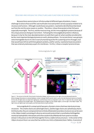
  • 1. MARK BOTIRIUS P a g e 1 | 9 DESCRIBE AND EXPLAIN THE STRUCTURES AND FUNCTIONS OF SEVERAL PROTEINS Because there seemstobe an infinitenumberof differenttypesof proteins,itwasa challenge toselectafewoutof the vastmultitude since eachproteinservesapurpose related toits structure and function. AlthoughIcouldchoose anyprotein,Iwantedtoidentifythose thatstood out fromthe rest forsome reasonor another. Therefore,Ichose three proteinsthatIfeltwere especiallymeaningful. The first,andthe mostexciting,are the immunoglobulinproteinsbecause of the unique processesthatgive rise tothem. Followingthe immunoglobulinproteinsisRubisco, because itisby far the most abundantproteinonearththat ispart of,what couldbe consideredto be the mostimportantbiological processonearth,photosynthesis. Formylastchoice I was goingto selecthemoglobindue toitsrole ineveryhumanbeingasthe life sustainingcarrierof oxygen,but that wouldhave leftme withthree globulinproteins,andfor variety,Iwantedatleastone protein that wasrelativelystationaryaspart of a membrane. Forthis,I chose a receptortyrosine kinase. Immunoglobulin IgG Figure 1. The picture ontheleft is from Austin Community College, andthepictureon the right is fromtheNationalCenter for Biotechnology Information(a nationaldatabase, similar to doing a BLASTsearch), which is partoftheNIH (National Institutes ofHealth). On the left, thelight chains aredepictedin lightgreen, andtheheavy chains are depicted in pink. The “C”stands for the constant region, and the “V” stands for thevariableregion. The antibody binds toantigens at the variable regions. On theright is the ribbon model. The light chains are light brown andturquois, and theheavy chains arepink and blue. ImmunoglobulinGiscomprisedof twopairs of proteinchainsthathave identical primary structuresi . The smallerchainsare calledlightchains,andthe largerchainsare calledheavychains. Architecturally,theyare composedalmostentirelyof βpleatedsecondarystructures(ascanbe seen inthe ribbonmodel) consistingof a diverse aminoacidcontentthatisnonethelessheldtogether mostlybyhydrophobicinteractionsandafew covalentdisulfidebondsfromcysteine residues (showninthe picture onthe left) thattogetherformsa stable tertiarystructure. Inthe centerof the ribbonstructure there isan area that hasneitheranα helix orβpleatedsheet.Rather,itconsistsof
  • 2. MARK BOTIRIUS P a g e 2 | 9 a flexible hinge,whichgivesthe moleculeelasticitythatservestoaugmentitsabilitytobindtoan antigen. Notice thatthe β pleatedsheetsof boththe heavyandlightchainsinthe ribbonmodel are connectedbysingle strandedloops. The loopslocatedatthe top rightand topleft of the molecule comprise the areasinthe variable regionswhere the antibodybindstoanantigen. Whenwe considerthe variousstructural aspectsof thismolecule,itisclearthat thismolecule isstructurally adaptedto functionasa bindingmolecule. The β pleatedsheetsgive itthe necessarystrengthand stability,andyetthe loopingsingle strandsgive itthe neededflexibilitytobindstronglytoan antigen. Flexibilityisalsofoundatthe centerof the molecule atthe hinge. What isso amazingabout thisantibodymolecule (andalsoaboutall antibodymoleculesin general) isthatif youpicture the molecule withoutitsvariable lightandheavyregionsonthe ends,it isclear that itis an excellentplatformforamolecule whose functionisto bindavarietyof other molecules. AsIhave just described,itisbothstructurallystable andyetflexible. Itissimilartohow a multi-tipscrewdriverhandle isanexcellentplatformtoturnvariousscrew types. If youneedto turn a standardscrew,you popon a standardtip. If you needtoturn a Phillip’sscrew,youpopona Phillip’shead. Incredibly, thisissortof whathappensinour immune systems.The difference isthat our immune systemsaren’tmakingtipstofita particularscrew,they make as manydifferentkinds of tipsas possible,inthe eventtheyrunintoa“pathogenscrew”that fitstheirparticulartip. The genesthatencode the variable regionsatthe endsof the “Y” onthe antibodymolecule are composedof manysegments. These segmentsare differentforlightandheavychains;however,the principle isthe same. Forexample,the lightvariablechainsegmentsare comprisedof two categories:V andJ. There are several different“V”segments,andseveral different“J”segments. Onlyone of each isbroughttogetherina developingBcell tocreate (literally) the gene forthe variable region. Inotherwords,the gene thatcodesforthe variable regioniscreatedinthe Bcell throughgenetic recombination. Itturnsout,naturehasbeen in thegenetic engineering business long beforemankind waseven a twinklein theevolutionary eye! In thisway,the immune system can theoreticallycreate anantibodywithinagivenpopulationforalmostanypathogeninexistence, by creatingdifferent“tips”(the variable regionsonthe ends) andattachingthemtoa standard “platform”(the constantregions). V1 V2 V3 J1 J2 J3 J4 C Variable Region ConstantRegion Figure 2. The top row represents the region of DNA that contains the various V,J, and C segments. Through DNA recombination,one of each of the V, J, and C segments is broughttogether to form a contiguous gene represented by the bottom row. Because each gene is different, they each code for a di fferent variableregion, which can bind to a different antigen. Rubisco V3 J2 C
  • 3. MARK BOTIRIUS P a g e 3 | 9 Because RuBisCo(ribulose 1, 5 bisphosphate carboxylase/ oxygenase)isthe onlyknown proteinthatcan fix CO2 in higherplants,itisarguablythe mostabundantproteinon earth. Despite itsimportant role inthe biochemistryof life, it isa rather slow and inefficientenzyme. On average,RubisCocanonly catalyze 3 to ten molecules each second (Cambridge CAPP, 2016). One reasonisbecause RuBisCo,asits unabbreviated name indicates,isbotha carboxylase andanoxygenase. Althoughitwill preferentially bindCO2,it doessoonly slightly,andsointhe presence of oxygenitnotonlyattaches carbon to RuBP,it will attach oxygenaswell,whichisa costlymove forthe plantbiochemically. Toprocessthe oxidizedRuBPmolecule(called 2- phosphoglycolate)the plantendsuplosingCO2. The primarystructure of thisproteinisalsohighlyvaried,andthe secondarystructure,as depictedinfigure 3,consistsof bothα helicesandβpleatedsheetsinadditiontoloops. Ananalysis of the secondarystructure revealsthatthe α helicesandβpleatedsheetslocatedinthe centerare hydrophobic,while the loopslocatedalongthe peripheryandinthe centerhole are mostly hydrophilic. ThisallowsRuBPaccesstothe active siteswhichare locatedonthe large subunitsof the molecule towardsthe center. These active sitesare constructedof β barrelssurroundedbyα helices. The βbarrels are the catalyticcenters of the active site that holda Mg2+ ionin the catalyticpocketby interactingwiththe polarchargedamino acidshistidine andlysine. Thisisa good example of the relationshipbetweenstructure andfunction. The creationof a six carbon intermediate sugarfromRuBPisaccomplishedviaa chargedmetal ion. Thisis the reasonthat we findchargedpolar aminoacidsin the catalyticsite. WhenCO2 entersthe active site, it attachesto the lysine residue,whichdestabilizesthe metal ion that resultsin a conformational change thatbringsthe RuBP molecule andCO2 moleculetogether,catalyzingthe reactionthat formsa six carbon sugar. (PNAS,2017) (Goodsell,2017) Tyrosine Kinase Receptor Figure 3. This is a ribbon model for the RuBisCo protein found in a spinach plant. Itis composed of 8 largesubunits and 8 small subunits. Figure 4. Ribulose 1,5 bisphosphate carboxylase/oxygenase active site
  • 4. MARK BOTIRIUS P a g e 4 | 9 Lastly,I chose a receptorproteinbecause it demonstratesnicelythe connectionbetween structure and function;since there isa distinctdifference betweenthosestructures, for example,thatare inside the membrane of a cell incontrast to those that are stickingout fromthe ends. Tyrosine kinase receptorsare so namedbecause theyare a signaling molecule that,whenboundtotheirparticular ligand,phosphorylate (enzymesthat phosphorylate othermoleculesare called kinases) theirtyrosine residues. RTK’sare amphipathicintegral membrane proteinsthat consistof three domains:anouterdomain that protrudesoutside of the cell andserves to bindligands,amiddle domainthatis situatedinside the cellmembrane,andan innerdomainthat protrudesintothe inside of the cell that servesto deliverthe signal received fromthe ligand. Thisisan excellentexampleof the connectionbetween structure and function. The outer endsof the molecule are incontact withthe aqueous environmentsthatexist bothwithinandwithout the cell. The middle section,hasa primary structure made up entirely of non-polaraminoacids, that have adoptedanα helical secondary structure. Thisnonpolar middle sectionisindirect contact withthe non-polar phospholipidtailsthat make up the intermembranespace. This,alongwithan α helical secondarystructure,securesthe proteininthe membrane. If you lookcarefullyatthe actual canonical sequence,youwillnotice thatthe tyrosine residuesare missing. Thatisbecause thisisthe actual sequence fromthe papershowninthe NCBI Figure 5. An NCBI rendered ribbonmodel of a tyrosine kinase receptor Figure 6 This is a tubular modelof the protein depicted in figure 5. The canonical sequence shown is the actual sequence ofthe protein, andthe highlighted areas depict the hydrophobic regionof the molecule to clearlyshow the connection betweenthe structure and function ofthe amino acids.
  • 5. MARK BOTIRIUS P a g e 5 | 9 picture infigure five,whichfocusedspecificallyonthe transmembrane region. The shortened sequence alsomade iteasierforme toput the aminoacid sequence inthe text,sothatI couldmore clearlydemonstrate the hydrophobicstructure –functionconnection. The overall receptorfunction of the molecule ismore clearlyshowninthe followingfigure. As can be seeninthe figure above,whenaligandbindstoreceptordomainsof the proteins, a conformational change bringsthe totwodimerstogether. Asa result,the twodimersactually phosphorylate eachother,activatingthem. The activatedkinasesthenphosphorylate another molecule,which,whenactivatedphosphorylatesyetanothermolecule downthe cascade. One possible resultof the conclusionof the cascade isthe activationof some transcriptionfactorthat influencesgeneticexpression. Itisno accidentthatthe aminoacidtyrosine isfoundinside the cell. Tyrosine, serine,andthreonineare almostexclusivelythe active residuesinthe phosphorylation activitiesof proteins. Why?Because these are the onlyaminoacidswithanOH group as part of theirside chains,andthe hydroxyl groupiswhere the phosphatesare added(orremoved). Likewise,manyof the extracellulardomainsof kinaseshave polarand/orchargedresidues. The reasonisbecause theyoftenneedtobe able tobindwithligandmoleculeswithanaffinitythatis greaterthan that of unchargedor nonpolarmolecules. EXPLAIN AND SHOW HOW THE REACTIONS OF THE CENTRAL METABOLISM (GLYCOLYSIS, TCA CYCLE, OXIDATIVE PHOSPHORYLATION) INTERACT WITH ONE ANOTHER Figure 7 The two blue structures are the twoRTKdomains pictured as pinkandblue infigure 5. Thispicture is from “Membranereceptors.com
  • 6. MARK BOTIRIUS P a g e 6 | 9 Aftercareful consideration,Ihave concludedthatglycolysisisthe mostlikelyplace tobegin, because itscarbonstartingmaterial (glucose) isnotprovidedbythe othertwopathways,andof the three,itappearsto have the fewestmoleculesfedintoitfromthe others. My biggestconcern,is that my answeriscohesive,organized,andclear. To ensure this,Ihave decidedthatthe bestwayis to addresseachreactionseparately,stepbystep,andasktwoquestions.1. What, if anything,did the othertwo reactionscontribute tothisstep? 2. What, if anything,doesthisstepcontribute to the othertwo? Glycolysis 1. Glycolysisbeginswithglucose asitsstartingmaterial. Glucose isphosphorylatedbythe enzyme hexokinase,usingamolecule of ATPinthe process. a. What, if anything,didthe othertworeactionscontribute tothisstep? The glucose came from sourcesoutside the three reactionsunderconsideration,sotheydidnot contribute the glucose. The ATP,theoretically,couldhave come fromanyof the three,since all three produce ATP. Since glycolysisalsoproducesATP,Iwouldsaythat mostlikely,the ATPcame fromglycolysisitself. b. What,if anything,doesthisstepcontributetothe othertwo? The product fromthisreaction(Glucose 6 phosphate) isnotusedineitherthe TCA or ETC reactions. Sonothingispassedonto the other two. Since the ETC needsADPto make ATP,it is possible thatthe ADPiscontributedtooxidativephosphorylation. 2. In the nextstepinglycolysis,fructose6phosphate isproducedfromglucose 6phosphate via the enzyme phosphoglucoseisomerase. The startingmaterial (glucose6phosphate) came fromthe previousstep,andthe productisnotusedin eitherthe TCA or ETC reactions. Therefore,the answertobothquestionsisnone. 3. Next,fructose 6 phosphate isphosphorylatedbythe enzyme phosphofructokinaseto produce fructose 1,6 bisphosphate. The answerstobothquestionsare the same as instep one,that is,none. 4&5. Next,fructose 1,6 bisphosphateissplitintotwothree carbonmolecules. One is glyceraldehyde 3-phospate,andthe otherisan intermediate metabolite,dihydroxyacetone phosphate. Dihydroxyacetone phosphate isthenisomerizedintoglyceraldehyde3- phosphate. Sooverall,thisreactionproduces2moleculesof G3P. It shouldbe noted,that the productsof thisreaction(G3P) are intermediatesinotherreactionsnotunder considerationhere,suchasthe Calvincycle. However,the answerstobothquestionsatthis pointisstill none. 6. In thisstep,bothmoleculesof G3Pare oxidizedbyNAD+ reducingtwomoleculesof NAD+ to NADH+ H+ . The productis 1,3 bisphosphoglycerate. a. What, if anything,didthe othertworeactionscontribute tothisstep? In the presence of oxygen,onlythe ETCproducesNAD+ . The othertwo reactions,glycolysis and TCA,consume it. In the absence of oxygen,however,the endproductof glycolysis,pyruvate,
  • 7. MARK BOTIRIUS P a g e 7 | 9 can be reducedtoproduce NAD+ . Therefore,the NAD+ couldhave come fromthe ETC or fermentation. b. What,if anything,doesthisstepcontributetothe othertwo? The NADH moleculescarry electronsthatare usedinthe ETC reactionsto drive protonsinto the intermembrane space of the mitochondriontoproduce aprotongradient. It ispossible,the H+ moleculesalsohelpcontributetothe gradient. 7. In thisstep3-phosphoglycerate isproducedfrom1,3-bisphosphoglycerate producingone twomoleculesof ATPfromADP(remember,everythingfromsteps4 & 5 involve twomolecules). I considerthese moleculesof ATPtobe whatwas neededtobringthe glycolysisbalance to0. Therefore, the answertoquestionsaandb are none. 8, 9, & 10. None of the intermediate productsinthesestepsare usedinthe othertworeactions. The onlythingworthnotingis thattwo more moleculesof ATPare producedfromADP. Since ADP resultsfrom the utilizationof energy,itisprettymuchubiquitous,andtherefore there isnoreal needtoconnectit to any particularreaction. Lastly,the endproductof glycolysis,pyruvate,isused inthe TCA reaction. The TCA Cycle A careful lookatthe TCA cycle revealsthatit isessentiallylittle more thananelectroncarrier producingreaction. The onlyexceptionisthatitusesone GDP molecule toproduce GTP. It neither producesor useseitherATPorADP. Furthermore,it’sintermediatemetabolitesare notusedby glycolysisorthe ETC. Therefore,we needtoonlysummarizeitsusesof NAD+ ,FADand GDP to account forits relationshiptoglycolysisandthe ETC. The TCA producesNADHfrom NAD+ inthe followingreactions: 1. Oxidizingpyruvate toproduce Acetyl CoA 2. Oxidizingisocitrate toproduce α ketoglutarate 3. Oxidizingα ketoglutarate toproduce succinyl CoA 4. Oxidizingmalate toproduce oxaloacetate All of the NAD+ for these reactionswassuppliedbythe ETC. Glycolysis(fermentation notwithstanding) andthe TCA reactionsdonotproduce NAD+ . The NADHproducedisusedbythe ETC. The TCA cycle alsoproducesthe electroncarrierFADH2 from FADwhenitoxidizessuccinate to produce fumarate. Again,the FADissuppliedbythe ETC,and the FADH2 isusedbythe ETC. Lastly,a molecule of GDPisphosphorylatedwhenamolecule of succinate isproducedfromsuccynil CoA. This isthe onlyplace that we findGDP,and it isusedextensivelythroughoutthe cell,notjust inthisreaction. Oxidative Phosphorylation
  • 8. MARK BOTIRIUS P a g e 8 | 9 The oxidative phosphorylationcycle consistsof aseriesof membrane boundelectron carriersthat are arrangedin orderfromthe one withthe greatestredox potential tothe one with the least. NADHand FADH2 give theirelectronsto the carrierswiththe greatestpotential thatcan accept theirelectrons. These carriersthenuse the energytoshuttleprotonsacrossthe membrane and thenpassthe electronstothe nextcarrier,whichdoesthe same. Finally,atthe endof the chain,the electronsare passedtoan oxygen. Therefore,the ETCusesthe NADH andFADH2 producedbyglycolysisandthe TCA reactionstomake NAD+ and FAD that isusedby glycolysisand the TCA. The protonsare usedby ATPsynthase tomake ATP fromADP. ADPis produced throughoutthe cell. The followingfiguresummarizestheseinteractions. Glycolysis The Citrus Acid Cycle Oxidative Phosphorylation NAD+ FAD ATP NADH FADH2 ATPGDP GTP NAD+ from fermentation
  • 9. MARK BOTIRIUS P a g e 9 | 9 References (2016, September25).RetrievedfromCambridge CAPP: https://cambridgecapp.wordpress.com/improving-photosynthesis/rubisco/ (2017, September25).RetrievedfromPNAS:http://www.pnas.org/content/109/46/18785.full#F4 Goodsell,D.(2017, September25).RetrievedfromPDB101:http://pdb101.rcsb.org/motm/11 i For example, the canonical sequencefor the heavy chain pictured in the ribbon model is: 1 qvqlvqsgaevkkpgasvkv scqasgyrfsnfvihwvrqa pgqrfewmgw inpyngnkef 61 sakfqdrvtf tadtsantay melrslrsad tavyycarvgpyswddspqd nyymdvwgkg 121 ttvivssastkgpsvfplap sskstsggta algclvkdyf pepvtvswns galtsgvhtf 181 pavlqssgly slssvvtvps sslgtqtyic nvnhkpsntk vdkkaepksc dkthtcppcp 241 apellggpsv flfppkpkdtlmisrtpevtcvvvdvshed pevkfnwyvd gvevhnaktk 301 preeqynsty rvvsvltvlh qdwlngkeyk ckvsnkalpa piektiskak gqprepqvyt 361 lppsrdeltk nqvsltclvk gfypsdiavewesngqpenn ykttppvlds dgsfflyskl 421 tvdksrwqqg nvfscsvmhealhnhytqks lslspgk