3. Toll-Like Receptors
Novus Biologicals & Innate Immunity: The Story Toll’d
UPDATED EDITION • 2014
We dedicate this book to TLR researchers worldwide, including our customers, collaborators, colleagues
and heretofore unmet scientists. We stand in awe of the researchers who came before us and set the
groundwork for the present day knowledge of TLRs and immunity overall. We thank present day research-
ers for their ongoing contributions to the expanding knowledge of the TLR field; their presence and publica-
tions are invaluable to moving TLR understanding forward.
We hope that our book will be an inspiration and useful guide for established and emerging TLR researchers
alike. Likewise, we hope that researchers in related and distant fields will gain insight into how TLRs might
be linked to their area of research.
We wish for students to use our book as a foundation for gaining additional information about how they, too,
might become involved in the exciting and promising arena of TLR research.
In closing, we hope that all of our readers will provide us with feedback, new publications, and suggestions
for our next edition of the TLR Handbook.
Editor: Lisa Heiden, PhD
Assistant Editor: Debashree Sahu
Other Contributors:
Javed Akhtar, Kody Andrew, Subhasis Chattopadhyay, Sebastian Krejewski, Hyun-Ku Lee, Prasanta Maiti, Satya Mishra,
Payton Quintel, Sanjay Raul, Jonathan Rosenberg, M. Simple, Gita Singh, Sujay Singh, Jason Stampfl, Peter Tobias,
Lauren Wardle, and other IMGENEX staff both in India and in the United States
Layout & Design: Cheryl Reyes
For additional information please visit www.novusbio.com
Novus Biologicals, LLC
8100 Southpark Way, A-8
Littleton, CO 80120
P: 1-888-506-6887
F: 303-730-1966
E: novus@novusbio.com
4. T L R H A N D B O O K & O V E RV I E W iii
a.k.a. also known as
aa amino acid(s)
Ab antibody
AD Alzheimer’s disease
APC antigen presenting cells
Ab Amyloid β peptides
B-CLL B-cell chronic lymphocytic leukemia
BCG Mycobacterium bovis bacillus Calmette-Mycobacterium bovis bacill Guérin
CAGR compound annual growth
CLL chronic lymphocytic leukemia
CTL cytotoxic T lymphocyte
DAMP damage associated molecular pattern
DC dendritic cell
dsDNA double stranded DNA
ECD extracellular domain, a.k.a ectodomain
ER endoplasmic reticulum
FADD Fas/Apo-1 associated death domain protein
hBD human β-defensin
HDP host-defense peptides
HMGB1 high-mobility-group box 1
H. pylori Helicobacter pylori
HPA hypothalamic-pituitary-adrenal
ICAM1 intercellular adhesion molecule 1
iDC immature dendritic cell
IFN interferon
IL-1R interleukin-1 receptor
ISS-ODN immunostimulatory sequence oligodeoxynucleotide
IP intraperitoneal injection
IRAK IL-1R associated kinase
IRF interferon regulatory factor
LPS lipopolysaccaride
LRR leucine rich repeats
mAb monoclonal antibody
Mature DC highly efficient professional APC
MSC mesenchymal stem/progenitor cell
MFI mean fluorescence intensity
MHC major histocompatibility complex
Abbreviations
5. 1IV T 3 0 3 - 7 3 0 - 1 9 5 0 F 3 0 3 - 7 3 0 - 1 9 6 6 E n o v u s @ n o v u s b i o . c o m w w w. n o v u s b i o . c o m
mDC myeloid DC
MyD88 myeloid differentiation factor 88
NLR Nod-like receptor
NO Nitric Oxide
pAb polyclonal antibody
PAMP pathogen associated molecular pattern
PBMC peripheral blood mononuclear cells
PMN polymorphonuclear neutrophils
pDC plasmacytoid DC
PRR pattern recognition receptor
RLR RIG-1 like receptor
S. aureus Staphylococcus aureus
SD standard deviation
SEAP secreted alkaline phosphatase
SLE systemic lupus erythematosus
SNP Single nuclear polymorphism
TAB1/TAB2 Tak binding proteins
TAK1 TGF-b activated kinase
TIR Toll/IL-1 receptor
TLR Toll like receptor
Tc2 T cytotoxic 2 cell, a.k.a. CD8+ T cytotoxic 2 cell
Teff effector T cell
Th T helper cell, a.k.a CD4+ T helper cell
Th1 T helper type 1 cell
Th2 T helper type 2 cell
Th3 Type 3 regulatory T cell, i.e. , Type 3 Treg, a.k.a T helper type 3 cell
Th17 T helper type 17 cell
Treg T regulatory cell, a.k.a. suppressor T cell
TID Type I diabetes
TIRAP TIR domain-containing adaptor protein, a.k.a. MAL (MyD88 adaptor like)
Tr1 Type 1 regulatory T cell, i..e., Type 1 Treg
TRAM a.k.a. TICAM2, TIRP
TRIF a.k.a. TICAM1
WB western blot
WT wildtype
Note: some of these abbreviations are defined in the text upon their first use. Other more common ones are defined only in this list.
Abbreviations
7. 2 T 3 0 3 - 7 3 0 - 1 9 5 0 F 3 0 3 - 7 3 0 - 1 9 6 6 E n o v u s @ n o v u s b i o . c o m w w w. n o v u s b i o . c o m
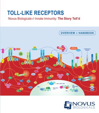
Figure 1. TLRSystemTM
portfolio of antibodies and related reagents for studying TLRs and signaling pathways in innate and adaptive immunity. Novus
offers researchers a comprehensive TLR product line and a wide range of supporting products through its TLRSystemTM
portfolio.
Fluorochrome Conjugates for TLR Phenotyping of DCs & T Cell Subsets by Flow Cytometry
Additional Applications: Immunohistochemistry • Western Blotting • ELISA
TH
2 Cell
TH
1 Cell
Treg
NF-κB
OX40L
CD80/
CD86
MHCII
TCR
CD28
OX40
CD40L
CD80/
CD86
CD28
CD40
TCR
TLR
CD80/
CD86
CD28
TCR
A system of CD antibodies, kits & reagents to study Toll-like Receptors (TLRs), Dendritic
Cells (DCs) and T Cell pathways shaping Innate and Adaptive Immune Responses
TLRs 1-13
Phospho TLRs
TRIF
TIRAP/Mal
TBK1
MD-2
NALPs 1-14
TLRs & Associated Proteins
pDC/iDC
CD207
BDCA-2/CD303
DC-SIGN/CD209
CD11b
CD11c
Dendritic Cells
CD3
CD4
CD8
CD25
CD40L
CD127/IL7R
GITR
GITRL
iNKT
FOXP3
FOXP3∆2
GPR83
T Cells
CD14
CD40
CD80
CD83
CD86
CD123
NF-κB Peptide Inhibitors
Phospho-IκBα ActivELISATM
Kit
NF-κB/p65 ActivELISATM
Kit
NF-κB SEAPorterTM
Assay Kit
NF-κB Pathways
ActivELISATM
Kits for
Cytokines, Chemokines +
Inflammatory Mediators
TNFα
IL-1α
IL-1β
PGE2
CD83+
/CD14-/low
iDCs
stained with hTLR8 (PE)
Caspase-1
IKKα
IκBα
p65
MyD88
NOD1
NOD2
TLR Agonists
Pam3
CSK4
Poly(I).Poly(C)
LPS
Flagellin
MALP-2
Imiquimod
R-848
CpG ODN
For a complete listing of our
Immunology & TLRSystemTM
Research
Tools visit www.novusbio.com
Isotype Control
TLR8 PE
8. T L R H A N D B O O K & O V E RV I E W 3
T
he Handbook grew out of researcher’s requests for scientific, protocol, and technical information for the rapidly
evolving TLR field. We have responded by developing this Handbook as a comprehensive resource that will
be useful for researchers, students and educators as well as the lay person or business professional who is
interested in learning more about TLR signaling.
Novus is a biotechnology company which develops and commercializes leading edge research reagents for the
worldwide Life Sciences community. Novus’ TLR portfolio of reagents includes antibodies, assays kits, peptide inhibitors,
protein expression systems and tissue microarrays. Our major areas of focus are immunology, stem cell biology, cell
signaling, cell death, phosphorylation, cancer, and infectious diseases.
Novus’ TLRSystemTM
is the most comprehensive portfolio of TLR, innate immunity, inflammation, and related
immunology reagents in the world. Many of the TLRSystemTM
products are highly cited in scientific publications, enabling
researchers to easily tap information about how these products are used by the scientific community. Representative
TLRSystemTM
products are shown in Figure 1. For a complete listing and additional information about TLRSystemTM
,
Screening Services, and the entire Novus product portfolio, please visit our website at www.novusbio.com.
This updated edition, reflects the rapidly developing TLR field and the emergence of Novus as the leading innovator and
source of TLR products and technology.
Preface - Update
9. 4 T 3 0 3 - 7 3 0 - 1 9 5 0 F 3 0 3 - 7 3 0 - 1 9 6 6 E n o v u s @ n o v u s b i o . c o m w w w. n o v u s b i o . c o m
T
he Toll-like receptors (TLRs) belong to a family
of innate immune receptors which also includes
Nod-like receptors (NLRs) and RIG-I like receptors
(RLRs) (reviewed in Takeuchi and Akira, 2009; and
Kawai and Akira, 2009). TLRs have taken center stage
as scientists have recognized that these receptors have
far-reaching effects beyond their well-known role of
pathogen recognition and activation of the innate immune
response.
Specifically, a large body of evidence has accumulated
indicating that TLRs have key roles in the development,
direction, and modulation of immune responses overall
(reviewed in Medzhitov, 2009 and Sabroe et al, 2008).
The extensive interplay between TLRs and immune/
inflammatory signaling networks modulates not only
immune responses, but is also tied to other fundamental
biological processes such as homeostasis and cell
survival/cell death.
TLRs are expressed in a variety of cell types, many
within the immune system where they have been linked
to different cellular activation
states, immune defense,
maintenance of homeostasis,
and various diseases (reviewed
in Mogensen, 2009 and Sabroe
et al, 2008). As sensors and
shapers of immune responses,
TLRs and related immunological
pathways are being aggressively
Overview of TLR Related
Scientific & Market Sectors
studied across research, diagnostic and therapeutic
market sectors. In the therapeutics sector, the
applications spectrum spans potential therapeutics and
vaccines based on the numerous TLR/immune signaling
networks and heretofore uncharted intervention points for
drug and vaccine development. Hence the therapeutic
market alone represents significant grant opportunity
and revenue for TLR related products (Makkouk and
Abdelnoor, 2009).
Table I represents some of the significant commercial
opportunity or overall areas for TLR related products in
each of the therapeutic, diagnostic and research market
sectors. From Table I, the term ‘addressable market’ is
defined as the total potential market measured in billions
of dollars of revenue per year. Hence the addressable
research market including the major areas in the Table
(bead arrays, ELISA, protein arrays, cell signaling, flow
Market Sector Therapeutic Diagnostic Research
Addressable Market Estimate $120 Billion $45 Billion $1.5 Billion
Key Market Areas by Sector Vaccines Immunodiagnostics Bead Arrays
Allergy Microbiology ELISA
Atherosclerosis Molecular Diagnostics Protein Arrays
Autoimmunity Hematology Cell Signaling
Sepsis Coagulation Flow Cytometry
Table I. Key addressable commercial markets for TLRs
Source: Data compiled by Redmont Marketing Associates (2009).
10. T L R H A N D B O O K & O V E RV I E W 5
cytometry) is estimated to be $1.5 billion. The direct
potential TLR and innate immunity reagents market is
estimated to be $315 miliion or ~21% of the overall $1.5
billion research market (Fig 2).
Surveys, reports and journals were utilized as sources of
information to help generate the information in Table I and
Figure 2, including Biocompare (2007 and 2009), Brown
et al (2007), Business Insights (2006), Frost & Sullivan
(2005), and Terradaily (2007). Estimations of the overall
direct potential market for the research areas are also
based on surveys and polls at immunology venues where
approximately 15-20% of individuals indicate interest or
use of TLRs or related reagent tools.
As indicated in Table 1 and Figure 2, the therapeutic,
diagnostic and research sectors are large and growing
markets, measuring in the multi-billion dollar range with
the therapeutic area being the largest. Therapeutics is
estimated to be $120 billion for tools and reagents that
are used in the pre-clinical pre-human drug discovery
areas. Tools and reagents represent ~27% of total R&D
expenditures in the therapeutic market sector according
to PhRMA’s 2009 Annual Report (PhRMA, 2009). The
direct potential market for TLRs and innate immunity
tools and reagents is estimated to be ~$35 billion,
which is ~29% (35/120) of the total tools and reagents
expenditures in the therapeutic sector.
TLR signaling is a complex process and defining the
nuances is key to the success and growth of all three
market sectors. Li et al (2009) developed a conceptual
model system which provides a useful framework for
elucidating nodes in the TLR signaling network (Fig
3). For example, probes and assays from the research
sector are essential for defining the molecules and
underlying processes comprising the signaling nodes.
Likewise, the knowledge gained from researchers is
key for developing biomarker screening systems in the
diagnostic sector as well as for identifying targets for
intervention in the therapeutic sector.
Figure 2. Direct potential research market of TLR & innate immunity
reagents. Areas of biology and platforms representing major uses of TLR
and related reagents in the market are calculated as percentages of their
respective overall addressable research market sizes shown in Table I.
ELISA
120
Cell
Signaling
60
Flow
Cytometry
60
Other including
Stem Cell
Research 15Protein
Arrays
35
Bead
Arrays
25
Direct Potential Research Market $315M
11. 6 T 3 0 3 - 7 3 0 - 1 9 5 0 F 3 0 3 - 7 3 0 - 1 9 6 6 E n o v u s @ n o v u s b i o . c o m w w w. n o v u s b i o . c o m
Figure 3. Key TLR signaling pathways. The TLR signaling network can be divided into 14 input receptors (TLRs 1-11, IL-1, NODs 1 and 2*) which
collectively signal through ten pathways (IRF3, IRF7, ROS, PI3K, IL-1 and MyD88, RIP1, NODs 1 and 2 and RIP2) to five output transcription objectives
(IRF3, IRF7, ROS, CRE/AP-1 and NF-kB). NF-kB is the most redundant transcriptional objective where it is a target for the majority of the pathways.
Four of the pathways (RIP1, NOD1, NOD2, and RIP2/TRIP6/TRAF2) are thought to signal only to NF-kB. Adapted from Li et al (2009).*NODs belong to
the Nod-like receptor (NRL) family of innate immune receptors. Whereas TLRs detect ligands exposed either in the extracellular milieu or in the lumen of
endocytic vesicles, NLRs detect analogous ligands in the cytosol (reviewed in Benko et al, 2008).
TLR3
TLR4
TLR7
TLR8
TLR9
TLR2
TLR3
IL-1
IL-1
TLR1
TLR2
TLR4
TLR5
TLR6
IL-1
TLR7
TLR8
TLR9
TLR10
TLR11
TLR3
TLR4
TLR5
NOD1 NOD2 TLR2
RECEPTORPATHWAYREAD-OUT
IRF3 IRF7 ROS CRE-AP-1 NF-kB
IRF3
IRF7
ROS
IL-1
MyD88
P13K
RIP1
NOD1
NOD2
RIP2TRAF6
TRAF2
Key TLR Signaling Pathways
Overview of TLR Related
Scientific & Market Sectors
12. T L R H A N D B O O K & O V E RV I E W 7
The mounting interest in TLRs is underscored by the
rise in the number of publications in PubMed [Fig 4
(Keywords: Toll-like receptors or TLR)] over the past
15 years. For example, there were 13 citations in 1996,
the year when Drosophilia Toll was identified as key to
fly immunity. There were just 14 citations the next year,
1997, when the first human Toll homologue (TLR) was
discovered. However, the publication rate jumped every
year thereafter and there were over 2700 citations in
Figure 4. TLR publications. This figure was generated using Keywords: Toll-like receptors or TLR as key words in a
PubMed search (http://www.ncbi.nlm.nih.gov/sites/entrez?db=PubMed ).
2008. Growth is very robust, with a three year publication
(2006-2009) compound annual growth rate (CAGR) of
over 20%.
In summary, the importance of studying Toll-like receptors
is clearly indicated and interest is expected to continue
to grow in each of the three market sectors: research,
diagnostics and therapeutics.
3500
3000
2500
3000
1500
1000
500
0
1996 1997 1998 1999 2000 2001 2002
2003 2004 2005 2006 2007 2008 2009
TLR publications
13. 8 T 3 0 3 - 7 3 0 - 1 9 5 0 F 3 0 3 - 7 3 0 - 1 9 6 6 E n o v u s @ n o v u s b i o . c o m w w w. n o v u s b i o . c o m
Concepts of Host Immunity Against Pathogens
O
rganisms are continuously exposed to infectious
agents, yet in most cases are able to avoid
infections and survive. How is this possible? It
is possible because organisms descriminate between
between self and nonself, enabling them to detect and
protect themselves against invading pathogens, which
are recognized as nonself. Self/nonself descrimination is
also essential for eliminating tumor and other abnormal
cells.
Self/nonself descrimination is an integral function of
the immune system (reviewed in Beutler, 2009). Two
main subsystems developed during evolution that act in
cooperation to protect against infection in vertebrates
(Table II and Fig 5). The most ancient is the “non-specific”
innate system found in all multicellular organisms. The
evolutionarily newer “antigen-specific” adaptive system is
present in all vertebrates except jawless fish. This feature
enables the vertebrate immune system to recognize
and remember specific pathogens, i.e., vertebrates
aquire immunity through pathogen exposure. Acquired
immunity, in turn, enables the organism to generate a
more rapid and finely tuned immune response each
time the pathogen is encountered. The adaptive system
is activated by the evolutionarily older innate immune
system. Whereas innate immunity is the sole mode of
pathogen defense in invertebrates, it is the first line of
defense in vertebrates.
Innate Adaptive
Response Immediate (hours): criti-
cal to adaptive immune
response
Delayed (days): clones
of responding cells need
3-5 days to develop
Cells Dendritic cells, granu-
locytes, macrophages,
monocytes, neutrophils,
natural killer cells
B and T cells
Receptors Germ-line encoded: pat-
tern recognition recep-
tors (PRRs) including
TLRs, NLRs, and RLRs
Randomly somatically
generated: B cell (BCR)
and T cell (TCR) antigen
receptors
Gene rearrangement
necessary for receptor
generation
Receptor
specificity
Broad: recognizes many
conserved pathogen
associated molecular
patterns (PAMPs)
Narrow: each recog-
nizes a unique epitope
Receptor
ligands
PAMPs
PAMPs are essential
pathogen entities that
are highly conserved
PAMPs are not found
in hosts
Unique epitopes
Epitopes reflect
pathogen
individuality
Collectively epitopes
recognize a wide num-
ber (~1018
) and variety
(proteins, peptides) of
molecular structures
Memory No memory Memory of prior
exposure
Evolution Conserved: plants-
animals
Vertebrates: jawed fish
–human
Table II. Classical Comparison of Innate and Adaptive Immunity
14. T L R H A N D B O O K & O V E RV I E W 9
CD4+
T Cell
CD8+
T Cell
NK/T
gδ T
T Cell
Dendritic Cell
Macrophage
NK
Neutrophil
Basophil
Mast Cell
B Cell
Y
Y
Y
Y Y
Eosinophil
Innate Immunity Adaptive Immunity
The innate immune system expresses a series of
conserved germ line encoded transmembrane receptors
called pattern recognition receptors (PRRs). PRRs
identify potential pathogens by recognizing microbial
motifs as ligands (reviewed in Kawai and Akira, 2009 and
Mogensen, 2009). The PRR ligand motifs are commonly
referred to as pathogen associated molecular patterns
(PAMPs). The original definition states that PAMPs have
the following characteristics:
I. Expressed by microbes but not host cells
II. Expression is essential for microbe survival
(integrity, function, or replication)
III. Structure is conserved among microbes of
a given class
IV. Ability to stimulate innate immunity
Figure 5. Cells of the innate and adaptive immune systems.
The classical PAMP-PRR dogma states that
I. PAMPs are recognized by the PRRs as
“molecular signatures” of infection. This is
because PAMPs are expressed only by microbes
and not by host self.
II. PAMP-PRR binding is an integral mechanism
used by the innate immune system to distinguish
microbial (PAMP) nonself from self.
III. PAMP-PRR binding activates PRR signaling
pathways resulting in antimicrobial responses,
expression of genes involved in inflammation, and
pathogen eliminaton.
IV. Microbial pathogens should theoretically not be
able to escape the strict surveillance of the innate
immune system. This is because their PAMPs
bind to PRRs, and pathogens are eliminated
through downstream PRR signaling responses.
15. 10 T 3 0 3 - 7 3 0 - 1 9 5 0 F 3 0 3 - 7 3 0 - 1 9 6 6 E n o v u s @ n o v u s b i o . c o m w w w. n o v u s b i o . c o m
Living Cell
DAMPs
Hidden-self
Apoptotic cell Necrotic cell
Cell Death
It should be noted that the term ‘PAMP’ is widely used
and has been accepted into the complex language
of immunology, a.k.a immunology lingo! However,
it is important to note that PAMPs are not unique to
pathogens because they are also expressed by microbes
that do not cause disease. Hence, there has been some
controversy about the use of the term ‘PAMP’ for referring
to PRR ligands. Some researchers use the more recently
coined more general term ‘MAMP’ (microbial associated
molecular patterns) to refer to TLR and other PRR
ligands in lieu of, or along with PAMP for this reason [see
Palazzo et al (2008) as an example]. We will continue
to use the term PAMP as it is more commonly used than
MAMP. However over time the terminology may shift
towards MAMP.
In addition to PAMPs, the current PAMP-PRR dogma
also recognizes the existence of danger or damage
associated molecular patterns (DAMPs) as PRR ligands.
DAMPs arise from molecules of non-microbial origin
that already exist in the host and are released from cells
during tissue injury or necrosis. DAMPs are also referred
to as “hidden-self” and are thought to activate TLRs in
an analogous manner to PAMPs. However, for various
reasons, much less is known about DAMPs than PAMPs
(reviewed in Cambi and Figdor, 2009). The overall
concept of the current DAMP/PAMP-PRR dogma is
shown in Fig 6. Both PAMPs and DAMPs will be referred
to in more depth throughout this handbook.
Concepts of Host Immunity Against Pathogens
16. T L R H A N D B O O K & O V E RV I E W 11
TLRs
PAMPs
Pathogens
Viruses
Bacteria
Fungi
DAMP Release
Nonself
Several distinct classes of PRRs developed during
evolution, recognizing different PAMP classes and
inducing various host defense signaling pathways.
Toll-like receptors (TLRs) are ancient, phylogenetically
conserved PRRs that play a crucial role in defending the
host against pathogenic microbes through the induction
of inflammatory cytokines and type I interferons (reviewed
in Kumar et al, 2009). Whereas it is well established that
TLRs are essential for innate immunity, it is increasingly
being recognized that TLRs are also important for
developing B and T cell mediated, pathogen-specific
adaptive immunity. However, the role of TLRs in adaptive
immunity is much more elusive and less well defined than
their essential function in innate immunity.
It is without question though, that TLRs are the most well
known and studied PRRs (Mogensen, 2009). Interest
in TLRs is expected to reach new heights as scientists
from diverse scientific disciplines seek to understand
their roles in both health and disease, and to leverage
their TLR signaling pathways for basic diagnostics and
therapeutics.
The remaining chapters of this handbook provide a
comprehensive overview of TLRs and the research
tools available for studying them. Examples of how
Novus’ TLRSystemTM
(Tools, Ligands, Antibodies,
and other Reagents) products are used for advancing
scientific understanding of innate and adaptive immune
mechanisms are shown throughout the handbook.
Detailed protocols are also provided in the Appendix.
Figure 6. Hidden–self (DAMPs) and microbials nonself (PAMPs) in PRR Activation. Initially, TLR ligands were thought to be exclusively of pathogen
or non-self origin and were coined PAMPs for pathogen associated molecular patterns. Hence TLRs provided a built in mechanism for the host innate
immune system to distinguish non-self (PAMPs) from self, eg they could bind to PAMPs but not self. Later it was discovered that TLRs could recognize
self, but under normal conditions this recognizable self was hidden. Hidden self emerges during tissue injury or cell death and components thought to be
ligands have been coined DAMPs for damage associated molecular patterns (reviewed in Bianchi, 2007). Source: Cambi and Figdor, 2009.
17. 12 T 3 0 3 - 7 3 0 - 1 9 5 0 F 3 0 3 - 7 3 0 - 1 9 6 6 E n o v u s @ n o v u s b i o . c o m w w w. n o v u s b i o . c o m
Milestones in the TLR Field
T
he TLR story as it is “Toll’d” could be said to begin
in 1985 when proteins inducing dorsal-ventral
polarity in fruit fly (Drosophila melanogaster)
embryos were discovered by Christiane Nusslein-
Volhard, et al., and termed “Toll.” She coined the term
Toll from the German for ‘weird’ because flies lacking Toll
develop in a weird way. Christiane Nusslein-Volhard and
her collaborators later won a Nobel Prize for discovering
the “Toll” signaling pathway and other components of
Drosophila embryogenesis.
However, the discovery of “Toll” turned out to have far
reaching implications beyond Drosophila development,
becoming a catalyst that sparked a new age in
immunology (reviewed in Beutler, 2009; Lemaitre, 2004;
and O’Neil, 2004). By the early 1990’s multiple labs had
identified similarity between Toll and the mammalian
interleukin-1 receptor (IL-1R). Their cytoplasmic domains
[now termed Toll-IL-1R (TIR) domains] and signaling
pathways were found to be highly conserved and
homologous. This was the merging point of the Toll field
with more conventional innate immunity research. Indeed,
in 1996 Toll was found to be integral to Drosophilia
immunity against fungal infections. This pivotal discovery
tied Toll to immunity and launched a furious quest for
homologous mammalian proteins.
Within one year, the first human homologue of the
Drosophila Toll was described and termed hToll. hToll,
now known as TLR4, was shown to activate immune
signaling pathways. Four additional Toll homologues were
reported in 1998, and hTolls became known as human
Toll-like receptors [hTLRs (the “h” was later dropped
from the name)]. Over the next decade tremendous
progress was made as TLRs were characterized in many
vertebrate species where they were found to detect
and defend against a variety of bacterial, viral, fungal,
and protozoal ligands. Even plants are now known to
express Toll-like molecules, called R proteins that are
also involved in disease resistance (reviewed in Rafiqi et
al, 2009).
Figure 7 shows a historical time line, and the overall
history of the TLR field can be said to trace back to
Mechnikov’s first description of phagoyctes in the late
1800’s. Mechnikov was one of immunology’s founding
fathers and won the Nobel prize in 1908 for his
discovery of cellular immunity. In his Nobel Prize lecture,
Mechnikov mentions microbial endotoxins and nucleic
acids as activators of phagocytes. Of course we now
know that microbe PAMPs bind to TLRs and activate
immune responses. However, Mechnikov would have had
to wait 90 years for the discovery of TLRs to incorporate
them into his lecture!
The discovery of TLRs revolutionized our understanding
of innate immunity and its relationship to adaptive
immunity. A renaissance of interest in innate immunity
was sparked, and TLRs have been pegged as a
prime contender for the most important discovery in
immunology over the past three decades (O’Neil, 2004).
The current dogma indicates that TLRs, as well as Toll
and plant homologs, evolved as sensing molecules
(PRRs) to provide defense against pathogens by
18. T L R H A N D B O O K & O V E RV I E W 13
Figure 7. Milestones in TLR research
recognizing PAMPs. PAMP-PRR interaction results in
activation of innate immune signaling pathways, leading
to pathogen elimination. Furthermore, TLR activation in
vertebrates also results in dynamic extensive interplay
between innate and adaptive immunity that shapes and
modulates adaptive immune responses.
However, the role of TLRs and other PRRs in immune
defense and immunity remains to be fully elucidated. Of
particular interest is defining the obligate links of PRRs
to the initiation of adaptive immunity, and developing a
comprehensive map of the signaling pathway interplay
between innate and adaptive immunity.
Translational medicine also ranks high on the TLR goals
list. The recognition that infectious diseases are more
readily and rapidly exhibiting global spread than ever
before in history is fueling the renaissance of interest
in innate immunity. For example, the World Health
Organization (WHO) declared H1N1 (swine flu) as the
Mechnikov describes
phagocytes:
discovered in
starfish and Greek
for “devouring cells”
Beeson identifies
endogenous pyrogen,
now known as IL-1,
a fever promoting
substance produced in
response to LPS
IL-1b cloned
Toll proteins described in
Drosophila: development
role established
NF-kB identified
in mammals
Drosophilia
Toll cloned
Concept of
“pattern recognition
receptors” put forth
NF-kB cloned
CD14 identified
as part of the
LPS receptor
Cytoplasmic domain homology
between Toll and IL-1R described,
and parallelism between their
signaling pathways found. Function
paradox: Toll in development, but
IL-1R in immunity. Reconciliation:
molecular ancestor dedicated to
development in one species (fly)
and inflammation (mammals) in
another?
kB motifs
associated with
anti-microbial
responses
Mouse
Toll (TLR)
discovered:
development
role presumed
Drosophilia Toll regulates
anti-fungal immune
response: development +
immunity role recognized
Medzhitov discovers hToll
(TLR): TLR linked to NF-kB
activation. Development role
presumed, immunity role
proposed .
4 more TLRs
described
TLR4 identified as LPS
receptor. Mammalian TLR
immune role established,
and mammalian TLR
development role set aside.
Toll-like,
plant disease
resistance “R”
genes described
Additional
TLRs and
ligands
described
Additional components
of TLR signaling
pathways identified
IL-1R cloned
TLRs linked
to Cancer,
Autoimmunity,
& Disease
IMGENEX launches
+ the TLR Handbook
The role of Toll was first established as developmental, then
developmental + immunity. Initially, TLRs were thought to play a
role in development like Toll. However, later it was discovered that
TLRs play a key role in immunity, but not development.
1883 1948 1984 1985 1986 1988 1989 1990 1991 1994 1996 1997 1998 1999-2004 2005-present
19. 14 T 3 0 3 - 7 3 0 - 1 9 5 0 F 3 0 3 - 7 3 0 - 1 9 6 6 E n o v u s @ n o v u s b i o . c o m w w w. n o v u s b i o . c o m
first influenza pandemic of the 21st century on June 11,
2009, only two months after the first identified outbreak
(synopsis in CNN, 2009). Global travel facilitated
the rapid H1N1 spread which resulted in world wide
fears about the virus. Since global travel has become
common place, understanding the relationships between
pathogens and immune defense mechanisms is critical.
The need for translational research to develop the
knowledge, technology and therapeutics to maintain and
improve world health has never been more urgent.
A case in point is VaxInnate’s (www.vaxinate.com)
recombinant H1N1 flu vaccine which was developed
in less than 3 weeks and reported positive preclinical
results on June 17, 2009, only 6 days after the WHO
announcement (Skidmore J, 2009). This vaccine is based
on a TLR-mediated immune enchancement mechanism;
it contains sequences from the TLR5 agonist flagellin
which interact with host TLRs to enhance immunological
potency.
In this context, Novus has recently launched their
TLRSystemTM
portfolio of high quality and frequently
published research reagents for TLRs and other
aspects of innate immunity. The availability of highly
specific and well-validated research reagents is key for
facilitating leading edge research and for translating
Milestones in the TLR Field
Figure 8. Immunofluorescence analysis of TLR9 in peritoneal
macrophages from WT and TLR9-/- mice using TLR9 mAb, clone
26C593.2 (NBP2-24729). Cells were counterstained with cholera toxin
(green) which labels membranes. The data shows that the TLR9 antibody
(red) labeled the WT but not the TLR9-/- macrophages. In the WT cells,
TLR9 was detected as punctate staining in sub-plasmalemmal vesicular
compartments.
This data is an example of specificity validation of the TLR9 Ab for TLR9
because the Ab labeled the WT but not the TLR9-/- macrophages.
Source: Tabeta et al (2006).
research findings into clinical strategies and applications.
Examples of published results using Novus’ TLRSystemTM
research reagents are shown in Figs 8 and 9. The figures
illustrate specificity validations of monoclonal (Fig 8) and
polyclonal (Fig 9) TLR9 Abs in WT and TLR9-/- mice.
20. T L R H A N D B O O K & O V E RV I E W 15
Publications are valuable tools for reserachers to
gain additional information not only about scientific
advancements, but also about the properties of
antibodies and other research reagents as well as
technical approaches. For example, both Figs 8 and
9 are good examples of how researchers can conduct
antibody specificity validations to evaluate and optimize
antibodies for their own model systems and techniques.
Additional examples of published TLRSystemTM
products
will be presented throughout this manual. We encourage
researchers to notify Novus with their publications and
we will add them to our technical literature. This gives
exposure to the research of individual scientists, and
enables the scientific community to stay up to date
with both relevant scientific and product application
information.
Figure 9. Western blot analysis of TLR9 in adrenal gland
tissue lysate from WT and TLR9-/- mice using TLR9 pAb
(IMG-431). WT mouse spleen was used as a positive control for
TLR9 expression. The data shows that the antibody detected
TLR9 in the WT but not the TLR9-/- tissue. This data is an
example of specificity validation of the TLR9 antibody for TLR9
because the antibody detected TLR9 in the WT but not the
TLR9-/- tissue. Source: Tran et al 2007.
A Beta Actin antibody was used to standardize protein loading.
The Western Blot Loading Control Kit (NBP2-25090) is
recommended and includes a Beta Actin polyclonal antibody
(NB100-56874) and other polyclonal antibodies [GAPDH (NB100-
56875), Beta Tubulin (IMG-5810), and Histone H2B (NB100-
56347)] are widely used as protein loading controls. The Kit also
contains an HRP-conjugated secondary antibody.
spleen adrenal gland
WT TLR9-/-
TLR9
IMG-431
113kDa
b-actin
42kDa
21. 16 T 3 0 3 - 7 3 0 - 1 9 5 0 F 3 0 3 - 7 3 0 - 1 9 6 6 E n o v u s @ n o v u s b i o . c o m w w w. n o v u s b i o . c o m
Mammalian TLRs
T
LR4, identified through Toll homology searches and
initially called hToll, was the first recognized human
Toll homologue (reviewed in Beutler, 2009). TLR4
was also the first TLR to have its specificity elucidated,
the Beutler laboratory identified it as the LPS endotoxin
receptor in 1998. Subsequent studies revealed multiple
structurally and functionally related proteins. Collectively,
these hToll/TLR proteins were found to recognize a broad
array of microbial structures and became known as TLR
family members.
It is interesting to note that despite the homology between
Drosophila Toll and TLRs, an orthologous relationship
does not exist between mammals and fruit flies. Hence,
it is thought that the TLR and Toll families developed
independently during evolution.
Collectively they recognized a broad array of microbial
structures and became known as TLR family members.
Despite the similarity between Toll and TLRs, an
orthologous relationship does not exist between
mammalian and fruit flies. Hence, it is thought that
TLR and Toll families developed independently during
evolution.
Most mammalian species have between ten and fifteen
types of TLRs. Ten functional TLRs (TLR1-10) have
been identified in human (Fig 10) (Kaisho T, 2006).
Human TLRs can be divided into five subfamilies based
on chromosomal localization, genomic and amino acid
sequence, and intracellular signaling adaptor molecules:
1. TLR2 (TLRs 1, 2, 6 and 10)
2. TLR3
3. TLR4
4. TLR5
5. TLR9 (TLRs 7-9)
Humans also encode a TLR11 gene, but it contains
several stop codons and protein is not expressed. Mouse
and rat TLR11 are functional, and it is thought that human
TLR11 function was lost during evolution (Reviewed in
Lauw et al, 2005).
Homologous forms of human TLRs have been found in
most other mammalian species as well as many other
vertebrate species (Reviewed in Roach et al, 2005).
However, certain TLRs are not expressed in all mammals.
For example, the C-terminal half of the mouse TLR10
gene is substituted with a retroviral insertion sequence
which is unrelated to the human TLR10 sequence and
is non-functional (Hasan et al, 2005). The rat genome,
however, encodes a complete TLR10 sequence.
Additionally, both mouse (Fig 10) and rat express TLR12
and TLR13 which are not encoded for in the human
genome (Roach et al, 2005). The fact other mammals
may express TLRs which are not found in humans and
vice versa can make it challenging to generalize findings
about innate immunity between humans and different
animal model systems.
22. T L R H A N D B O O K & O V E RV I E W 17
Figure 10. Phylogenetic tree of human and murine TLRs. Solid lines: A phylogenetic analysis of amino acid structures was used to establish
connections between human (h) and murine (m) TLRs. Dotted arrows: representative ligands. Murine TLR8 does not appear to recognize
individual ligands known to activate hTLR8. Murine TLR8 may recognize TLR8 ligands in combination. However, this remains to be fully elucidated
(adapted from Kaisho and Akiro, 2006).
23. 18 T 3 0 3 - 7 3 0 - 1 9 5 0 F 3 0 3 - 7 3 0 - 1 9 6 6 E n o v u s @ n o v u s b i o . c o m w w w. n o v u s b i o . c o m
TLRs: Structure, Ligands & Expression
STRUCTURE
T
he TLR family is a member of interleukin-1 receptor (IL-1R)/TLR superfamily. This superfamily was delineated in
1998 as a family of type I transmembrane proteins that contain a TIR intracellular domain (Fig 11). All TLRs have
a common basic structure (reviewed in Akira and Takeda, 2004):
1. N-terminal extracellular or ecto domain (ECD)
containing leucine rich repeat (LRR) modules and
a 60 aa domain rich in cysteines. LRRs make
up the majority of the ECD where there is 19-25
repeated tandem modules. LRRs contain the
highly conserved segment LxxLxLxxNxL, in which
“L” is Leu, Ile, Val, or Phe; “N” is Asn, Thr, Ser, or
Cys; and “x” is any amino acid. Each LRR module
is 20-30 aa long, and the modules are sandwiched
between the LRRNT (cysteine clusters on the
N-terminal side of LRRs) and LRRCT (cysteine
clusters on the C-terminal side of LRRs) modules.
As far as is known, it is just the ECD that is directly
involved in pathogen (ligand) recognition.
2. Transmembrane domain
3. C-terminal intracellular globular domain containing
a conserved region which has high homology with
mamallian IL-1R family members. This region is
called the Toll/IL-1 receptor (TIR) domain. The TIR
domain contains 200 aa including three conserved
regions (Boxes 1-3) that are essential for signal
transduction. Although highly conserved, the TLR
TIR domains are not identical.
The LRR modules and the TIR domain are the two
core domains of the TLRs. The LRRs are involved in
ligand binding where they play an integral role in ligand
specificity and TLR dimerization. Both TLR dimerization
and the TIR domains are thought to be required for signal
transduction.
Both extracellular and intracellular membrane-bound
(endosomal) PAMPs bind to LRR domains and activate
downstream TLR signaling pathways via the interaction of
TIR domains with adaptor molecules. This leads to NF-kB
pathway activation, which in turn induces inflammatory
responses such as the production of pro-inflammatory
cytokines.
It is interesting to note that the TLR signaling cascades
are much more well defined than the structural
mechanisms that confer LRR pathogen recognition
specificity. This is because, in part, obtaining the crystral
structure of the LRRs for the TLRs has proven to be very
difficult to achieve. However, recent advances, including
the hybrid LRR technique, are helping to elucidate TLR-
ligand structures. The following crystal structures have
now been described (reviewed in Carpenter and O’Neill,
2009) and are illustrated in Figure 12:
24. T L R H A N D B O O K & O V E RV I E W 19
Figure 11. Structural comparison of TLR and interleukin-1 receptor (IL-1R). Adapted
from Akira and Takeda (2004). TLRs and IL-1R have highly homologous cytoplasmic
TIR domains, both containing three regions known as boxes 1,2 and 3. However, their
extracellular regions vary significantly. TLRs have tandem LRR repeats whereas IL-1R has
three immunoglobulin-like (Ig-like) extracellular domains.
1. TLR1/2: The crystal structure of the
extracellular domains of TLR1 and TLR2
bound to the Pam3CSK4 ligand was
described in 2007 by Jin et al. This group
found that TLR1 and TLR2 resolved
as monomers in the absence of bound
ligand. However, the TLRs formed stable
heterodimers when Pam3CSK4 was
added. PAM3CSK4 consists of three lipid
chains; two inserted within a hydophobic
pocket in TLR2, and one inserted into
a narrow hydrophobic channel in TLR1.
Hydrophobic, hydrogen-bonding and
ionic interactions between TLR1 and
TLR2 added stability to the TLR1-TLR2
heterodimer/Pam3CSK4 complex.
2. TLR3: The first structure of TLR3 was
described in 2005 by two independent
groups, Bell et al and Choe et al. Both
groups showed that TLR3 has a heavily
glycosylated horseshoe-shaped structure.
However, the groups differed in their
prediction of where the dsRNA ligand
would bind. The crystal structure of TLR3
bound to dsRNA was subsequently solved
by Lui et al in 2008. This group found that
TLR3 ECDs are monomeric in solution
and that TLR3 dimerization occurs upon
dsRNA ligand binding. Binding to dsRNA
occurs on N-terminal (LRR1-LRR3) and
C-terminal (LRR19-LRR21) sites of the
S
S
S
S
S
IL-1R
NH2
NH2
LRRs
Ig-like domain
Cystene-rich domain
TIR domain
Box1
Box2
Box3
COOH COOH
TLRs 1-13
TLRs 1-13
25. 20 T 3 0 3 - 7 3 0 - 1 9 5 0 F 3 0 3 - 7 3 0 - 1 9 6 6 E n o v u s @ n o v u s b i o . c o m w w w. n o v u s b i o . c o m
LRR modules. These are convex surfaces of the
ECD which lie on opposite sites of the dsRNA.
The binding between the two C-terminal TLR3
domains brings stability to the TLR3 dimer, and
also brings the TIR domains into contact with one
another. These features are thought to allow the
downstream signaling cascade to occur.
3. TLR4: The crystal structure of the TLR4-MD2
complex binding to the LPS antagonist Eritoran
was described in 2007 by Kim et al. Eritoran is
a structural analog of the lipid A portion of LPS.
TLR4 requires its co-receptor MD2 to recognize
LPS. The crystal structure showed that all four acyl
chains of Eritoran bound to a hydrophobic pocket
in MD2, but that there was no apparent direct
interaction of Eritoran with TLR4.
The lack of direct TLR4 interaction remained a
puzzle until 2009, when Park et al solved the
crystal structure of TLR4-MD2 binding to LPS.
This group showed that TLR4 and MD2 associate
without LPS. However, dimerization of the TLR4-
MD2 complex with another TLR4-MD2 complex
only occurs following binding of LPS. The structure
showed that LPS bound to two symmetrically
arranged copies of TLR4 and MD2. TLR4 forms
hydrophobic and hydrophilic interactions with LPS
which is required for TLR4 dimerization to occur.
It is interesting to note that LPS associates
with the Phe126 and Leu86 loops of MD2.
This association has helped to understand the
mechanisms underlying Eritoran’s function as a
TLR4 antagonist. When Eritoran binds to MD2, the
Phe126 loop of MD2 is exposed. This inhibits TLR4
dimerization, thereby preventing the activation
of TLR4 downstream signaling pathways. Hence,
knowledge of TLR/ligand structures is critical for
understanding inhibitor mechanisms, like Eritoran,
which block ligand binding and thereby inhibit TLR
activation.
Solving TLR/ligand crystal structures is key to elucidating
the molecular basis of dimerization, and has significantly
advanced our knowledge about how TLR signaling
is initiated. This knowledge is important for drug and
vaccine design, designing TLR inhibitors, as well as for
elucidating autoimmune and other disease mechanisms.
See the chapter ‘TLR Signaling Pathways’ which
illustrates how the TLR3 Novus antibody (IMG-315A)
was instrumental in identifying a deletion in the ECD that
caused structural changes and abolished TLR3 function,
but did not prevent protein expression.
Likewise, solving TLR/adaptor structures will be critical
for advancing our understanding of TLR signaling. TIR-
TIR domain interactions also occur between TLRs and
their adaptor proteins. Therefore, elucidating the crystal
structures of TLR/adaptor TIR-TIR domain interactions
will be particularly important for defining the mechanisms
linking TLR ligand binding to downstream signaling
events. This knowledge will provide additional points in
the TLR pathway which can be potentially manipulated by
inhibitors or other compounds.
TLRs: Structure, Ligands & Expression
26. T L R H A N D B O O K & O V E RV I E W 21
Fig 12. Structural interactions between TLRs and their ligands. Crystral structures of TLRs bound to their ligands indicate TLR-TLR homo or
heterodimer configuration with the formation of a common ‘M’ shaped architecture. The C-termini of the TLR ECDs converge between the TLRs in the
dimer. This is thought to enable interactions between TIR domains to occur and initiate downstream signaling events. Adapted from Carpenter and
O’Neil, 2009.
TIR TIR
Cap Cap
Cap Cap
Cap
Cap
Cap
Cap Cap
Cap Cap
Cap
MD2 MD2
TIR TIR
TRIF TRIF
TRAF3
NAP1
TBK1
IKKε
IRF3 IRF3
TIR TIR
MAL
IRAK4
IRAK1 IRAK2
IKKβIKKα
NEMO
IκBα
NF-κB
MAL
RIP1
TRIF MyD88
TRAM
MyD88
TAB2 TAB3
TAK1
TRAF6
IFN-β
TNFα
Rantes
IL-6
TLR3 dimers TLR4 dimers TLR2/TLR1 or TLR6
dsRNA
LPS
Pam3
CSK4
LIGANDS:Interferons
Proinflammatory
Cytokines/Chemokines
IRF3
NF-κB
27. 22 T 3 0 3 - 7 3 0 - 1 9 5 0 F 3 0 3 - 7 3 0 - 1 9 6 6 E n o v u s @ n o v u s b i o . c o m w w w. n o v u s b i o . c o m
LIGANDS
T
LRs are essential for allerting the innate immune system to the presence of a myriad of pathogens, including
protozoa, bacteria, fungi, and viruses, as well as to synthetic analogs of natural products (reviewed in Kumar,
2009). They accomplish this through recognizing the PAMPs of pathogens as ligands. PAMPs then activate
TLRs, setting the stage for pathogen elimination through downstream TLR signaling pathways and subsequent immune
system responses. PAMPs, referred to as exogenous TLR ligands, are molecularly diverse and include nucleic acids,
protein, lipids and polysaccharides (Table III).
Table III. Exogenous (PAMP) TLR Ligands
TLR LIGAND SOURCE REFERENCE
TLR1
(associated with TLR2)
Glycosylphosphatidylinositol
(GPI)-anchored proteins
Parasite JI 167:416-423 (2001)
Lipoarabinomannan Mycobacterial cell wall JI 163:3920-3927 (1999)
Outer surface lipoprotein Borrelia burgdorferi Nat Med 8:878–884 (2002)
Pam3CSK4 Synthetic lipopeptide JI 172:2739-2743 (2004)
Soluble factors Neisseria meningitidis cell wall JI 165:7125–7132 (2000)
Triacylated lipopeptides Gram positive and gram negative
bacteria
JI 169:10–14 (2002)
TLR2
(associated with TLR2)
Atypical LPS L. interrogans Nat Immunol 2:346-352 (2001)
P. gingivalis Int Immunol 14:1325-1332 (2002)
CMV envelope proteins Human cytomegalovirus J Virol 77:4588-4596 (2003)
Diacylated and triacylated
lipopeptides, lipoproteins
Many pathogens JI 169:10–14 (2002)
Glycoinositolphospholipids Trypanosoma cruzi JI 163:6748–6755 (1999)
Glycolipids Spirochetes JI 167:987–994 (2001)
GPI anchors Trypanosoma cruzi JI 167:416–423 (2001)
HSV-1 Herpes simplex virus 1 PNAS 101:1315-1320 (2004)
Lipoarabinomannan Mycobacteria PNAS 96:14459–14463 (1999)
TLRs: Structure, Ligands & Expression
28. T L R H A N D B O O K & O V E RV I E W 23
TLR LIGAND SOURCE REFERENCE
TLR2
(associated with TLR2)
LPS Gram negative bacteria Science 282:2085–2088 (1998)
H. pylori JBC 278:32552–32560 (2003)
Spirochetes J Am Soc Nephrol 15:854–867 (2004)
Lipoteichoic acid Gram positive bacteria J Biol Chem 274:17406–17409 (1999)
Mannuronic acid polymers Pseudomonas aeruginosa JI 164:2064–2069 (2000)
Outer membrane porins N. gonorrhoeae, H. pylori JI 168:1533-1537 (2002)
Outer membrane protein A Klebsiella Nat Med 8:878–884 (2002)
Outer surface lipoprotein Borrelia burgdorferi Nat Med 8:878–884 (2002)
Soluble factors Neisseria meningitides JI 165:7125–7132 (2000)
Wild-type H protein Measles virus J Virol 76:8729-8736 (2002)
Zymosan Yeast JI 171:417–425 (2003)
TLR3 dsRNA Viruses Nature 413:732–738 (2001)
ssRNA Viruses J Clin Invest 110:1175–1184 (2002)
Poly(I).Poly(C) Synthetic analogue of double
stranded viral RNA (West Nile virus)
JI 176:1348 (2006)
TLR4 Fusion protein RSV (respiratory syncytial virus) Nat Immunol1:398–401 (2000)
LPS Gram negative bacteria Science 282:2085–2088 (1998)
Lipoteichoic acids Gram positive bacteria Gene 231:59–65 (1999)
Mannuronic acid polymers Pseudomonas aeruginosa JI 164:2064–2069 (2000)
Murine retroviral envelope
protein
Murine mammary tumor virus
(MMTV)
PNAS 99:2281–2286 (2002)
Taxol Plants (antitumor agent) JBC 275:2251–2254 (2000)
TLR5 Flagellin Motile bacteria JI 170:5165–5175 (2003)
TLR6 Diacetylated lipopeptides Mycoplasma fermentans JI 169:10–14 (2002)
Lipoteichoic acid Bacteria PNAS 97:13766–13771 (2000)
Modulin Staphylococcus JI 166:15–19 (2001)
Outer surface protein A Borrelia burgdorferi JI 167:987–994 (2001)
Zymosan Yeast PNAS 97:13766–13771 (2000)
TLR7 Imidazoquinolines (imiquimod,
resiquimod, loxoribine (a gua-
nosine analogue), bropirimine),
antiviral compounds
Synthetic compounds PNAS 100:6646–6651 (2003)
Hemmi H et al Nat Immunol 3:196–200
(2002)
ssRNA Viruses PNAS 101:5598–5603 (2004)
TLR8
(inactive in mice)
Imidazoquinolines (imiquimod,
resiquimod, loxoribine, bropiri-
mine), antiviral compounds
Synthetic ligands JI 36:1815–1826 (2006)
ssRNA Influenza virus, vesicular stomatitis
virus
Science 303:1526–1529 (2004)
Guanosine and uridine-rich
ssRNA oligonucleotides
HIV-1 PNAS 100:6646–6651 (2003)
Table III. Exogenous (PAMP) TLR Ligands
29. 24 T 3 0 3 - 7 3 0 - 1 9 5 0 F 3 0 3 - 7 3 0 - 1 9 6 6 E n o v u s @ n o v u s b i o . c o m w w w. n o v u s b i o . c o m
TLRs are also activated by several nonpathogenic
organisms as well as by self (host) proteins and nucleic
acids, which are referred to as endogenous TLR ligands
or DAMPs (Table IV). Under normal physiological
conditions, DAMPs are typically not produced by host
cells or are otherwise concealed from the immune
system. This avoids spurious activation of TLRs by self.
However, molecules with TLR stimulatory activity (i.e.,
DAMPs) are thought to be produced and released by
dying cells during tissue inflammation, oxidative stress,
necrosis or other conditions that disrupt homeostasis.
The emerging concept is that TLR DAMP ligands act as
signals to the host that an immediate immune response is
required, akin to PAMP signals (Fig 6). Damaged tissue
and necrotic cell components, immune complexes, heat
shock proteins, uric acid crystals, surfactant Protein
A, mammalian genomic DNA, and extracellular matrix
breakdown products have all been proposed to act as
DAMPs (Table IV). The degree to which “self’ or DAMPs
can act as TLR ligands remains to be fully elucidated, in
part because defining their role can be compounded by
technical challenges (reviewed in Tsan and Gao, 2007):
Caveats in endogenous ligand evaluation
1. There is a significant possibility of mycoplasma
or endotoxin contamination, particularly LPS in
ligand preparations. Hence, observed effects can
potentially be from the putative DAMP, from PAMP
contamination or a combination of both.
2. Low levels of endotoxin can be difficult to detect
by conventional approaches, and can therefore be
a major problem in interpreting results. Thus, it is
important for researchers to assess whether LPS
or other PAMP contamination could be wholly or
partially responsible for observed effects of putative
DAMPs.
3. The exact amount of LPS in ligand preparations is
not always rigorously quantified or even reported
in publications. Thus, researchers should take
this into consideration when evaluating results
in the literature regarding DAMPs. This is
because observed results could have been due
to or tainted by the presence of PAMPs. That is,
DAMP preparations can contain elusive PAMP
contamination.
TLRs: Structure, Ligands & Expression
30. T L R H A N D B O O K & O V E RV I E W 25
Table IV. Endogenous (DAMP) TLR Ligands
TLR LIGAND SOURCE REFERENCE
TLR9 Unmethylated CpG oligode-
oxynucleotides
Viruses, bacteria, protozoa Nature 408:740–745 (2000)
Viral genomic DNA HSV-2 J Exp Med 198:513-520 (2003)
TLR10
(found in humans but not
mice)
Unknown
TLR11
(found in mice; human form is
truncated and thought to be
inactive)
Uropathogenic bacteria Toxoplasma gondii, E. coli, Stapy-
lococcus, Enterococcus, Proteus,
Psedomas
Science 303:1522–1526 (2004)
Profilin Science 308:1626–1629 (2005)
TLR12
(found in mice but not
humans)
Unknown
TLR13
(found in mice but not
humans)
Unknown
Table III. Exogenous (PAMP) TLR Ligands
TLR LIGAND REFERENCES
TLR1
(associated with TLR2)
Unknown
TLR2
(associated with TLR2)
Heat shock proteins Curr Top Microbiol Immunol 270:169–184 (2002)
HMGB1 J BiolChem 279:7370–7377 (2004)
Necrotic cells JI 166:7128–7135 (2001)
Oxygen radicals JBC 276:5197–5203 (2001)
Urate crystals Arthritis Rheum 52:2936–2946 (2005)
TLR3 mRNA JBC 279:12542–12550 (2004)
TLR4 Beta-defensin 2 Science 298:1025–1029 (2002)
Extravascular fibrinogen/fibrin JI 167:2887–2894 (2001)
Fibronectin extra domain A JBC 276:10229–10233 (2001)
Gp96 JBC 277:20847–20853 (2002)
Heat shock proteins Curr Top Microbiol Immunol 270:169–184 (2002)
HMGB1 JBC 279:7370–7377 (2004)
Hsp22 JI 176:7021–7027 (2006)
Hsp60 JI 164:558–561 (2000)
Hsp70 JBC 277, 15028–15034 (2002)
31. 26 T 3 0 3 - 7 3 0 - 1 9 5 0 F 3 0 3 - 7 3 0 - 1 9 6 6 E n o v u s @ n o v u s b i o . c o m w w w. n o v u s b i o . c o m
TLR LIGAND REFERENCES
TLR4 Lung surfactant protein A JI 168:5989–5992 (2002)
Minimally modified (oxidized) low density lipopro-
teins (mmLDL)
JBC 278:1561–1568 (2003)
Pancreatic elastase BBRC 323:192–196 (2004)
Polysaccharide fragments of heparan sulfate JI 168:5233–5239 (2002)
Soluble hyaluronan J Exp Med 195:99–111 (2002)
Alpha A-crystallin JI 176:7021–7027 (2006)
TLR5 Unknown
TLR6 Unknown
TLR7 RNA immune complexes J Exp Med 202:1131–1139 (2005)
TLR8
(inactive in mice)
Unknown
TLR9 CpG chromatin-IgG complexes Nature 416:603–607 (2002)
DNA immune complexes J Exp Med 202:1131–1139 (2005)
TLR10
(found in humans but not mice)
Unknown
TLR11
(found in mice; human form is truncated
and thought to be inactive)
Unknown
TLR12
(found in mice but not humans)
Unknown
TLR13
(found in mice but not humans)
Unknown
Table IV. Endogenous (DAMP) TLR Ligands
TLRs: Structure, Ligands & Expression
32. T L R H A N D B O O K & O V E RV I E W 27
Excessive activation of TLR signaling pathways by either
PAMPs or DAMPs is thought to lead to significant tissue
injury, sepsis, chronic inflammation, and inappropriate
autoimmune responses (reviewed in Rock and Kono,
2008). This process contributes to the pathogenesis of
many chronic diseases, including:
1. Asthma (reviewed in Willart and Lambrecht, 2008)
2. Autoimmune diseases (reviewed in McGonagle et
al, 2007 and Foell et al, 2008)
3. Alzheimer’s disease (reviewed in Salminen et al,
2009)
4. Cancer (reviewed in Huang et al, 2008)
Elucidating the mechanisms and consequences of
aberrent recognition of DAMPs or “self” is key to
developing therapeutics targeting TLR pathways. TLR
pathway targeted therapeutics based on management
of DAMP activation will be potentially useful for treating
pathological autoimmune responses, allergies, and
diseases.
It is well recognized that ligand-induced TLR activation
is a double-edge sword. On the one hand, exogenous
threats initiate a quick anti-microbial innate immune
response through the PAMP-TLR system which signals
adaptive immunity to mount a protective response. On
the other hand, excessive TLR activation can result in
too much inflammation leading to tissue damage and cell
death. The injured and dying cells release DAMPs which
in turn can activate TLR pathways, causing more tissue
damage and DAMP release, thereby perpetuating cycles
of inflammation.
Nevertheless, DAMP-TLR activation is thought to be
important in restoring homeostasis after cell death.
For example, when the immune system is activated
by necrosis it leads to necessary clean up or necrotic
cell removal (reviewed in Rock and Kono, 2008). This
process is orchestrated by the release of danger signals
from injured or dying cells that alert the host to cell
death. Some of the released signals are DAMPs that
activate TLRs on dendritic cells, thereby promoting the
generation of immune responses leading to elimination of
cellular debris. This can be considered akin to pathogen
elimination, eg DAMP + TLR leads to cellular debris
elimination whereas PAMP + TLR leads to pathogen
elimination.
33. 28 T 3 0 3 - 7 3 0 - 1 9 5 0 F 3 0 3 - 7 3 0 - 1 9 6 6 E n o v u s @ n o v u s b i o . c o m w w w. n o v u s b i o . c o m
EXPRESSION
H
istorically TLR expression has been most extensively studied in the immune system. In general, TLRs are highly
expressed in immune competent cells, including macrophages, dendritic cells, neutrophils, mucosal epithelial
cells and dermal endothelial cells. However, TLRs have also now been identified in numerous other cell types
and anatomical tissue locations, where they are expressed either constitutively or induced during infection.
TLRs: Structure, Ligands & Expression
Tables V-VIII provide an overview of TLR
expression in various cells [Tables V (protein)
and VI (mRNA)] and tissues [Tables VII (protein)
and VIII (mRNA)], compiled from the published
literature. Protein and mRNA expression are
displayed in separate Tables for clarity. This is
because publications often report either protein
or mRNA expression, and compiling information
together would add a level of ambiguity to
understanding what is known about TLR expression
patterns.
Although TLR expression patterns and their underlying
regulating mechanisms likely have profound biological
significance, many aspects of TLR expression and
regulation remain poorly characterized (Mogensen,
2009). This is in part because the expression patterns of
TLRs are complex both within and outside of the immune
system. For example, little may be known in a given
model system about how TLR expression changes in
response to infection, inflammation or other mediators, or
even during the course of normal cell culture conditions.
It is increasingly being recognized that donor variability,
and the plasticity of TLR expression during cell
differentiation, maturation, and various physiological,
pathological, and infectious states are among the factors
contributing to the complexity of TLR expression. For
example, Figure 13 shows that TLR10 was detected in
pre B and mature B cell lines, but not in an early pre B
cell line (Reh-6).
Figure 13. Western blot analysis of TLR10 using IMG-386 mAb (clone
158C1114) in human ex vivo cells and cell lines. B cells were purified
from whole blood or tonsil. pDCs (plasmacytoid DC) were purified from
tonsil and matured (activated) overnight with IL-3 and CD40L-expressing
L cells. TLR10 expression was detected in all samples except the T98G
gliobastoma cell line and the Reh-6 early pre B cell line. Source: Hasan
et al, 2005.
34. T L R H A N D B O O K & O V E RV I E W 29
TLR CELL TYPE NOVUS
ANTIBODIES USED
APPLICATION REFERENCES
TLR1 B cells (Tonsillar B
lymphocytes)
FACS, IF JI 173:7426–7434 (2004)
Infect Immunol 72:7202–7211
(2004)
J Leukoc Biol 69:474–481 (2001)
Eosinophils NB100-56563 WB, FACS Am J Respir Cell Mol Biol 7:85-
96 (2007)
Immature DCs FACS JI 166:249–255 (2001)
Mature DCs FACS Infect Immunol 72:7202–7211
(2004)
Monocytes FACS JI 166:249–255 (2001)
Myeloid DCs FACS JI 31:3388–3393 (2001)
Neutrophils NB100-56563 WB, FACS Am J Respir Cell Mol Biol 7:85-96
(2007)
Plasmacytoid DCs FACS Infect Immunol 72:7202–7211
(2004)
T cells FACS PNAS 101:3029–3034 (2004)
TLR2 293/TLR2
transfected cells
NB100-56722 FACS Immunol, 114:83 (2005)
B cells (Tonsillar B
lymphocytes)
NB100-56058 FACS Immunol Lett 117:26-34 (2008)
JI 173:7426–7434 (2004)
Infect Immunol 72:7202–7211
(2004)
Basophils FACS JI 168:4701–4710 (2002)
CD16+56+cells NB100-56058 FACS Immunol Lett 117:26-34 (2008)
CD4+T cells NB100-56058 FACS Immunol Lett 117:26-34 (2008)
CD8+T cells NB100-56058 FACS Immunol Lett 117:26-34 (2008)
Eosinophils NB100-56058 FACS Am J Respir Cell Mol Biol 7:85-96
(2007)
Granulocytes NB100-56722 FACS J Leukoc Biol 69:474-481 (2001)
HeLa cells NB100-56728 WB JBC 277:17263-17270 (2002)
HMEC(LPS-treated,
LPS-stimulated)
NB100-56722 WB, IHC JI 166: 2018-2024 (2001)
HOK-16B NB100-56058 FACS Infection Immunity 77: 1044-1052
(2009)
Human airway epi-
thelial cell lines
NB100-56720 WB JI 177:1330-1337 (2006)
Immature DCs FACS JI 166:249–255 (2001)
Macrophages NB100-56722 IF/ICC J Leukoc Biol 69:474-481 (2001)
Mast cells WB JI 167:2250–2256 (2001)
J Leukoc Biol 70:977–984 (2001)
Table V. TLR Protein Expression in Cells and Cell Lines
35. 30 T 3 0 3 - 7 3 0 - 1 9 5 0 F 3 0 3 - 7 3 0 - 1 9 6 6 E n o v u s @ n o v u s b i o . c o m w w w. n o v u s b i o . c o m
TLR CELL TYPE NOVUS
ANTIBODIES USED
APPLICATION REFERENCES
TLR2 Monocytes NB100-56728,
NB100-56722
WB, FACS, IP JI 171:6680-6689 (2003)
Surgery 140:170-178 (2006)
Immunol, 114:83 (2005)
Myeloid DCs FACS Eur J Immunol 31:3388–3393
(2001)
Neutrophils NB100-56058,
NB100-56726
FACS J Respir Cell Mol Biol 7:85-96
(2007)
J Leukoc Biol 69:474–481 (2001)
JI 168:4701–4710 (2002)
PBMC NB100-56722,
NB100-56058
FACS Vet Immunol Immunopathol
124:184-191 (2008)
Platelets NBP2-24909 FACS Blood 107:637–641 (2006)
Immunol Cell Biol 83: 196-198
(2005)
RAW 2647. cells NB100-56573 WB JBC 279:55127-55136 (2004)
SRIK-NKL cells NB100-56722 IF/ICC Leukemia Res 29:813-820 (2005)
T cells NB100-56058 FACS PNAS 101:3029–3034 (2004)
J Immunol 184: 2814-2824 (2010)
THP-1 cells NB100-56726 FA JI 175:4641-4646 (2005)
Treg cells NB100-56058 FACS Immunol Lett 117:26-34 (2008)
TLR3 Eosinophils NBP2-24899 FACS Am J Respir Cell Mol Biol 7:85-96
(2007)
HT29 human colon
adenocarcinoma
cell line
NBP2-24875 IF JI 175:376-384 (2005)
Immature DCs FACS JI 166:249–255 (2001)
J774 macrophages NB100-56571 FACS J Am Soc Nephrol 17:141-149
(2006)
Mature DCs FACS JI 164:5998–6004 (2000)
Table V. TLR Protein Expression in Cells and Cell Lines
TLRs: Structure, Ligands & Expression
36. T L R H A N D B O O K & O V E RV I E W 31
TLR CELL TYPE NOVUS
ANTIBODIES USED
APPLICATION REFERENCES
TLR3 Mesangial cell lines NB100-56571 FACS J Am Soc Nephrol 16:1326-1338
(2005)
Monocytes NBP2-24875 FACS Surgery 140:170-178 (2006)
Neutrophils NBP2-24875,
NBP2-24899
WB, FACS Am J Respir Cell Mol Biol 7:85-96
(2007)
FJI 174:3633-3642 (2005)
PBMC NBP2-24875 FACS Vet Immunol Immunopathol
124:184-191 (2008)
T cells FACS PNAS 101:3029–3034 (2004)
THP-1 NBP2-24902 FACS Infection Immunity 78: 1364-1375
(2010)
TLR4 B cells (Tonsillar B
lymphocytes)
NB100-56059 FACS Immunol Lett 117:26-34 (2008)
JI 173:7426–7434 (2004)
Infect Immunol 72:7202–7211
(2004)
Basophils FACS JI 168:4701–4710 (2002)
B-CLL cells from a
leukemia patient
NB100-56727 FACS Nitric Oxide 19:138-145 (2008)
Caco-2 cells NB100-56566 FACS Infect Immun doi: 10.1128/
IAI.01406-09 (2010)
CD16+56+cells NB100-56059 FACS Immunol Lett 117:26-34 (2008)
CD4+T cells NB100-56059 FACS Immunol Lett 117:26-34 (2008)
CD8+T cells NB100-56059 FACS Immunol Lett 117:26-34 (2008)
Cholangiocyte cell
lines
NB100-56723 IF JBC 282:28929-28938 (2007)
Endothelial cells NBP2-24821 FACS Am J Pathol 166: 185-196 (2007)
Eosinophils NB100-56059 FACS Am J Respir Cell Mol Biol 7:85-96
(2007)
TLR4 transfected
HEK 293T cells
NB100-56723 WB, IP JI 177:322-332 (2006)
HT29 human colon
adenocarcinoma
cell line
NB100-56723 IF JI 175:376-384 (2005)
Immature DCs FACS JI 166:249–255 (2001)
Leukocytes NBP2-24450 FACS JI 179:4444-4450(2007)
Macrophages NBP2-24450,
NBP2-24865
FACS, Fluorescent microscopy Med Mycol 45:708-719 (2007)
Blood 99:3427–3431 (2002)
Gastroenterol 122:1987–2000
(2002)
Mast cells WB JI 167:2250–2256 (2001)
J Leukoc Biol 70:977–984 (2001)
Table V. TLR Protein Expression in Cells and Cell Lines
37. 32 T 3 0 3 - 7 3 0 - 1 9 5 0 F 3 0 3 - 7 3 0 - 1 9 6 6 E n o v u s @ n o v u s b i o . c o m w w w. n o v u s b i o . c o m
TLR CELL TYPE NOVUS
ANTIBODIES USED
APPLICATION REFERENCES
TLR4 Monocytes NB100-56723, NB100-56566 FACS Surgery 140:170-178 (2006)
JI 177:5077-5087 (2006)
PLoS Med 2006 Sep;3(9):e358
Myeloid DCs FACS Eur J Immunol 31:3388–3393
(2001)
Neutrophils NB100-56059 FACS Am J Respir Cell Mol Biol 7:85-96
(2007)
J Leukoc Biol 69:474–481 (2001)
JI 168:4701–4710 (2002)
PBMC NB100-56723,
NB100-56059
FACS JBC 282:1039-1050 (2007)
Int Immunol 20: 829-840 (2008)
Platelets NB100-56062 FACS Immunol Cell Biol 83: 196-198
(2005); Blood 107:637–641
(2006)
Splenocytes NBP2-24821 FACS Am J Pathol 166: 185-196 (2007)
SRIK-NKL cells NB100-56723 IF/ICC Leukemia Res 29:813-820 (2005)
T cells FACS PNAS 101:3029–3034 (2004)
THP-1 cells NB100-56727,
NB100-56566
Functional Assay, FACS JBC 280:4279-4288 (2005)
Infection Immunity 78: 1364-1375
(2010)
Treg cells NB100-56059 FACS Immunol Lett 117:26-34 (2008)
TLR5 Caco-2 cells NBP1-97728 FACS Infect Immun doi: 10.1128/
IAI.01406-09 (2010)
CD14+ monocytes NB200-571 FACS JI 175:8051-8059 (2005)
CD4+ T cells NB200-571, NBP2-24787 FACS, WB JI 175:8051-8059 (2005)
Dendritic cells NB200-571 FACS JI 180:3399-3405 (2008)
Eosinophils NBP2-24787, NB200-571 WB, FACS Am J Respir Cell Mol Biol 7:85-96
(2007)
Immature DCs FACS JI 166:249–255 (2001)
Table V. TLR Protein Expression in Cells and Cell Lines
TLRs: Structure, Ligands & Expression
38. T L R H A N D B O O K & O V E RV I E W 33
TLR CELL TYPE NOVUS
ANTIBODIES USED
APPLICATION REFERENCES
TLR5 Lymphatic endothe-
lial cells
NB200-571 FACS JI 180:3399-3405 (2008)
Macrophages NBP2-24784 FACS Med Mycol 45:708-719 (2007);
Blood 99:3427–3431 (2002);
Gastroenterol 122:1987–2000
(2002)
Monocyte-derived
dendritic cells
NB200-571 FACS JI 175:8051-8059 (2005)
Monocytes FACS JI 166:249–255 (2001)
Myeloid DCs FACS Eur J Immunol 31:3388–3393
(2001)
Neutrophils NBP2-24787, NB200-571 WB, FACS Am J Respir Cell Mol Biol 7:85-
96 (2007)
JI 174:3633-3642 (2005)
PBMC NBP2-24787, NBP1-97728 FACS Veterinary Immunol Immuno-
pathol 124:184-191 (2008)
RAW 264.7 cells NB200-571 FACS JI 177:7122-7130 (2006)
T cells FACS PNAS 101:3029–3034 (2004)
TLR6 B cells (Tonsillar B
lymphocytes)
FACS, IF JI 173:7426–7434 (2004)
Infect Immunol 72:7202–7211
(2004)
J Leukoc Biol 69:474–481 (2001)
Dendritic cells NBP2-24971 FACS JI 180:3399-3405 (2008)
Eosinophils IMG-304A, NBP2-24971 WB, FACS Am J Respir Cell Mol Biol 7:85-96
(2007)
Immature DCs FACS JI 166:249–255 (2001)
Langerhans cells NB100-56536 FACS J Leukoc Biol 83:1118-1127
(2008)
Lymphatic endothe-
lial cells
NBP2-24971 FACS JI 180:3399-3405 (2008)
Mast cells WB JI 167:2250–2256 (2001)
J Leukoc Biol 70:977–984 (2001)
Mature DCs FACS Infect Immunol 72:7202–7211
(2004)
Monocytes NB100-56536 FACS Rheumatol Int 28(5):401-406
(2008)
Neutrophils NB100-56536, NBP2-24971 WB, FACS Am J Respir Cell Mol Biol 7:85-96
(2007)
J Leukoc Biol 69:474–481 (2001)
JI 168:4701–4710 (2002)
Plasmacytoid DCs FACS Infect Immunol 72:7202–7211
(2004)
Table V. TLR Protein Expression in Cells and Cell Lines
39. 34 T 3 0 3 - 7 3 0 - 1 9 5 0 F 3 0 3 - 7 3 0 - 1 9 6 6 E n o v u s @ n o v u s b i o . c o m w w w. n o v u s b i o . c o m
TLR CELL TYPE NOVUS
ANTIBODIES USED
APPLICATION REFERENCES
TLR6 Platelets NB100-56536 FACS Immunol Cell Biol 83: 196-198
(2005)
T cells FACS PNAS 101:3029–3034 (2004)
TLR7 B cells (Tonsillar B
lymphocytes)
FACS, Fluorescent microscopy JI 173:7426–7434 (2004)
Infect Immunol 72:7202–7211
(2004)
J Leukoc Biol 69:474–481 (2001)
Bone marrow-de-
rived dendritic cells
NBP2-24906 IP JCB 177:265-275 (2007)
Eosinophils NB100-56682, NB100-56588,
NBP2-24760
WB, FACS Am J Respir Cell Mol Biol 7:85-96
(2007)
J Leukocyte Bio 85: 1-9 (2009)
Fetal Cardiac
Fibroblasts
NBP2-24906 IHC-P J Immunol 184: 2148-2155 (2010)
HCV-infected liver
cells
NB100-56588 IHC-P PNAS 103:1828-1833 (2006)
Huh-7 hepatoma
cells
NB100-56588 FACS PNAS 103:1828-1833 (2006)
J774 macrophages NBP2-24906 FACS J Am Soc Nephrol 17:141-149
(2006)
Liver carcinoma cells NB100-56588 IHC-P PNAS 103:1828-1833 (2006)
Macrophages NB100-56588 FACS J Immunol 184: 2148-2155 (2010)
MBT-2, TCCSUP,
T24, and J82 cancer
cell lines
NBP2-24906 WB J Urol 177:2347-2351 (2007)
Monocytes FACS, Fluorescent microscopy JI 166:249–255 (2001)
Myeloid DCs FACS Eur JI 31:3388–3393 (2001)
J Exp Med 195:1507–1512
(2002)
Table V. TLR Protein Expression in Cells and Cell Lines
TLRs: Structure, Ligands & Expression
40. T L R H A N D B O O K & O V E RV I E W 35
TLR CELL TYPE NOVUS
ANTIBODIES USED
APPLICATION REFERENCES
TLR7 Neutrophils NBP2-24906, NB100-56682,
NB100-56588
WB, FACS Am J Respir Cell Mol Biol 7:85-
96 (2007)
JI 174:3633-3642 (2005)
NKL human leukemia
cell line
NBP2-24906 WB JI 175:1636-1642 (2005)
Normal fibroblasts NB100-56588 IHC-P PNAS 103:1828-1833 (2006)
Plasmacytoid DCs FACS Infect Immunol 72:7202–7211
(2004)
Squamous cell carci-
noma tumor cells
NBP2-24906 IF/ICC J Exp Med 205:2221-2234 (2008)
SRIK-NKL cells NBP2-24906 IF/ICC Leukemia Res 29:813-820 (2005)
TLR8 B cells (Tonsillar B
lymphocytes)
FACS, IF Infect Immunol 72:7202–7211
(2004)
J Leukoc Biol 69:474–481 (2001)
Eosinophils NBP2-24917, NBP2-24972 WB, FACS Am J Respir Cell Mol Biol 7:85-96
(2007)
Huh-7 hepatoma
cells
NBP2-24972 FACS PNAS 103:1828-1833 (2006)
Neonatal cord blood
CD25+ Tregs
NBP2-24917, NBP2-24972,
NBP2-24818, NBP2-24817
FACS J Immunol Methods 319:1-5
(2007)
Macrophages NBP2-24972 FACS J Immunol 184: 2148-2155 (2010)
Mast cells WB JI 167:2250–2256 (2001)
J Leukoc Biol 70:977–984 (2001)
Monocytes NBP2-24917 FACS, IF Surgery 140:170-178 (2006)
JI 166:249–255 (2001)
J Leukoc Biol 69:474–481 (2001)
Myeloid DCs NBP2-24818 FACS Eur J Immunol 31:3388–3393
(2001)
J Exp Med 195:1507–1512
(2002)
J Immunol 184: 2518-2527 (2010)
Neonatal cord or
adult peripheral
blood cells
NBP2-24817 FACS Blood 108:1284-1290 (2006)
Neutrophils NBP2-24917, NBP2-24972 WB, FACS Am J Respir Cell Mol Biol 7:85-96
(2007)
J Leukoc Biol 69:474–481 (2001)
JI 168:4701–4710 (2002)
NK92 human
lymphoma cell line
NBP2-24917 WB JI 175:1636-1642 (2005)
NKL human leukemia
cell line
NBP2-24917 WB JI 175:1636-1642 (2005)
PBMC NBP2-24972 FACS J Immunol 184: 2710-2717 (2010)
Table V. TLR Protein Expression in Cells and Cell Lines
41. 36 T 3 0 3 - 7 3 0 - 1 9 5 0 F 3 0 3 - 7 3 0 - 1 9 6 6 E n o v u s @ n o v u s b i o . c o m w w w. n o v u s b i o . c o m
TLR CELL TYPE NOVUS
ANTIBODIES USED
APPLICATION REFERENCES
TLR8 Platelets NBP2-24817 FACS Immunol Cell Biol 83: 196-198
(2005)
Squamous cell carci-
noma tumor cells
NBP2-24917 IF/ICC J Exp Med 205:2221-2234 (2008)
T cells FACS PNAS 101:3029–3034 (2004)
TLR9 TLR9 transfected
cells
NBP2-24908, NBP2-24907 FACS lmmunol 114:83 (2005)
Vet Immunol Immunopathol 124:
209-219 (2008)
B cells (Tonsillar B
lymphocytes)
FACS, Fluorescent microscopy JI 173:7426–7434 (2004)
Infect Immunol 72:7202–7211
(2004)
J Leukoc Biol 69:474–481 (2001)
B-CLL cells from a
leukemia patient
NBP2-24908 FACS Nitric Oxide 19:138-145 (2008)
Eosinophils NBP2-24729, NBP2-24908 WB, FACS Am J Respir Cell Mol Biol 7:85-96
(2007)
Huh-7 hepatoma
cells
NBP2-24907 FACS PNAS 103:1828-1833 (2006)
J774 macrophages FACS Nephrol Dial Transplant 21:3062-
3073 (2006)
Monocytes NBP2-24729 FACS, IF Immunol 114:83 (2005)
JI 173:7426–7434 (2004)
Myeloid DC’s NBP2-24908 FACS, IF/ICC J Immunol 184: 2518-2527 (2010)
J Immunol 180: 4067-4074 (2008)
Neutrophils NBP2-24729, NBP2-24908 WB, FACS Am J Respir Cell Mol Biol 7:85-96
(2007)
J Leukoc Biol 69:474–481 (2001)
JI 168:4701–4710 (2002)
PBMC NBP2-24729 FACS Vet Immunol and Immunopathol
124:184-191 (2008)
Table V. TLR Protein Expression in Cells and Cell Lines
TLRs: Structure, Ligands & Expression
42. T L R H A N D B O O K & O V E RV I E W 37
TLR CELL TYPE NOVUS
ANTIBODIES USED
APPLICATION REFERENCES
TLR9 Plasmacytoid DCs NBP2-24908 FACS Infect Immunol 72:7202–7211
(2004)
J Virol 83: 1952-1961 (2008)
Platelets NBP2-24907 FACS Immunol Cell Biol 83: 196-198
(2005)
SRIK-NKL cells NBP2-24729 IF/ICC Leukemia Res 29:813-820 (2005)
T cells NBP2-24729 WB, FACS PNAS 101:3029–3034 (2004)
Clin Vaccine Immunol 17: 342-
353 (2010)
TLR10 TLR10 transfected
cells
WB JI 174:2942-2950 (2005)
B cells (Tonsillar B
lymphocytes)
FACS, IF JI 173:7426–7434 (2004)
Infect Immunol 72:7202–7211
(2004)
J Leukoc Biol 69:474–481 (2001)
Human Cell Differen-
tiation Molecules
FACS J Immunol Methods 319:1-5
(2007)
Neutrophils FACS J Leukoc Biol 69:474–481 (2001)
JI 168:4701–4710 (2002)
Non-Treg (CD4+/
CD25-) cells
FACS JI 179:1893-1900 (2007)
Plasmacytoid DCs FACS Infect Immunol 72:7202–7211
(2004)
Raji FACS JI 179:1893-1900 (2007)
Treg (CD4+/
CD25++) cells
FACS, WB JI 179:1893-1900 (2007)
Table V. TLR Protein Expression in Cells and Cell Lines
43. 38 T 3 0 3 - 7 3 0 - 1 9 5 0 F 3 0 3 - 7 3 0 - 1 9 6 6 E n o v u s @ n o v u s b i o . c o m w w w. n o v u s b i o . c o m
Table VI. TLR mRNA Expression in Cells and Cell Lines
TLRs: Structure, Ligands & Expression
TLR CELL TYPE NOTES REFERENCES
TLR1 B cells (Activated, Memory (CD19+CD27+)
Tonsillar B cells)
Moderate Expression Curr Opin Immunol 17:230–236 (2005)
Blood 102:956–963 (2003)
B cells (Naïve/Resting, CD19+CD27- or
Tonsillar B cells)
Low Expression Curr Opin Immunol 17:230–236 (2005)
Blood 102:956–963 (2003)
CD4+CD25 − Teff Moderate Expression J Exp Med 197:403–411 (2003)
Immunol 122:149–156 (2007)
Int Immunopharmacol 9:528–533 (2009)
CD4+CD25+ Treg Moderate Expression J Exp Med 197:403–411 (2003)
Immunol 122:149–156 (2007)
Intl Immunopharmacol 9:528–533 (2009)
Eosinophils High Expression JI 171:3977-3982 (2003)
Hep G2 cells No or Weak Expression S Biol Pharm Bull 28:886–892 (2005)
Immature DCs High Expression JI 166:249–255 (2001)
Macrophages High Expression JI 164:5998–6004 (2000)
JI 168:4531-4537 (2002)
Mast cells High Expression JI 164:5998–6004 (2000)
Mature DCs Faint Expression JI 166:249–255 (2001)
Monocytes High Expression JI 168:4531-4537 (2002)
JI 166:249–255 (2001)
J Exp Med 194:863-869 (2001)
Myeloid DCs High Expression Eur JI 31:3388–3393 (2001)
Nat Immunol 5:987–995 (2004)
Neutrophils High Expression Med Mycol 99999:1-12 ( 2009)
Blood 102:2660–2669 (2003)
JI 171:3977-3982 (2003)
NK cells High Expression JI 168:4531-4537 (2002)
J Infect Dis 188:1562–1570 (2003)
JI 164:5998–6004 (2000)
Plasmacytoid DCs Moderate Expression Blood 102:956–963 (2003)
Polymorphonuclear leukocytes High Expression JI 164:5998–6004 (2000)
44. T L R H A N D B O O K & O V E RV I E W 39
TLR CELL TYPE NOTES REFERENCES
TLR2 A549 Moderate Expression Mol Immunol 44:2850–2859 (2007)
B cells (Activated, Memory (CD19+CD27+)
Tonsillar B cells)
Very Low Expression Curr Opin Immunol 17:230–236 (2005)
Blood 102:956–963 (2003)
B cells (Naïve/Resting, CD19+CD27- or
Tonsillar B cells)
Very Low Expression Curr Opin Immunol 17:230–236 (2005)
Blood 102:956–963 (2003)
Basophils High Expression JI 168:4701–4710 (2002)
CD4+CD25 − Teff Moderate Expression J Exp Med 197:403–411 (2003)
Immunol 122:149–156 (2007)
Int Immunopharmacol 9:528–533 (2009)
CD4+CD25+ Treg Moderate Expression J Exp Med 197:403–411 (2003)
Immunol 122:149–156 (2007)
Int Immunopharmacol 9:528–533 (2009)
Eosinophils High Expression JI 171:3977-3982 (2003)
HepG2 cells High Expression S Biol Pharm Bull 28:886–892 (2005)
Immature DCs High Expression JI 166:249–255 (2001)
KG-1 Moderate Expression Mol Immunol 44:2850–2859 (2007)
Langerhans cells High Expression J Invest Dermatol 122:95–102 (2004)
Macrophages High Expression Blood 99:3427–3431 (2002)
Jl 164:5998–6004 (2000)
JI 168:4531-4537 (2002)
Mast cells High Expression JI 164:5998–6004 (2000)
Monocytes High Expression JI 168:4531-4537 (2002)
JI 166:249–255 (2001)
J Exp Med 194:863-869 (2001)
Myeloid DCs High Expression Eur JI 31:3388–3393 (2001)
R Nat Immunol 5:987–995 (2004)
Neutrophils High Expression Med Mycol 99999:1-12 (2009)
Blood 102:2660–2669 (2003)
JI 171:3977-3982 (2003)
NK cells High Expression JI168:4531-4537 (2002)
J Infect Dis 188:1562–1570 (2003)
Jl 164:5998–6004 (2000)
Plasmacytoid DCs No or Weak Expression Blood 102:956–963 (2003)
PMLs Moderate Expression JI 168:4531–4537 (2002)
TLR3 A549 Very Low Expression Mol Immunol 44:2850–2859 (2007)
B cells (Activated, Memory (CD19+CD27+)
Tonsillar B cells)
Very Low Expression Curr Opin Immunol 17:230–236 (2005)
Blood 102:956–963 (2003)
B cells (Naïve/Resting, CD19+CD27- or
Tonsillar B cells)
Very Low Expression Curr Opin Immunol 17:230–236 (2005)
Blood 102:956–963 (2003)
CD4+CD25 − Teff Moderate Expression Immunol 122:149–156 (2007)
Inl Immunopharmacol 9:528–533 (2009)
Int Immunol 19:193–201 (2007)
Table VI. TLR mRNA Expression in Cells and Cell Lines
45. 40 T 3 0 3 - 7 3 0 - 1 9 5 0 F 3 0 3 - 7 3 0 - 1 9 6 6 E n o v u s @ n o v u s b i o . c o m w w w. n o v u s b i o . c o m
Table VI. TLR mRNA Expression in Cells and Cell Lines
TLRs: Structure, Ligands & Expression
TLR CELL TYPE NOTES REFERENCES
TLR3 CD4+CD25+ Treg No or Weak Expression J Exp Med 197:403–411 (2003)
Immunol 122:149–156 (2007)
Int Immunopharmacol 9:528–533 (2009)
H1299 Very Low Expression Mol Immunol 44:2850–2859 (2007)
HepG2 cells High Expression S Biol Pharm Bull 28:886–892 (2005)
KG-1 Moderate Expression Mol Immunol 44:2850–2859 (2007)
Macrophages Moderate Expression JI 164:5998–6004 (2000)
JI 168:4531-4537 (2002)
Mature DCs High Expression JI 166:249–255 (2001)
JI 164:5998–6004 (2000)
Monocytes No or Weak Expression JI 168:4531-4537 (2002)
JI 166:249–255 (2001)
J Exp Med 194(6):863-869 (2001)
Myeloid DCs High Expression JI 31:3388–3393 (2001)
R Nat Immunol 5:987–995 (2004)
Neutrophils Weak Expression Blood 102:2660–2669 (2003)
JI 171:3977-3982 (2003)
NK cells High Expression JI 164:5998–6004 (2000)
JI 168:4531-4537 (2002)
J Infect Dis 188:1562–1570 (2003)
Plasmacytoid Dendritic cells No or Weak Expression J Exp Med 194(6):863-869 (2001)
Raji Very Low Expression Mol Immunol 44:2850–2859 (2007)
TLR4 A549 High Expression Mol Immunol 44:2850–2859 (2007)
B cells (Activated, Memory (CD19+CD27+)
Tonsillar B cells)
Very Low Expression Curr Opin Immunol 17:230–236 (2005)
Blood 102:956–963 (2003)
B cells (Naïve/Resting, CD19+CD27- or
Tonsillar B cells)
Very Low Expression Curr Opin Immunol 17:230–236 (2005)
Blood 102:956–963 (2003)
Basophils High Expression JI 168:4701–4710 (2002)
CD4+CD25 − Teff Moderate Expression J Exp Med 197:403–411 (2003)
Immunol 122:149–156 (2007)
Intl Immunopharmacol 9:528–533 (2009)
46. T L R H A N D B O O K & O V E RV I E W 41
TLR CELL TYPE NOTES REFERENCES
TLR4 CD4+CD25+ Treg High expression J Exp Med 197:403–411 (2003)
Immunol 122:149–156 (2007)
Int Immunopharmacol 9:528–533 (2009)
Endothelial cells High expression JI 164:5998–6004 (2000)
Eosinophils High expression JI 171:3977-3982 (2003)
H1299 High Expression Mol Immunol 44:2850–2859 (2007)
HepG2 cells No or Weak Expression S Biol Pharm Bull 28:886–892 (2005)
Immature DCs High Expression JI 166:249–255 (2001)
KG-1 High Expression Mol Immunol 44:2850–2859 (2007)
Langerhans cells High Expression J Invest Dermatol 122:95–102 (2004)
Macrophages High Expression Blood 99:3427–3431 (2002)
JI 164:5998–6004 (2000)
JI 168:4531-4537 (2002)
MCF-7 Faint Expression Mol Immunol 44:2850–2859 (2007)
MDA-MB-231 Moderate Expression Mol Immunol 44:2850–2859 (2007)
Monocytes High Expression JI 168:4531-4537 (2002)
JI 166:249–255 (2001)
J Exp Med 194:863-869 (2001)
Myeloid DCs Weak Expression Eur J Immunol 31:3388–3393 (2001)
R Nat Immunol 5:987–995 (2004)
Neutrophils High Expression Med Mycol 99999:1-12 ( 2009)
Blood 102:2660–2669 (2003)
JI 171:3977-3982 (2003)
NK cells Moderate Expression JI 168:4531-4537 (2002)
J Infect Dis 188:1562–1570 (2003)
Plasmacytoid DCs No or Weak Expression Blood 102:956–963 (2003)
Raji Moderate Expression Mol Immunol 44:2850–2859 (2007)
TLR5 B cells (Activated, Memory (CD19+CD27+)
Tonsillar B cells)
Very Low Expression Curr Opin Immunol 17:230–236 (2005)
Blood 102:956–963 (2003)
B cells (Naïve/Resting, CD19+CD27- or
Tonsillar B cells)
Very Low Expression Curr Opin Immunol 17:230–236 (2005)
Blood 102:956–963 (2003)
CD4+CD25 − Teff Moderate Expression J Exp Med 197:403–411 (2003)
Immunol 122:149–156 (2007)
Inl Immunopharmacol 9:528–533 (2009)
CD4+CD25+ Treg High Expression J Exp Med 197:403–411 (2003)
Immunol 122:149–156 (2007)
Int Immunopharmacol 9:528–533 (2009)
HepG2 cells No or Weak Expression S Biol Pharm Bull 28:886–892 (2005)
Immature DCs High Expression JI 166:249–255 (2001)
JI 164:5998–6004 (2000)
Macrophages Moderate Expression JI 164:5998–6004 (2000)
JI 168:4531-4537 (2002)
Table VI. TLR mRNA Expression in Cells and Cell Lines
47. 42 T 3 0 3 - 7 3 0 - 1 9 5 0 F 3 0 3 - 7 3 0 - 1 9 6 6 E n o v u s @ n o v u s b i o . c o m w w w. n o v u s b i o . c o m
TLR CELL TYPE NOTES REFERENCES
TLR5 Monocytes High Expression JI 168:4531-4537 (2002)
JI 166:249–255 (2001)
J Exp Med 194:863-869 (2001)
Myeloid DCs Moderate Expression Eur J Immunol 31:3388–3393 (2001)
R Nat Immunol 5:987–995 (2004)
Neutrophils High Expression Blood 102:2660–2669 (2003)
JI 171:3977-3982 (2003)
NK Cells High Expression JI 164:5998–6004 (2000)
JI 168:4531-4537 (2002)
J Infect Dis 188:1562–1570 (2003)
Plasmacytoid DCs No or Weak Expression Blood 102:956–963 (2003)
TLR6 B cells (Activated, Memory (CD19+CD27+)
Tonsillar B cells)
High Expression Curr Opin Immunol 17:230–236 (2005)
Blood 102:956–963 (2003)
B cells (Naïve/Resting, CD19+CD27- or
Tonsillar B cells)
Low Expression Curr Opin Immunol 17:230–236 (2005)
Blood 102:956–963 (2003)
CD4+CD25 − Teff Moderate Expression J Exp Med 197:403–411 (2003)
Immunol 122:149–156 (2007)
Int Immunopharmacology 9:528–533
(2009)
CD4+CD25+ Treg Moderate Expression J Exp Med 197:403–411 (2003)
Int Immunopharmacol 9:528–533 (2009)
Eosinophils High Expression JI 171:3977-3982 (2003)
HepG2 cells High Expression S Biol Pharm Bull 28:886–892 (2005)
Macrophages Moderate Expression JI 164:5998–6004 (2000)
JI 168:4531-4537 (2002)
Mast cells High Expression JI 164:5998–6004 (2000)
Monocytes High Expression JI 168:4531-4537 (2002)
JI 166:249–255 (2001)
J Exp Med 194(6):863-869 (2001)
Myeloid DCs High Expression JI 31:3388–3393 (2001)
R Nat Immunol 5:987–995 (2004)
Neutrophils High Expression Blood 102:2660–2669 (2003)
JI 171:3977-3982 (2003)
Table VI. TLR mRNA Expression in Cells and Cell Lines
TLRs: Structure, Ligands & Expression
48. T L R H A N D B O O K & O V E RV I E W 43
TLR CELL TYPE NOTES REFERENCES
TLR6 NK cells Low Expression JI 168:4531-4537 (2002)
J Infect Dis 188:1562–1570 (2003)
Plasmacytoid DCs High Expression Blood 102:956–963 (2003)
TLR7 B cells (Activated, Memory (CD19+CD27+)
Tonsillar B cells)
High Expression Curr Opin Immunol 17:230–236 (2005)
Blood 102:956–963 (2003)
B cells (Naïve/Resting, CD19+CD27- or
Tonsillar B cells)
Low Expression Curr Opin Immunol 17:230–236 (2005)
Blood 102:956–963 (2003)
CD4+CD25 − Teff Moderate Expression J Exp Med 197:403–411 (2003)
Immunol 122:149–156 (2007)
Int Immunopharamcol 9:528–533 (2009)
CD4+CD25+ Treg High Expression J Exp Med 197:403–411 (2003)
Immunol 122:149–156 (2007)
Int Immunopharamcol 9:528–533 (2009)
Eosinophils High Expression JI 171:3977-3982 (2003)
HepG2 cells No or Weak Expression S Biol Pharm Bull 28:886–892 (2005)
Macrophages Moderate Expression JI 164:5998–6004 (2000)
JI 168:4531-4537 (2002)
Mast cells (Skin-derived) Moderate Expression JI 173:531–541 (2004)
Monocytes Moderate Expression JI 168:4531-4537 (2002)
JI 166:249–255 (2001)
Monocytic DCs Low Expression JI 168:4531–4537 (2002)
R Nat Immunol 5:987–995 (2004)
Myeloid DCs Low Expression JI 31:3388–3393 (2001)
J Exp Med 195:1507–1512 (2002)
Neutrophils Moderate Expression Blood 102:2660–2669 (2003)
JI 171:3977-3982 (2003)
Blood 102:2660–2669 (2003)
Plasmacytoid DCs High Expression Blood 102:956–963 (2003)
Eur J Immunol 31:3388–3393 (2001)
TLR8 B cells (Activated, Memory (CD19+CD27+)
Tonsillar B cells)
Moderate Expression Curr Opin Immunol 17:230–236 (2005)
Blood 102:956–963 (2003)
B cells (Naïve/Resting, CD19+CD27- or
Tonsillar B cells)
Low Expression Curr Opin Immunol 17:230–236 (2005)
Blood 102:956–963 (2003)
CD4+CD25 − Teff Moderate Expression J Exp Med 197:403–411 (2003)
Immunol 122:149–156 (2007)
Int Immunopharamcol 9:528–533 (2009)
CD4+CD25+ Treg High Expression J Exp Med 197:403–411 (2003)
Immunol 122:149–156 (2007)
Int Immunopharamcol 9:528–533 (2009)
HepG2 cells No or Weak Expression S Biol Pharm Bull 28:886–892 (2005)
Macrophages Moderate Expression JI 164:5998–6004 (2000)
JI 168:4531-4537 (2002)
Monocytes High Expression JI 168:4531-4537 (2002)
JI 166:249–255 (2001)
Table VI. TLR mRNA Expression in Cells and Cell Lines
49. 44 T 3 0 3 - 7 3 0 - 1 9 5 0 F 3 0 3 - 7 3 0 - 1 9 6 6 E n o v u s @ n o v u s b i o . c o m w w w. n o v u s b i o . c o m
TLR CELL TYPE NOTES REFERENCES
TLR8 Monocytic DCs High Expression JI 168:4531–4537 (2002)
R Nat Immunol 5:987–995 (2004)
Myeloid DCs High Expression Eur JI 31:3388–3393 (2001)
R Nat Immunol 5:987–995 (2004)
Neutrophils Moderate Expression Blood 102:2660–2669 (2003)
JI 171:3977-3982 (2003)
NK cells Low Expression JI 168:4531-4537 (2002)
J Infect Dis 188:1562–1570 (2003)
Plasmacytoid DCs No or Weak Expression Blood 102:956–963 (2003)
TLR9 A549 Moderate Expression Mol Immunol 44:2850–2859 (2007)
B cells (Activated, Memory (CD19+CD27+)
Tonsillar B cells)
Very High Expression Curr Opin Immunol17:230–236 (2005)
Blood 102:956–963 (2003)
B cells (Naïve/Resting, CD19+CD27- or
Tonsillar B cells)
Low Expression Curr Opin Immunol17:230–236 (2005)
Blood 102:956–963 (2003)
CD4+CD25 − Teff Moderate Expression Immunol 122:149–156 (2007)
Int Immunopharamcol 9:528–533 (2009)
Int Immunol 19:193–201 (2007)
CD4+CD25+ Treg Moderate Expression Immunol 122:149–156 (2007)
Int Immunopharamcol9:528–533 (2009)
Int Immunol 19:193–201 (2007)
CD8+ (lymphoid) cDCs Moderate Expression Eur JI 33:827–833 (2003)
CD8− (myeloid) cDCs Moderate Expression Eur JI 33:827–833 (2003)
Immunol Cell Biol 83:92–95 (2005)
Eosinophils High Expression JI 171:3977-3982 (2003)
H1299 High Expression Mol Immunol 44:2850–2859 (2007)
HepG2 cells High Expression S Biol Pharm Bull 28:886–892 (2005)
KG-1 High Expression Mol Immunol 44:2850–2859 (2007)
Langerhans cells High Expression J Invest Dermatol 122:95–102 (2004)
Macrophages Moderate Expression JI 164:5998–6004 (2000)
JI 168:4531-4537 (2002)
Mast cells (Skin-derived) Moderate Expression JI 173:531–541 (2004)
Table VI. TLR mRNA Expression in Cells and Cell Lines
TLRs: Structure, Ligands & Expression
50. T L R H A N D B O O K & O V E RV I E W 45
TLR CELL TYPE NOTES REFERENCES
TLR9 MCF-7 Faint Expression Mol Immunol 44:2850–2859 (2007)
MDA-MB-231 Moderate Expression Mol Immunol 44:2850–2859 (2007)
Monocytes Weak Expression JI 168:4531-4537 (2002)
JI 166:249–255 (2001)
Myeloid DCs Weak Expression Eur JI 31:3388–3393 (2001)
R Nat Immunol 5:987–995 (2004)
Neutrophils High Expression Blood 102:2660–2669 (2003)
NK cells High Expression JI 168:4531-4537 (2002)
J Infect Dis 188:1562–1570 (2003)
Plasmacytoid DCs High Expression Blood 102:956–963 (2003)
Eur J Immunol 31:3388–3393 (2001)
Raji Moderate Expression Mol Immunol 44:2850–2859 (2007)
TLR10 B cells (Activated, Memory (CD19+CD27+)
Tonsillar B cells)
Very High Expression Curr Opin Immunol 17:230–236 (2005)
Blood 102:956–963 (2003)
B cells (Naïve/Resting, CD19+CD27- or
Tonsillar B cells)
Low Expression Curr Opin Immunol 17:230–236 (2005)
Blood 102:956–963 (2003)
CD4+CD25 − Teff No or Weak Expression Immunol 122:149–156 (2007)
Int Immunopharamcol9:528–533 (2009)
Int Immunol 19:193–201 (2007)
CD4+CD25+ Treg Moderate Expression JI 179:1893–1900 (2007)
Immunol 122:149–156 (2007)
Int Immunopharamco1l9:528–533 (2009)
Eosinophils High Expression JI 171:3977-3982 (2003)
HepG2 cells No or Weak Expression S Biol Pharm Bull 28:886–892 (2005)
Macrophages Moderate Expression JI 164:5998–6004 (2000)
JI 168:4531-4537 (2002)
Monocytes Weak Expression JI 168:4531-4537 (2002)
JI 166:249–255 (2001)
Myeloid DCs Moderate Expression Eur J Immunol 31:3388–3393 (2001)
Neutrophils Moderate Expression Blood 102:2660–2669 (2003)
JI 171:3977-3982 (2003)
Plasmacytoid DCs Moderate Expression JI 168:4531–4537 (2002)
Blood 102:956–963 (2003)
Table VI. TLR mRNA Expression in Cells and Cell Lines
51. 46 T 3 0 3 - 7 3 0 - 1 9 5 0 F 3 0 3 - 7 3 0 - 1 9 6 6 E n o v u s @ n o v u s b i o . c o m w w w. n o v u s b i o . c o m
TLRs: Structure, Ligands & Expression
Table VII. TLR Protein Expression in Tissues
TLR TISSUE NOVUS Abs APPLICATION REFERENCES
TLR1 Female reproductive system IHC Hum Reprod 20:1372–1378 (2005)
Male reproductive tract NB100-56563 WB Biol Reprod 76:958-964 (2007)
TLR2 Active pouchitis IHC Surg Today 36:287–290 (2006)
Aortic tissue, aortic valve
interstitial cells
NB100-56722,
NBP2-24909
IHC, WB, FACS J Am College Cardiol 53:491-500 (2009)
Circulation 109:2801-2806 (2004)
Colonic epithelium IHC Immunol 115:565–574 (2005)
Female reproductive system IHC Hum Reprod 20:1372–1378 (2005)
Ileum IHC J Histochem Cytochem 56(3):267-274 (2008)
Keratinocytes NB100-56722 IF/ICC J Invest Dermatol 121: 1389-1396 (2003)
Large intestinal epithelium IHC, WB JI 164:966–972 (2000)
Male reproductive system NB100-56720 WB Endocrinol 14:4408-4412 (2008)
l Biol Reprod 76:958-964 (2007)
Ovary IHC-P Cancer Immunol Immunother 58(9):1375-1385 (2009)
Pulmonary epithelium IHC Histochem Cell Biol 119:103–108 (2003)
Rectum IHC J Histochem Cytochem 56(3):267-274 (2008)
Renal epithelium IF JI 178:6252–6258 (2007)
Small intestinal epithelium IHC, WB JI 164:966–972 (2000)
TLR3 Bone marrow NBP2-24899 FACS Immunobiol 112:1832-1843 (2008)
Cervical epithelium Functional Assay Biol Reprod 74:824–831 (2006)
Colonic epithelium NBP2-24875 IHC-P Immunol 115:565–574 (2005)
Female reproductive system IHC Hum Reprod 20:1372–1378 (2005)
Ileal mucosa IHC Surg Today 36:287–290 (2006)
Kidney NB100-56571,
NBP2-24875
IHC-P, WB Am Soc Nephrol 16:1326-1338 (2005)
Clinical Can Res 13: 5703-5709 (2007)
Large intestinal epithelium IHC, WB Infect Immun 68:7010–7017 (2000)
Ovary IHC-P Immunol Immunother 58(9):1375-1385 (2009)
Pulmonary epithelium FACS, WB JI 176:1733–40 (2006)
Small intestinal epithelium IHC Infect Immun 68:7010–7017 (2000)
Synovial membrane cells NBP2-24899 FACS JI 181:8002-8009 (2008)
52. T L R H A N D B O O K & O V E RV I E W 47
TLR TISSUE NOVUS Abs APPLICATION REFERENCES
TLR3 Tonsils NBP2-24902 FACS Respir Res 7:36 (2006)
TLR4 Active pouchitis IHC Surg Today 36:287–290 (2006)
Aortic valve interstitial cells NB100-56062 WB, FACS Journal Am College of Cardiol 53:491-500 (2009)
Bone marrow NB100-55951,
NB100-56560
FACS, FA Immunobiol 112:1832-1843 (2008), JI 180:1080-1087
(2008)
Brain (microglia cells) NB100-56560 FACS JI 174:6467-6476 (2005)
Colonic epithelium IHC, WB Immunol 115:565–574 (2005)
Female reproductive system IHC Hum Reprod 20:1372–1378 (2005)
Gastric epithelium IHC Clin Exp Immunol 136:521–526 (2004)
Ileum IHC Histochem Cytochem 56:267-74 (2008)
Keratinocytes NB100-56723 IF/ICC J Invest Dermatol 121: 1389-1396 (2003)
Large intestinal epithelium IHC, WB Infect Immun 68:7010–7017 (2000)
Lymph node NBP2-24821 IHC-Fr JI 179:4444-4450 (2007)
Ovary IHC-P Cancer Immunol Immunother 58(9):1375-1385 (2009)
Pulmonary epithelium IHC, WB, IF Am J Respir Cell Mol Biol 30:777–83 (2004)
Rectum IHC J Histochem Cytochem 56(3):267-74 (2008)
Renal epithelium IHC Infect Immun 72:3179–86 (2004)
Respiratory epithelium WB, FACS Guillot, L et al JBC 279:2712–2718 (2004)
Small intestinal epithelium IHC Infect Immun 68:7010–7017 (2000)
Ureter and Bladder
epithelium
IHC Infect Immun 72:3179–86 (2004)
TLR5 Bone marrow NBP2-24787 WB JI 180:1382-1389 (2008)
Female reproductive system IHC Hum Reprod 20:1372–1378 (2005)
Gastric epithelium IHC Clin Exp Immunol 136:521–526 (2004)
Ileal mucosa IHC Surg Today 36:287–290 (2006)
Intestine NB200-571 IHC-P JI 174:6137-6143 (2005)
Keratinocyte NBP2-24787 IHC-Fr JI 174:6137-6143 (2005)
Male reproductive system NBP2-24784,
NBP2-24827
WB, FACS Endocrinology 14:4408-4412 (2008)
Biol Reprod 76:958-964 (2007)
Ovary IHC-P Cancer Immunol Immunother 58(9):1375-1385 (2009)
Psoriasis NBP2-24787 IHC-Fr JI 174:6137-6143 (2005)
Pulmonary epithelium IHC, WB, IF Am J Respir Cell Mol Biol 30:777–83 (2004)
Skin NBP2-24787 IHC-Fr JI 174:6137-6143 (2005)
Temporal arteries NBP2-24787 WB Circulation Res 104: 488-495 (2009)
Tonsils NBP1-97728 FACS Respir Res 7:36 (2006)
TLR6 Female reproductive system IHC Hum Reprod 20:1372–1378 (2005)
Male reproductive system WB Endocrinol 14:4408-4412 (2008)
Biol Reprod 76:958-964 (2007)
TLR7 Bone marrow NB100-56588 FACS Immunobiol 112:1832-1843 (2008)
Table VII. TLR Protein Expression in Tissues
53. 48 T 3 0 3 - 7 3 0 - 1 9 5 0 F 3 0 3 - 7 3 0 - 1 9 6 6 E n o v u s @ n o v u s b i o . c o m w w w. n o v u s b i o . c o m
TLR TISSUE NOVUS Abs APPLICATION REFERENCES
TLR7 DMD muscle fibers NB100-56588 IHC-Fr Neurol 65:826-834 (2005)
Kidney NBP2-24906 IHC-Fr J Am Soc Nephrol 17:141-149 (2006)
Lung epithelial NBP2-24906 IF/ICC Am J Respir Crit Care Med doi: 10.1164/rccm.200908-
125OC (2010)
Male reproductive tract NBP2-24906 WB Biol Reprod 76:958-964 (2007)
Pulmonary epithelium Am J Respir Cell Mol Biol 30:777–83 (2004)
TLR8 Bone marrow NBP2-24972 FACS Immunobiol 112:1832-1843 (2008)
Pulmonary epithelium IHC Respir Res 6:1 (2005)
Synovial membrane cells NBP2-24972 FACS JI 181:8002-8009 (2008)
TLR9 Bone marrow NBP2-24908 FACS Immunobiol 112:1832-1843 (2008)
Cervical epithelium Functional Assay Biol Reprod 74:824–831 (2006)
Colonic epithelium NBP2-24908 WB, FACS Clin Exp Immunol 141:298–306 (2005)
Int Immunol 8: 961-970 (2008)
Gastric epithelium IHC Clin Exp Immunol 136:521–526 (2004)
Kidney NBP2-24729 IHC-Fr, IHC-P Nephrol Dial Transplant 21:3062-3073 (2006)
Nephrol Dial Transplant doi: 10.1093/ndt/gfq058 (2010)
Lung NBP2-24729 IHC-P J Immunol 184: 3145-3156 ( 2010)
Male reproductive tract NBP2-24729 WB Biol Reprod 76:958-964 (2007)
Pulmonary epithelium FACS JI 173:1219–1223 (2004)
Small intestinal epithelium FACS, IF Infect Immun 75:2572–2579 (2007)
Synovial membrane cells NBP2-24908 FACS JI 181:8002-8009 (2008)
Tonsils NBP2-24908 FACS Respir Res 7:36 (2006)
TLR10 Male reproductive tract WB Biol Reprod 76:958-964 (2007)
Lymph node FACS JI 179:1893-1900 (2007)
TLR11 Brain IF J Neuroinflammation 5:53 (2008)
Male reproductive tract WB Biol Reprod 76:958-964 (2007)
TLR12 Brain IF J Neuroinflammation 5:53 (2008)
Male reproductive tract NBP2-24833 WB Biol Reprod 76:958-964 (2007)
TLR13 Brain IF J Neuroinflammation 5:53 (2008)
Table VII. TLR Protein Expression in Tissues
TLRs: Structure, Ligands & Expression
54. T L R H A N D B O O K & O V E RV I E W 49
Table VIII. TLR mRNA Expression in Tissues
TLR RNA Reference
TLR1 Cervical epithelium Biol Reprod 74:824–831 (2006)
Epidermis Immunol 114:531–541 (2005)
Fallopian tubes, endometrium and cervix Infect Immun 72:5799–5806 (2004)
Large intestinal epithelium Gastroenterol 126:1054–1070 (2004)
Lung (pulmonary epithelium) Cell Mol Biol 30:777–783 (2004)
Ovary PNAS 95: 588-593 (1998)
Primary keratinocytes Immunol 114:531–541 (2005)
Renal epithelium JI 169:2026–2033 (2002)
Small intestinal epithelium PNAS 95: 588-593 (1998)
Spleen PNAS 95: 588-593 (1998)
Ubiquitous PNAS 95: 588-593 (1998)
TLR2 Cervical epithelium Biol Reprod 74:824–831 (2006)
Epidermis Immunol 114:531–541 (2005)
Fallopian tubes, endometrium and cervix Infect Immun 72:5799–5806 (2004)
Kidney (bladder, renal, and ureter epithelium) Immunol 169:2026–2033 (2002)
Large intestinal epithelium JI 167:1609–1616 (2001)
Lung (pulmonary epithelium) Cell Mol Biol 30:777–783 (2004)
Primary keratinocytes Immunol 114:531–541 (2005)
Small intestinal epithelium Gastroenterol Nutr 45:187–193 (2007)
Spleen, lung, heart, brain, muscle Blood 91:4020–4027 (1998)
TLR3 Cervical epithelium Biol Reprod 74:824–831 (2006)
Epidermis Immunol 114:531–541 (2005)
Fallopian tubes, endometrium and cervix Infect Immun 72:5799–5806 (2004)
Large intestinal epithelium Gastroenterol 126:1054–1070 (2004)
Lung (pulmonary epithelium) Cell Mol Biol 30:777–783 (2004)
Primary keratinocytes Immunol 114:531–541 (2005)
Renal epithelium JI 169:2026–2033 (2002)
Spleen, lung, heart, brain, muscle Blood 91:4020–4027 (1998)
Ureter and Bladder epithelium Cell Microbiol 3:153–158 (2001)
TLR4 Fallopian tubes, endometrium, cervix Infect Immun 72:5799–5806 (2004)
Heart Clin Invest 104:271–280 (1999)
Kidney (bladder, renal, and ureter epithelium) JI 169:2026–2033 (2002)
Lung (pulmonary epithelium) Cell Mol Biol 30:777–783 (2004)
Large intestinal epithelium JI 177:3273–3282 (2006)
Placenta PNAS USA 95:588–593 (1998)
Small intestinal epithelium JI 177:3273–3282 (2006)