Test & Scans
•
6 gostaram•622 visualizações

The document provides information about various tests and scans performed during pregnancy, including: - Ultrasound scans which use sound waves to visualize the fetus and are done routinely to check fetal growth and development. - 4D ultrasounds which show clear, moving 3D images of the fetus. - Amniocentesis which tests amniotic fluid for genetic disorders by inserting a needle into the uterus under ultrasound guidance. - Anomaly scans done in the second trimester to check for major fetal abnormalities by examining organs and structures. - Marker tests which analyze maternal blood for protein levels that can indicate chromosomal issues like Down syndrome. - The importance of reporting any vaginal

Recomendados
Mais conteúdo relacionado
Mais procurados
Mais procurados (20)
Semelhante a Test & Scans
Semelhante a Test & Scans (20)
Último
Último (20)
Test & Scans
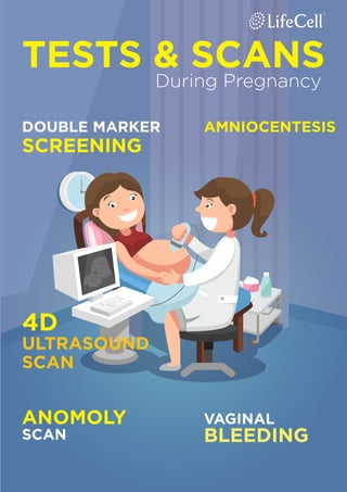
- 1. TESTS & SCANS DOUBLE MARKER SCREENING AMNIOCENTESIS 4D ULTRASOUND SCAN VAGINAL BLEEDING ANOMOLY SCAN During Pregnancy
- 3. Pregnancy Ultrasound WHY YOU NEED IT? When pregnant, all expectant mothers would like to know about the well-being of their babies. It is necessary to go for routine health check-ups during pregnancy to learn about the baby’s movements and health. Ultra- sound scan is one such routine medical check-up performed on pregnant women to ensure the baby’s health. Here is all about ultrasound scan and its importance during pregnancy: What is Ultrasound Scan? The practice of examining pregnant women using ultrasound is known as obstetric sonography. The ultrasound scan is a standard painless procedure used to visualize the fetus inside the uterus using sound waves to create images of the organs, bones and structures. The sound waves usually bounce off the baby as echoes which are turned into images on the moni- tor. As bones and other hard tissues reflect more echoes, they appear white in the image while soft tissues appear grey. The sound waves pass through fluids like amniotic fluid and appear dark due to absence of echoes.
- 4. Why is it required? The pregnancy ultrasound scan is required for various purposes such as: • To detect any abnormality in the growth of the fetus • To check the heartbeat of the baby • Detect if you are pregnant with one baby, twins or more • Check out the cause of any bleeding during pregnancy • Find out the date of pregnancy by measuring the size of the baby • To check if the embryo is implanted in a normal position or outside the uterus (fallopian tube) • To ensure that all the baby organs are normal • Diagnose any developmental abnormality in the baby such as Spina bifida • Assess the baby’s growth rate by comparing over several scan results • Check the amount of amniotic fluid and the position of the placenta • To detect the gender of the baby • To assess the risk of Down syndrome and other abnormalities during pregnancy. When is it performed? • The first pregnancy ultrasound scan is performed around 8-13 weeks to confirm the pregnancy and to arrive at the due date. • The second ultrasound scan is performed between week 18 and week 21 to see if the baby is normal and without any structural abnormality. • The doctor might recommend for a growth scan in the third trimester between week 28 and week 40.
- 5. 4DUltrasound Scans: A New Trend in Pregnancy Care Having an ultrasound scan during pregnancy is a part of prenatal care to check the growth and normal development of the baby inside the womb. These scans can detect any developmental ab- normality in the baby so that necessary actions can be performed. Apart from diagnosis, ultrasound scans are also used the see the baby before its official arrival. 4D ultrasound scan shows a clear video of the baby moving inside the uterus which can be made into a DVD.
- 6. What is 4D Ultrasound Scan? A 4D ultrasound scan is the moving 3D images of the baby inside the uterus, having time as the fourth dimension. It clearly shows the movements and the facial features of the baby in real time like a video. How is 4D scan different from 2D & 3D? The difference between 2D, 3D and 4D scan lies mostly in the com- puter software advancement rather than the scanning mechanism. The 2D scan can show the baby’s internal organ while the 3D and 4D scan shows only the external features. The 2D scan has grey blurry lines that show the baby’s movement while 3D scan shows clear images of the baby like a photo. The 4D scan on the other hand shows clear movements of the baby where one can see the facial ex- pressions, body movements, shape of the nose, mouth, eyes etc. When is the right time to have a 4D scan? It is advisable to wait for 28 to 32 weeks in order to have a clear 4D scan result. However, it is important to consult your doctor about the scan, its advantages and disadvantages before making a decision.
- 7. AMNIOCENTESIS Why is it needed during pregnancy? Amniocentesis also called as Amniotic fluid test is a prenatal test to detect various medical conditions in the fetus. Amniotic fluid is a watery substance surrounding the fetus which provides protection and information about the baby’s health inside the uterus. During the test, a fine thin needle is inserted into the abdomen and a sample of amniotic fluid is drawn from the uterus under ultrasound guidance without hurting the baby. This sample will be sent to lab for testing on various genetic disorders.
- 8. Why is it performed? Amniocentesis is performed to test the presence of birth defects such as Down syndrome (chromosomal disorder), sickle cell anemia, muscular dystrophy, cystic fibrosis, spina bifida, Tay-Sachs etc. It is usually performed if the expectant mother shows the following signs: • Family history of above mentioned birth defects • Pregnancy after 35 years of age • Previous child with birth defects • Abnormal ultrasound When is it performed? It is usually performed between week 16 and week 18 to ensure the absence of birth defects in baby. Sometimes, the test is also per- formed in the last trimester to check the growth and maturity of the fetal lung. The test results might take up to 12 -15 days for processing. The amniocentesis results are 99.4% accurate and it is completely harmless for the mother and the baby inside the womb. How is it performed? During the test, a small area of the abdomen is cleansed to avoid infection and local anesthesia will be given to ease the pain if re- quired. Through ultrasound, the doctor will locate the position of the fetus and the placenta and then safely insert a long thin needle in to the uterus through abdomen without disturbing the baby. With ultra- sound guidance, the doctor can insert the needle in to the uterus far away from the baby. A small amount of amniotic fluid will be drawn and sent to the lab for testing. You may feel the amniocentesis pain like a minor menstrual cramp during the procedure and it may last for few more hours. If the pain prolongs or if you experience any other symptom like bleeding or vaginal discharge, consult your gynecolo- gist.
- 9. Wellbeing of the baby is a constant thought in the minds of expect- ant mothers. Doctors recommend an ultrasound scan to ensure the healthy development and normal growth of the baby. In the second trimester, you can expect level II ultrasound scan prescribed by your doctor. What Is Anomaly Scan? This a detailed scan usually carried out in the second trimester to check for major abnormalities. It is also called as Level II ultrasound scan. By the 20th week, the individual organs of the baby are clearly seen so this scan will show the growth rate, safe development of internal organs and external features like face, legs, arms etc. When is it performed? The Anomaly scan is usually performed in the second trimester be- tween 18 and 22 weeks. Doctors may ask for more scans in the second trimester if the anomaly scan reveals any problem in pregnan- cy. ANOMALY SCAN Level II Ultrasound Scan
- 10. WHY IS IT PERFORMED? The scan is mainly performed to ensure that the baby’s growth is normal without any abnormality. The Level II anomaly scan helps to detect the following: 1. The scan measures the baby to see if the growth pattern is as expected. 2. The number of babies inside the womb. In most cases, twins are not detected untill week 20. 3. The shape and structure of the head are examined to rule out severe brain problems. 4. The length and cross section of the spine is checked to see if the bones are aligned and covered by skin at the back. 5. All 4 chambers of the heart, valves connected to it, heart beat and major arteries & veins are examined. 6. The stomach is observed to see if the baby could swallow and process the amniotic fluid (seen as a small black bubble). 7. Both the kidneys and the urinary bladder of the baby were checked. 8. Check if the baby’s abdominal wall covers the internal organs in the front. 9. Baby’s face is checked for cleft lip and other facial abnormalities. 10. The arms, hands, legs and feet were also checked to see if they have developed normally. The scan takes about 30 minutes and you are allowed to see while it is performed. If you would like to know about the Level II anomaly scan, talk to your gynecologist and have it done if your doctor recom- mends it.
- 11. Pregnant women have to undergo various tests during all the three trimesters to ensure normal development of the baby and to avoid complications during labour. Based on health condition, previous his- tory of pregnancy and age of the expectant mother, gynaecologists will recommend double, triple and quad marker screening tests. How Is The Test Performed? Initially, pregnant women undergo ultrasound scan and if there are any anomalies detected, the doctor would suggest for a double, triple or multiple marker tests. In these tests, a small amount of mother’s blood sample is taken and tested for the presence of marker sub- stances originated from the foetus, placental tissue or combination of both. The test result will indicate if the growing child is normal or have any chromosomal abnormality by measuring the level of the sub- stances. Importance Of PROTEIN MARKER TESTS
- 12. DOUBLE MARKER TEST TRIPLE MARKER TEST • Beta hCG – Human Chorionic Gonadotropin marker • PAPP – A – Pregnancy Associated Plasma Protein marker • UE3 – Unconjugated protein MULTIPLE MARKER TEST (QUAD SCREENING) This test measure 2 sub- stances in blood - beta hCG and PAPP - A Performed between 10-13 weeks This test measure 3 sub- stances in blood - beta hCG, Alpha feto-protein and UE3 Performed between 14-20 weeks To check for Down syndrome, Trisomy 18 and neural tube defects (spina bifida) Beta hCG - High levels indicate Down syndrome & low levels indicate Edward's syndrome Alpha Feto-protein - Low level indicates Down syndrome and Edward's syndrome & high levels indicate neural tube defects UE3 - Low levels indicate Down syndrome and Edward's syndrome This test measure 3 substances in blood - beta hCG, Alpha feto-protein and UE3 Performed between 15-19 weeks To check for Down syn- drome and neural tube defects (spina bifida) Beta hCG - High levels indicate Down syndrome & low levels indicate Edward's syndrome Alpha Feto-protein - Low level indicates Down syndrome and Edward's syndrome & high levels indicate neural tube defects UE3 - Low levels indicate Down syndrome and Edward's syndrome Inhibin A - high level indicates Down syndrome To check for Down syndrome, Trisomy 18 (Edward's syndrome) and chromosomal abnormalities Beta hCG-High levels indicate Down syndrome & low levels indicate Edward's syndrome PAPP-A-Low levels indicate Down syndrome and Edward's syndrome DOUBLE/TRIPLE/MULTIPLE MARKER TESTS
- 13. Importance Of VAGINAL BLEEDING IN EARLY PREGNANCY: Vaginal bleeding is a common symptom experienced by many moms-to-be during the first trimester of pregnancy. Pregnancy bleeding occurs in 2 out of 10 women during the early stages and in most cases it is not associated with miscarriage or ectopic pregnancy. Generally, first time pregnant mothers confuse the symptoms of spotting with bleeding, so it is necessary to check with your medical practitioner. How spotting is different from bleeding? Spotting is when you see a few drops of blood now and then which may not enough to cover a panty liner whereas bleeding is a heavy flow of blood which requires a sanitary pad or panty liner to avoid staining of the clothes. Spotting occurs when the fertilized egg implants itself to the uterus wall which occurs between 6- 12 days after you got conceived. Se- rious bleeding in early pregnancy needs to be checked immediately so if you spot heavy flow of blood, contact your gynecologist or visit the near- est hospital to get treatment
- 14. Does spotting causes any harm? Light bleeding during early pregnancy may occur due to various reasons so it is advisable to let your gynecologist know about your health condition. If you have a normal pregnancy confirmed through ultrasound, inform your doctor during the next visit. If you have not had an ultrasound scan and sense spotting, it is crucial to consult your doctor as early as possible. Sometimes spotting occurs if the fetus grows outside the uterus (ectopic pregnancy) which is not safe for the mother.
- 15. Causes of vaginal bleeding It is possible to experience bleeding in pregnancy due to the following reasons: • Implantation of fertilized egg in the uterus • Infection in the vagina • Sexual intercourse • Hormonal changes etc. Apart from these, there are other reasons for early vaginal bleeding which are as follows: Infection - Any kind of infection in the vagina and cervix due to sexually transmitted diseases may cause bleeding in the first trimester. It is important to use protection while having sex in all the three trimesters. Change in the cervix – During intercourse, there is possibility of change in the cervix which may result in bleeding. However, this type of bleeding is nothing to worry about. However, if there is heavy bleeding then it might be a sign for miscarriage, ectopic pregnancy or molar pregnancy (pregnancy not formed properly). It is necessary to visit the hospital if you experience heavy bleeding in pregnancy, bleeding with pain and dizziness or abnormal pain in the belly and pelvis region.
- 16. WHAT TO EXPECT ON FIRST TRIMESTER ULTRASOUND SCAN? Being pregnant for the first time is exciting and hectic at the same time, like going on a roller coaster ride. Pregnant women may have million questions on their mind about the growth and well being of the baby along with the symptoms of pregnancy. This article will help you understand the importance of ultrasound scan performed in the first trimester. Ultrasound scan is one of the routine tests in pregnancy which determines the normal development of the baby and helps to identify any abnormalities, if present. This is a painless procedure which doesn’t have any known side effects to the mother and the baby. How is it performed? During ultrasound scan, a small amount of gel will be applied on your abdomen and the physician will run the transducer over it. This will send high frequency dure normally takes around 25-30 minutes and the image can be printed or copied as a video.
- 17. Why is it performed? 1. To arrive at the pregnancy due date and confirm it by measuring the size of the fetus. This will help pregnant women who have irregular menstrual cycle or who can’t recollect the date of their last period. 2. To check the heart beat of the baby and confirm the normal development. 3. To check the number of babies in the uterus and if there are twins, the scan will help to identify whether they are identical or not. 4. To detect if there are any fetal abnormalities.