
Recomendados
Mais conteúdo relacionado
Mais procurados
Mais procurados (20)
Semelhante a Vertical jaw relation in Complete Dentures- Kelly
Semelhante a Vertical jaw relation in Complete Dentures- Kelly (20)
Mais de Kelly Norton
Mais de Kelly Norton (13)
Último
Último (20)
Vertical jaw relation in Complete Dentures- Kelly
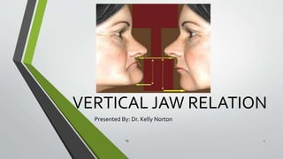
- 1. VERTICAL JAW RELATION Presented By: Dr. Kelly Norton 65 1
- 2. INTRODUCTION • Recording of jaw relations in the treatment of edentulous patients aims at facilitating the adaptation of the complete denture to the masticatory system to give them an optimal and comfortable function. • To achieve this goal, the recording must include an approximate vertical dimension of occlusion, stable occlusal contacts in harmony with the existingTMJ and masticatory muscle functions. . 65 2
- 3. Definition Maxillomandibular relationship - GPTVIII • Any spatial relationship of the maxillae to the mandible; • Any one of the infinite relationships of the mandible to the maxillae 365
- 4. Classification of Jaw relations • Orientation jaw relation. • Vertical jaw relation. • Horizontal jaw relation. 465
- 5. Classification • The vertical jaw relation can be classified as follows: 1)Vertical dimension at rest-VDR 2)Vertical relation at occlusion-VDO 565
- 6. Definition Vertical dimension, GPTVIII The distance between two selected anatomic or marked points (usually one on the tip of the nose and the other upon the chin), one on a fixed and one on a movable member 665
- 7. Principle The single most important factor in deciding the vertical dimension is: the mandibular musculature and occlusal stops or occlusion rims from the teeth. 765
- 8. 65 8 PROPONENT THEORY HUNTER(1771) Lies in the middle of extremes of motion by which all the muscles and ligaments are equally relaxed. WALLISCH(1906) All muscle action eliminated Mandible passively suspended Opposing teeth no contact NISWONGER(1934) Constancy concept of face height -1st investigator to study extensively the rest position of mandible by recording measurements on patients. -Rest position is a neutral position of the mandible since the opening and closing muscles are in a state of equilibrium. -Jaw relator-A gauge to measure the vertical dimension of the face Thompson and Brodie (1942) Thomson believed that the rest position is determined by a balance of tension in the musculature which suspends the mandible and that the rest position is not affected by the presence or absence of teeth.They stated, “The proportions of face as far as vertical height is concerned, are constant through out life HISTORY
- 9. 65 9 Leof (1950) -Stressed that muscle tone rather than muscle length controls the rest position, and that muscle tone can vary. -Muscle tone can be increased by exercise and decreased by excessive rest. Meyer M Silverman (1952-1953) Presented a physiologic phonetic method of measuring the vertical dimensions of the closest speaking space Toolson LSmith D. Clinical measurement and evaluation of vertical dimension. The Journal of Prosthetic Dentistry. 2006;95(5):335-339.
- 10. 65 10 Zarb, Bolender – Two hypothesis about rest position 1. Active mechanism This position is assumed only when the muscles that close the jaws and that open the jaws are in a state of minimal contracture to maintain the posture of the mandible. 2. Passive mechanism Elastic elements of the musculature, and not any muscle activity, balance the influence of gravity.
- 11. Physiologic rest position GPT-VIII • 1:The mandibular position assumed when the head is in an upright position and the involved muscles, particularly the elevator and depressor groups, are in equilibrium in tonic contraction, and the condyles are in a neutral, unstrained position • 2:The position assumed by the mandible when the attached muscles are in a state of tonic equilibrium.The position is usually noted when the head is held upright • 3:The postural position of the mandible when an individual is resting comfortably in an upright position and the associated muscles are in a state of minimal contractual activity 1165
- 12. Significance of Physiological Rest position • Bone to bone relation • Fairly constant throughout the life in absence of any pathosis. • Can be recorded and measured within acceptable limits. • Used to determine theVDO. 1265 Textbook of complete dentures : Charles M. Heartwell Jr., Arthur O Rahn, 5th Edition
- 13. Vertical dimension of occlusion, GPTVIII Occlusal vertical dimension The distance measured between two points when the occluding members are in contact. 1365
- 14. Vertical Dimension at Rest • Definition: - • The distance between two selected points (one of which is on the middle of the face or nose and the other of which is on the lower face or chin) measured when the mandible is in the physiologic rest position-GPT-8 • It is essential to record the vertical dimension at rest as it acts as a reference point during recording the vertical dimension at occlusion. • TheVD at rest should be recorded at the physiological rest position of the mandible. 65 14
- 15. Inter-relationship VDR-VDO=Freeway space or the interocclusal rest space Interocclusal rest space- GPTVIII The difference between the vertical dimension of rest and the vertical dimension while in occlusion. It ranges from 2-4 mm in vertical direction at the position of the 1st premolar 1565
- 16. Syllabus of complete dentures : Charles M. Heartwell Jr., Arthur O Rahn, 4th Edition 1665 Factors considered for rest position Gravity Neuromuscular disturbances make measurements without delay calm, cool and relaxed No one method for determining rest position can be accepted as being valid for all patients. Several methods are available to confirm this record
- 17. Classification of the methods • Mechanical : • 1. Ridge relation A.Incisive papilla to mandibular incisors B. Parallelism of the ridges • 2. Measurement of the former dentures • 3. Preextraction records A. Profile radiographs B. Casts of teeth in occlusion C. Facial mesurements 1765 Physiological 1. Physiological Rest Position 2. Phonetic and Esthetics as guides 3. Swallowing threshold 4.Tactile Sense
- 18. • 1.Incisive papilla to mandibular incisors • Incisive papilla – stable landmark • Distance : Papilla -Incisal edges of mandibular anterior teeth : 4 mm Papilla -Incisal edges of maxillary central incisors : 6 mm Mean vertical overlap : 2 mm • Individual variations • Not relevant in patients with severe resorption 65 18 RIDGE RELATIONS MECHANICAL METHODS
- 19. • Paralleling and a 5 degree opening in the posteriors is acceptable as suggested by Sears. Marked resorption of the ridges makes this rule void. • Natural, provided there has been no abnormal change in the alveolar process such as a previous advanced periodontal disease or gross supra-eruptions. • Most people – ridges are not parallel Teeth lost at different times Resorption pattern 65 19 PARALLELISM OFTHE RIDGES
- 20. • Measurements are made from the borders of with a Boley gauge. • Correlated with the observations of the patient’s face • Distance is too short or long – Corresponding change can be made. 65 20 MEASUREMENT OFTHE FORMER DENTURES • Problems: • Loss of the ridges under the dentures results in an increase in interocclusal distance. • Patients can shift the mandible, or the denture can be shifted on its support to accommodate for errors in occlusion. • Inaccurate adaptation of the denture base to the support results in displacement of the denture
- 21. These data reveal the progressive changes which occur when the natural teeth are extracted. They provide information about – • Occlusal vertical dimension • Anteroposterior angle of occlusal plane • Position and inclination of maxillary central incisors • Horizontal and vertical overlap of each tooth. • Length and width of teeth. (A pre-extraction profile record. -J. Prosth. Dent. 45: 479, 1981) 65 21 PREEXTRACTION RECORDS
- 22. 1. PROFILE RADIOGRAPHS • Much used in research of vertical dimension of occlusion • Lateral skull radiographs before and after extraction are compared. Disadvantages: • Radiation risks – So cannot be considered for routine clinical use. • Considerable time • Unreliable- -Inaccuracies that exist in the technique -Inaccuracies in the method of comparing measurements 65 22
- 23. 2.PROFILE PHOTOGRAPHS • Made with the teeth in maximum occlusion • Enlarged to life size • Measurements of anatomic landmarks on the photograph are compared with measurements using the same anatomic landmarks on the face. • These measurements can be compared when the records are made and again when the artificial teeth are tried in. • Disadvantages : Angulation of the photos might differ. Photo enlargements cause inaccuracies. 65 23
- 24. 3.PROFILE SILHOUETTES • Lead wire adaptation along the midline helps preparing a cardboard cutout, which is preserved after extraction. • Repositioned to the face after the vertical dimension has been established at the initial recording and/or when the artificial teeth are tried in. (A pre-extraction profile record. -J. Prosth. Dent. 45: 479, 1981) 65 24
- 25. 4. CASTS OFTEETH IN OCCLUSION • Practical method • Measurements: • - Incisive papilla and crest of the lower ridge -Extended height of upper and lower buccal frena -Hamular notch and retromolar pad • Indicate the amount of space required between the ridges for teeth of this size. • Valuable in patients whose ridges are not sacrificed during the removal of teeth or resorbed during a long waiting period for denture construction. 65 25
- 26. 5. RESIN FACE MASKS: SWENSON (1959)- • Acrylic resin face masks are made before the extractions and later, when the patient is edentulous, fitted on the face to see whether the vertical dimension has been restored properly. Disadvantages: • Requires a great deal of time • Extensive experience with the use of facial impressions and casts. • Different topography of face in erect and recumbent posture. 65 26 Swenson’s Complete Dentures, Boucher, Editor, Fifth edition
- 27. 6.FACIAL MEASUREMENTS a. To record the relation of the head to the central incisors vertically and anteroposteriorly by placement of a face bow with auditory meatus plugs and with spectacle suspension. b.To record the distance from the chin to the tip of the nose by means of a pair of calipers or dividers before the teeth are extracted. 65 27
- 28. c.Willis guage. • Pupil of the eye to rima oris = anterior nasal spine to inferior border of mandible. 28 J Prosthet Dent 2004;91:59-66. 65
- 29. d) Equal- thirds concept : The face can be divided into equal thirds – the forehead, the nose and the lips and chin. 2965
- 30. PHYSIOLOGICAL METHODS 1.PHYSIOLOGICAL REST POSITION • Indication of the appropriate vertical dimension at rest • Rest vertical dimension – Occlusal vertical dimension= Interocclusal distance • Interocclusal distance : 2-4mm when observed at the position of first premolars. • May not be an exact guide65 30
- 31. • METHOD : -Patient relaxed, with trunk upright and the head unsupported. -After insertion of the occlusal rims the patient should be asked to swallow and let the jaw relax. -The lips are parted to reveal how much space is present between the occlusal rims. -The interocclusal distance should be 2- 4mm at the premolar region. 65 31
- 32. ->4mm- occlusal vertical dimension is too small. -< 2mm- occlusal vertical dimension is too great. - Occlusion rims are adjusted until adequate interarch space is obtained and patient comfort and phonetic and esthetic considerations are satisfactory. 65 32
- 33. 2.PHONETICS • Listening to speech sound production and observing the relationships of teeth during speech. 65 33
- 34. 1. CH, S, AND J – • Bring the anterior teeth close together. • Lower incisors should move forward to a position nearly directly under and almost touching the upper incisors. • If anterior teeth touch when these sounds are made or if the teeth click together during speech, the vertical dimension is too great. 65 34
- 35. 2. Have the patient repeat the name ‘Emma’ or ‘ Om’ until he is aware of the contacting lips as the first syllable ‘m’ is pronounced.When the patient has rehearsed this procedure, ask him to stop all jaw movement when the lips touch.At this time measure between the two points of reference. • 3. Engage the patient in a conversation that will divert his attention from conscious participation in the procedure.A pause in the speech, followed by relaxation as indicated by a drop of mandible, is an indication for another measurement. 65 35
- 36. 4.CLOSEST SPEAKING SPACE: SILVERMAN • Measures the vertical dimension when the mandible and muscles involved are in physiologic function of speech. • The occlusion rims are placed in the mouth and the height is adjusted until a minimum of 2 mm space exists when the patient pronounces the letter “S”. • Closest speaking space – vary from 0-10mm • Disadvantage: Patient who has an 8-10mm closest speaking space will require other means for determination of the vertical dimension. 65 36
- 37. 5.THE ‘F’ OR ‘V’ AND ‘S’ SPEAKING ANTERIOR TOOTH RELATION: POUND AND MURRELL ‘f’ and ‘v’ sounds - The incisal edges of the maxillary anterior teeth create a seal on the moist area of the vermilion border of the lower lip. 65 37 • ‘s’ -The position of the mandibular anterior teeth is determined when the patient says words beginning with ‘s’.When the ‘s’ sounds are articulated, the mandible moves forward. The incisal edges of the anterior teeth do not make contact.
- 38. 3.ESTHETICS • Labial surfaces of the occlusion rims should be contoured to replace or restore the tissue support provided by the natural structures. • If lips are not properly supported anteriorly- tendency to increase the vertical dimension is great- leads to increased lower face height. • Recent evidence – this method is unreliable65 38
- 39. 4.FACIAL EXPRESSION • In normal related jaws, the lips will be even antero-posteriorly and in slight contact. • Patient with a retruded mandible has uneven lip position and the two are not in contact. Vice versa is observed in case of prognathic mandibles. • Skin around the eyes and over the chin will be relaxed. • Relaxation around the nares reflects unobstructed breathing. 65 39
- 40. 5.SWALLOWING THRESHOLD • Powell and Zander(1965), Boucher (1955) and Shanahan (1955) • Teeth come together with a very light contact at the beginning of the swallowing cycle. • Highest point of jaw during deglutition = VDO Technique: • Build cones of wax on the lower denture base in such a way that they contact the upper occlusion rim when the jaws are opened too wide.65 40
- 41. • The flow of the saliva is stimulated by food, such as a piece of candy. • Repeated action of swallowing the saliva will gradually reduce the height of the wax cones to allow the mandible to reach the level of occlusal vertical dimension. • Length of the time to complete this action and the relative softness of the wax cones will affect the results. • No consistency in the final vertical positioning of the mandible has been found. 65 41 (The consistency of the the swallowing technique in determining occlusal vertical relation in edentulous patients - J Prosth. Dent., 36: 159, 1976)
- 42. 6.TACTILE SENSE PATIENT A. PERCEIVED COMFORT • Instruct the patient to stand erect and open the jaws wide until strain is felt in the muscles. • When this opening becomes uncomfortable , ask him to close slowly until the jaws reach a comfortable , relaxed position. 65 42
- 43. B) Lytle’s Neuromuscular perception - Lytle RB in 1964 • It relies on patient’s perception of different vertical height. • A central bearing device is attached to accurately adapted record base • Bearing pin is adjusted beyond the rest position, pin is then lowered by half turn. Patient has to signify over- closure. • Pin is raised again till excess opening is seen. • Appropriate vertical relation is judged by the patient. • Disadvantage : • it cannot be used in patients with poor neuromuscular coordination. • Presence of foreign objects in the palate and the tongue space. • Conflicting results on the precision of this method 43 Lytle RB,Vertical relation of occlusion by the patient's neuromuscular perception,Volume 14, Issue 1,January–February 1964, Pages 12–21 65
- 44. 65 44 7. POWER POINT ( BOOS BIMETER , 1940): • Attach the bimeter to an accurately adapted mandibular record base. • Attach a metal plate in the vault of an accurately adapted maxillary record base to provide a central bearing point. • Adjust the vertical distance by turning the cap. • The gauge indicates the pounds of pressure generated during closure at different degrees of jaw separation. • When the maximum power point is determined, lock the set nut. • Make plaster registrations and transfer the cast to an articulator. • Disadvantage: Such a device offers no more accuracy than Niswonger’s or Silverman’s method.
- 45. 8.WAX OCCLUSION RIMS • 1.Establish the vertical dimension at rest • 2.Maxillary occlusion rim contoured to the tentative occlusal plane is not altered unless absolutely necessary. When they must be , reduce the rim distal to the cuspid and retain the guides for positioning the anterior teeth. • 3.Make the interocclusal distance approximately 3-4 mm less than the interocclusal distance at rest position. • 4.Thinly coat the maxillary occlusion rim with petrolatum. 65 45
- 46. • 5.Soften a roll of baseplate wax in a waterbath at 130o F and contour it in a triangular shape with the base on the occlusion rim and attach it to the occlusal surface of the mandibular occlusion rim. • 6.Seat the mandibular record base in the mouth and place the tips of the index fingers bilaterally on the buccal flanges in the area of the second bicuspids to assure that the record base is stable when the jaw is moved. 65 46
- 47. • 7.Request the patient to retrude the mandible and close on the back teeth but to stop closing the jaw when he feels that the closure is sufficient. • 8. Allow the wax to harden before removing the tentative record from the mouth. 65 47
- 48. • 9.Reinsert the record and have the patient close to maximum occlusion. Measure the distance between the points of reference and compare with the measurements made with the mandible at rest. - If the measurement is less than the measurement at rest and the baseplate wax is not penetrated through to make occlusion rim contact, the record is acceptable.65 48
- 49. Niswonger’s method • The camper’s plane or the ala- tragus line and the inter- pupilliary line are the hallmarks of this procedure. • An upright position leads the planes to be parallel to the floor. • The marks are made on the tip of the nose and the most stable area on the chin. 4965
- 50. • The distance between the marks is recorded after the patient is asked to swallow and relax. • Subsequently occlusal rims are fabricated so that when they occlude, have a measurement 1/8” less than the original measurement. • This 1/8” average gives a freeway space of 2 to 4 mm. 5065
- 51. Preparing for evaluation • Relate the mandibular cast to the face-bow mounted maxillary cast and attach the mandibular cast to the articulator with plaster. • Orient the plane of occlusion and arrange the anterior and posterior artificial teeth to meet the functional and esthetic requirements in centric occlusion. • Contour the wax in the form of the finished denture.65 51
- 52. Evaluating vertical dimension Patient’s tactile sense: • Place the trial dentures in the patient’s mouth. • Instruct the patient to open and close until the teeth contact. • Ask the patient if the teeth appear to touch too soon, if the jaws seem to close too far before they touch, or if the teeth feel just right. • This method is not very effective with senile patients or with those who have impaired neuromuscular coordination. 65 52
- 53. Swallowing followed by relaxing 1.With the dentures in place instruct the patient to wipe the lips with the tip of the tongue, swallow and let the shoulders drop in a relaxed position. • If the teeth are together it can indicate that no interocclusal distance exists. 2.Two small cones of a soft wax are placed, one in each central sulcus of the mandibular first molars. • Encourage the patient to swallow several times. • If the vertical dimension of occlusion is correct, the wax will be penetrated and reduced to tooth contact. 65 53
- 54. Phonetics • Three, thirty-three :There should be enough space for the tip of the tongue to protrude between the anterior teeth. • Fifty-five : Incisal edges of the maxillary central incisors should contact the vermilion border of the lower lip at the junction of the moist and dry mucosa. • Emma and Mississippi :Teeth should not contact.65 54
- 55. Effects of increased vertical dimension • Discomfort to the patient. • Trauma and pain under the basal seat areas of dentures:The jarring effect of the teeth coming into contact sooner than expected may not only cause discomfort but in most cases it will also cause pain owing to the bruising of the mucosa • Loss of free way space : Muscular fatigue of any one or group of muscles of mastication. In turn results in annoyance from the inability to find comfortable resting position. • Clicking sound :When occlusal vertical dimensions is increased, opposing cusp will frequently meet each other producing an embarrassing clicking sound. 5565
- 56. • Appearance : Elongated appearance and at rest the lips are parted; Patient tries to close them together producing an expression of strain. • Bone resorption : Due to continuous pressure on the residual alveolar ridge it undergoes rapid resorption. • Loss of retention and stability : Leverages are caused due to premature contacts, further loss of ridge leads to loss of retention and stability. • Generalized Hyperemia : Space between the teeth is essential when mandible is at rest. If no space is present between the teeth in denture, it may result in generalized hyperemia. 5665
- 57. Effects of decreased vertical relation 57 Inefficiency : Pressure which is possible to exert with teeth in contact decreases considerably with over closure because the muscles of mastication acting from attachments have been brought closer together. Cheek, Tongue and lip biting : Loss of muscular tone, as well as reduced vertical height, the flabby cheek tend to become trapped between the teeth during mastication. Appearance (Denture look) : The general effect of over closure on facial appearance is of increased age because of closure approximation of nose to chin, soft tissue sag and fall in and the lines on the face are deepened. 65
- 58. Angular cheilitis (perleche): A reduced vertical dimension results in a crease at the corners of the mouth beyond the vermilion border and the deep fold thus formed becomes bathed in saliva thus leading to infection and soreness. Pain in temporomandibular joint : Over closure may cause pain in temporomandibular joint probably due to strain of the joint and associated ligaments. 5865
- 59. Costen’s syndrome (Mild catarrhal deafness): There will be a tendency to push the tongue towards the throat, adjacent tissues will be displaced, which may in turn result in occlusion of Eustachian tubes which would interfere with function of ear which may cause ear discomfort and impaired hearing. • Tinnitus or snapping noises in joint. • Tenderness to palpation overT.M.J. • Dryness of the mouth. • Various neurologic symptoms such as burning or picking sensation of the tongue. Prognathism : Over the years as a result of resorption of ridges and abrasion of denture teeth, there is a loss of occlusal vertical dimension.So the lower jaw over- closes in a forward and upward direction.Then the patient may appear prognathic. 59 Weinberg L, Role of condylar position inTMJ dysfunction-pain syndrome, J Prosthet Dent,Vol 41, 6, Jun 1979, Pages 636–643 65
- 60. REFERENCES 65 60
- 61. Reestablishment of OcclusalVertical Dimension in Complete DentureWearing inTwo Stages 65 61 Case Reports in Dentistry. 2015;2015:1-5. A 65-year-old woman came to a Dental School, Brazil, for assessment of a new complete denture. The complete dentures were fabricated 23 years ago and her principal complaint was poor esthetics and ear pain.
- 62. The Study of the Effect of Altering theVertical Dimension of Occlusion on the Magnitude of Biting Force • Aim:To develop a device capable of measuring the biting force generated during maximum biting, during a change inVD and to determine, the relationship between theVDO and the biting force. • Materials and Methods: Indigenously fabricated electronic gnathodynamometer was used to record biting force of 10 individuals at alteredVD.The range of alteration was chosen from increased 7.5 mm to decreased 4.5 mm and establishedVDO as base line. • Results: Bite force was maximum at theVDO in edentulous subjects. Maximum biting force recorded atVDO was reduced with subsequent increase or decrease inVD. • Clinically highest biting force could act as an aid in determining and verifyingVDO for edentulous patients 65 62 Journal of International Oral Health 2015; 7(11):1-5
- 63. Reliability of determining vertical dimension of occlusion in complete dentures: A clinical study. • Objectives :1.To assess the reliability of the conventional methods in obtaining vertical dimension. 2.To analyse changes in morphologic face height after extraction. 3.To assess the reliability of measuring base of nose to chin distance in obtaining vertical dimensions. • Results: Nose-chin measurement has a significant corelation with cephalometric measurements, hence it is found to be a very effective pre extraction aid in determining vertical dimension. However, reliability of pre extraction records in long period of edentulousness is limited.The conventional methods used to determine the vertical dimension are not reliable 65 63 The Journal of Indian Prosthodontic Society. 2006;6(1):38.
- 64. Conclusion • Many methods of assessing and recording vertical jaw relations in edentulous patients have been presented and evaluated. • Since there is no precise scientific method of determining the correct vertical relations, the registration of vertical relations depends upon the clinical experience and judgment of the dental surgeon himself. • Several methods should be used to verify the vertical dimension and there is no one single best method to do so. 6465
- 65. REFERENCES AND CROSS REFERENCES 1. Prosthodontic treatment for edentulous patients : Boucher 9th Edition 2. Textbook of complete dentures : Charles M. Heartwell Jr., Arthur O Rahn, 4th and 5th Edition 3. Marin D, Leite A, de Oliveira Junior N, Compagnoni M, Pero A, Arioli Filho J. Reestablishment of Occlusal Vertical Dimension in Complete DentureWearing inTwo Stages. Case Reports in Dentistry. 2015;2015:1-5. 4. Gosavi SS, Ghanchi M, Patil S, Sghaireen MG, AliAH, Aber AM.The Study of the Effect of Altering the Vertical Dimension of Occlusion on the Magnitude of Biting Force. Journal of International Oral Health. 2015 Nov 1;7(11):110. 65 65
- 66. REFERENCES AND CROSS REFERENCES 5. BhatVGopinathan M. Reliability of determining vertical dimension of occlusion in complete dentures: A clinical study.The Journal of Indian Prosthodontic Society. 2006;6(1):38. 6. Irving M. Sheppard, Stephen M. Sheppard, “Vertical dimension measurements”, JPD 1975, 34(3) : 269 – 277 7. A.J.Turell; “Clinical assessment of vertical dimension”,JPD 1972, 28(3) : 238 – 246 8. Silvermann MM; “The speaking method in measuring vertical dimension”,JPD 1953, 3(2) : 193 – 199 9. Swerdlow H;Vertical Dimension literature review,J Prosthet Dent March April 1965,Vol 15, no. 2. 241-247. 65 66
- 67. 65 67
Notas do Editor
- Opening muscles; suprahyoid and infrahyoid: lateral pterygoid, geniohyoid, digastric, mylohyoid, platysma etc Closing musclews: masseter medial pterygoid and temporal muscles
- The position of the mandible is influenced by gravity; so the patient should be seated upright or standing with the head erect, looks straight ahead when jaw relation records are made Patient should be calm, cool and relaxed when the jaw relations are recorded Neuromuscular disturbances make the records difficult. With these patients the operator must be very considerate and cool. The dentist should be prepared to make measurements without delay when the position is assumed because the rest position is not to be maintained for a duration of time.
- Small variations of vertical dimension are not so critical because the adaptive capacity of the masticatory system usually is great.
- Not to be confused with freeway-space of the physiologic method reported by Niswonger(1934) and Thompson(1946). Establishes vd when muscles are at rest and the mandible is at rest position
- Inadequate lip support results in a flat upper lip with loss of vermillion border, loss of muscular function and loss of dominance of upper lip over lower lip will give a denture look.
- After determining the OVD through the association of the metric, phonetic, and esthetic methods, the need of 14 mm for the reestablishment of the OVD was demonstrated in the current prostheses.
- Background: Determination of the correct vertical dimension of occlusion (VDO) for the edentulous patients is one of the most important steps in making dentures for optimum esthetics and functions. Knowledge of the magnitude, direction, and distribution of the masticatory forces are of special value in designing a prosthesis, which has high masticatory effi ciency and which transfers a minimum load to the supporting tissue. Impression compound occlusal rims with 3 U-shaped bioacryl sheets on upper occlusal rims and 2 bioacryl sheets on lower occlusal rims were adjusted on Hanau articulator to recorded VDO. The range of alteration was chosen from increased 7.5 mm to decreased 4.5 mm to that of established VDO as base line. To increase VD by 7.5 mm three additional “U” shaped biocryl sheets were placed on the lower occlusal rim
- distance between the base of the nose and chin was measured with the help of vernier Lateral cephalograms were made with teeth in occlusion to give the vertical dimension of occlusion [Figure 3]. The cephalometric points and planes considered was Nasion (N), Gnathion (Gn), Point A, Point B, Pogonion and Mandibular plane. The total face height was obtained as the linear measurement between Nasion and Gnathion After extraction records were made, with the occlusal rims The patients were asked to pronounce the letter ‘M’ twice or thrice and then asked to relax. This was also further verified by noting the closest speaking space. The patients were asked to pronounce words with sibilants like ‘six’, ‘sixty six’ and the amount of space between the rims were observed. The patients were engaged in conversation and the speaking space was observed. A minimum of 1 mm space was acceptable. Facial aesthetics was also observed