
Recomendados
Mais conteúdo relacionado
Mais procurados
Mais procurados (20)
Destaque
Destaque (20)
Semelhante a Basic ap chapter 7 powerpoint 2017
Semelhante a Basic ap chapter 7 powerpoint 2017 (20)
Mais de Kathy Richards
Último
Último (20)
Basic ap chapter 7 powerpoint 2017
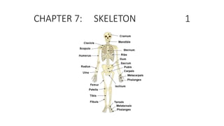
- 1. CHAPTER 7: SKELETON 1
- 2. Skeletal System: 2 • Skeletal System: • rigid internal framework of the body. • consists of the bones, cartilages, and ligaments. • Bones support the weight of the body, allow for body movements, and protect internal organs. • Cartilage provides flexible strength and support for body structures such as the thoracic cage, the external ear, and the trachea and larynx. At joints of the body, cartilage can also unite adjacent bones or provide cushioning between them. • Ligaments are the strong connective tissue bands that hold the bones at a moveable joint together and serve to prevent excessive movements of the joint that would result in injury. • Providing movement of the skeleton are the muscles of the body, which are firmly attached to the skeleton via connective tissue structures called tendons. As muscles contract, they pull on the bones to produce movements of the body. • Thus, without a skeleton, you would not be able to stand, run, or even feed yourself!
- 3. Skeletal System: 3 • Each bone body serves a particular function, and therefore bones vary in size, shape, and strength based on these functions. • For example, the bones of the lower back and lower limb are thick and strong to support your body weight. • Similarly, the size of a bony landmark that serves as a muscle attachment site on an individual bone is related to the strength of this muscle. Muscles can apply very strong pulling forces to the bones of the skeleton. To resist these forces, bones have enlarged bony landmarks at sites where powerful muscles attach. • This means that not only the size of a bone, but also its shape, is related to its function.
- 4. Skeletal System: 4 • Bones are also dynamic organs that can modify their strength and thickness in response to changes in muscle strength or body weight. Thus, muscle attachment sites on bones will thicken if you begin a workout program that increases muscle strength. • Similarly, the walls of weight-bearing bones will thicken if you gain body weight or begin pounding the pavement as part of a new running regimen. • In contrast, a reduction in muscle strength or body weight will cause bones to become thinner. This may happen during a prolonged hospital stay, following limb immobilization in a cast, or going into the weightlessness of outer space. Even a change in diet, such as eating only soft food due to the loss of teeth, will result in a noticeable decrease in the size and thickness of the jaw bones.
- 5. Skeletal System 5 • The skeletal system: • All of the bones, cartilages, and ligaments of the body that support and give shape to the body and body structures. • Skeleton consists of the bones of the body. • Adults: 206 bones in the skeleton. • Younger individuals have higher numbers of bones because some bones fuse together during childhood and adolescence to form an adult bone. • Primary functions of the skeleton: to provide a rigid, internal structure that can support the weight of the body against the force of gravity, and to provide a structure upon which muscles can act to produce movements of the body. • The lower portion of the skeleton is specialized for stability during walking or running. • In contrast, the upper skeleton has greater mobility and ranges of motion, features that allow you to lift and carry objects or turn your head and trunk.
- 6. Skeletal System: 6 • In addition to providing for support and movements of the body, the skeleton has protective and storage functions. • protects the internal organs, including the brain, spinal cord, heart, lungs, and pelvic organs. • Bones of the skeleton serve as the primary storage site for important minerals such as calcium and phosphate. The bone marrow found within bones stores fat and houses the blood-cell producing tissue of the body. • The skeleton is subdivided into two major divisions—the axial and appendicular.
- 7. The Axial Skeleton 7 • The axial skeleton forms the vertical, central axis of the body and includes all bones of the head, neck, chest, and back. It serves to protect the brain, spinal cord, heart, and lungs. It also serves as the attachment site for muscles that move the head, neck, and back, and for muscles that act across the shoulder and hip joints to move their corresponding limbs
- 8. The axial skeleton supports the head, neck, back, and chest and thus forms the vertical axis of the body. It consists of the skull, vertebral column (including the sacrum and coccyx), and the thoracic cage, formed by the ribs and sternum. The appendicular skeleton is made up of all bones of the upper and lower limbs. 8
- 9. Axial Skeleton 9 • The axial skeleton • forms the vertical, central axis of the body • includes all bones of the head, neck, chest, and back. (trunk) • serves to protect the brain, spinal cord, heart, and lungs. • serves as the attachment site for muscles that move the head, neck, and back, and for muscles that act across the shoulder and hip joints to move their corresponding limbs.
- 10. Axial Skeleton 10 • The axial skeleton of the adult • consists of 80 bones, including the skull, the vertebral column, and the thoracic cage. • The skull is formed by 22 bones. Also associated with the head are an additional seven bones, including the hyoid bone and the ear ossicles (three small bones found in each middle ear). • The vertebral column consists of 24 bones, each called a vertebra, plus the sacrum and coccyx. • The thoracic cage includes the 12 pairs of ribs, and the sternum, the flattened bone of the anterior chest
- 11. The Appendicular Skeleton 11 • The appendicular skeleton: • includes all bones of the upper and lower limbs • plus the bones that attach each limb to the axial skeleton. • 126 bones in the appendicular skeleton of an adult.
- 12. SKULL 12
- 13. Bones of the Skull 13 • Parietal bone: • forms most of the upper lateral side of the skull • Temporal bone • forms the lower lateral side of the skull • Frontal bone: single bone that forms the forehead. • Occipital bone: single bone that forms the posterior skull and posterior base of the cranial cavity.
- 14. Sutures of the Skull 14 • A suture: an immobile joint between adjacent bones of the skull. • The narrow gap between the bones is filled with dense, fibrous connective tissue that unites the bones. • The long sutures located between the bones of the brain case are not straight, but instead follow irregular, tightly twisting paths. • These twisting lines serve to tightly interlock the adjacent bones, thus adding strength to the skull for brain protection.
- 15. Facial Bones of the Skull 15 • The facial bones of the skull form the upper and lower jaws, the nose, nasal cavity and nasal septum, and the orbit. • Facial bones include 14 bones, with six paired bones and two unpaired bones. • The paired bones are the maxilla, palatine, zygomatic, nasal, lacrimal, and inferior nasal conchae bones. • The unpaired bones are the vomer and mandible bones. • Although classified with the brain-case bones, the ethmoid bone also contributes to the nasal septum and the walls of the nasal cavity and orbit.
- 16. Maxillary Bone 16 • Maxillary bone: referred to simply as the maxilla (plural = maxillae), is one of a pair that together form the upper jaw, much of the hard palate, the medial floor of the orbit, and the lateral base of the nose. • The curved, inferior margin of the maxillary bone that forms the upper jaw and contains the upper teeth is the alveolar process of the maxilla. Each tooth is anchored into a deep socket called an alveolus. • On the anterior maxilla, just below the orbit, is the infraorbital foramen. This is the point of exit for a sensory nerve that supplies the nose, upper lip, and anterior cheek. • On the inferior skull, the palatine process from each maxillary bone can be seen joining together at the midline to form the anterior three-quarters of the hard palate. • The hard palate is the bony plate that forms the roof of the mouth and floor of the nasal cavity, separating the oral and nasal cavities.
- 17. Zygomatic Bone 17 • The zygomatic bone: • known as the cheekbone.
- 18. Nasal Bone 18 • The nasal bone: • one of two small bones that articulate (join) with each other to form the bony base (bridge) of the nose. • They also support the cartilages that form the lateral walls of the nose. • These are the bones that are damaged when the nose is broken.
- 19. Mandible 19 • Mandible: • forms the lower jaw and is the only moveable bone of the skull. • At the time of birth, the mandible consists of paired right and left bones, but these fuse together during the first year to form the single U-shaped mandible of the adult skull.
- 20. Orbit 20 • The orbit: • bony socket that houses the eyeball and contains the muscles that move the eyeball or open the upper eyelid. • Each orbit is cone-shaped, with a narrow posterior region that widens toward the large anterior opening. • To help protect the eye, the bony margins of the anterior opening are thickened and somewhat constricted. • The medial walls of the two orbits are parallel to each other but each lateral wall diverges away from the midline at a 45° angle. This divergence provides greater lateral peripheral vision. • The walls of each orbit include contributions from seven skull bones. • The frontal bone forms the roof and the zygomatic bone forms the lateral wall and lateral floor. • The medial floor is primarily formed by the maxilla, with a small contribution from the palatine bone. • The ethmoid bone and lacrimal bone make up much of the medial wall and the sphenoid bone forms the posterior orbit.
- 21. Nasal Septum and Conchae 21 • The nasal septum consists of both bone and cartilage • The anterior nasal septum is formed by the septal cartilage, a flexible plate that fills in the gap between the perpendicular plate of the ethmoid and vomer bones. • This cartilage also extends outward into the nose where it separates the right and left nostrils. • The septal cartilage is not found in the dry skull. • Nasal conchae are bony plates that curve downward as they project into the space of the nasal cavity. They serve to swirl the incoming air, which helps to warm and moisturize it before the air moves into the delicate air sacs of the lungs. This also allows mucus, secreted by the tissue lining the nasal cavity, to trap incoming dust, pollen, bacteria, and viruses.
- 22. Paranasal Sinuses 22 • The paranasal sinuses: hollow, air-filled spaces located within certain bones of the skull • All of the sinuses communicate with the nasal cavity and are lined with nasal mucosa. • serve to reduce bone mass and thus lighten the skull, and they also add resonance to the voice. • second feature is most obvious when you have a cold or sinus congestion. These produce swelling of the mucosa and excess mucus production, which can obstruct the narrow passageways between the sinuses and the nasal cavity, causing your voice to sound different to yourself and others. • This blockage can also allow the sinuses to fill with fluid, with the resulting pressure producing pain and discomfort.
- 23. Paranasal Sinuses 23 • The paranasal sinuses are hollow, air-filled spaces named for the skull bone that each occupies. • The most anterior is the frontal sinus, located in the frontal bone above the eyebrows. • The largest are the maxillary sinuses, located in the right and left maxillary bones below the orbits. • The most posterior is the sphenoid sinus, located in the body of the sphenoid bone, under the sella turcica. • The ethmoid air cells are multiple small spaces located in the right and left sides of the ethmoid bone, between the medial wall of the orbit and lateral wall of the upper nasal cavity.
- 24. Vertebral or Spinal Column, Spine 24 • It consists of a sequence of vertebrae, each of which is separated and united by an intervertebral disc. • Together, the vertebrae and intervertebral discs form the vertebral column. • It is a flexible column that supports the head, neck, and body and allows for their movements. • It also protects the spinal cord, which passes down the back through openings in the vertebrae
- 25. Three Regions of Vertebrae 25 • The adult vertebral column consists of 24 vertebrae, plus the sacrum and coccyx. • The vertebrae are divided into three regions: • cervical C1–C7 vertebrae (7) • thoracic T1–T12 vertebrae(12) • lumbar L1–L5 vertebrae. (5) • Coccyx, or tailbone, results from the fusion of four small coccygeal vertebrae. • sacral and coccygeal fusions do not start until age 20 and are not completed until middle age.
- 26. Curvatures of the Vertebral Column 26 • The vertebral column is curved, with two primary curvatures (thoracic and sacrococcygeal curves) and two secondary curvatures (cervical and lumbar curves).
- 27. Curvatures of the Spine 27 • The adult vertebral column does not form a straight line • four curvatures along its length. • These curves increase the vertebral column’s strength, flexibility, and ability to absorb shock. • When the load on the spine is increased, by carrying a heavy backpack for example, the curvatures increase in depth (become more curved) to accommodate the extra weight. They then spring back when the weight is removed.
- 28. Curvatures of the Vertebral Column 28 • The four adult curvatures are classified as either primary or secondary curvatures. • Primary curves are retained from the original fetal curvature. In the adult, this fetal curvature is retained in two regions of the vertebral column as the thoracic curve, which involves the thoracic vertebrae, and the sacrococcygeal curve, formed by the sacrum and coccyx.
- 29. Curvature of the Spine 29 • Secondary curvatures develop after birth. • A secondary curve develops gradually after birth as the child learns to sit upright, stand, and walk. Secondary curves are concave posteriorly, opposite in direction to the original fetal curvature. The cervical curve of the neck region develops as the infant begins to hold their head upright when sitting. Later, as the child begins to stand and then to walk, the lumbar curve of the lower back develops. In adults, the lumbar curve is generally deeper in females.
- 30. Curvatur Disorders of the Spine 30 • Disorders associated with the curvature of the spine include: • kyphosis (an excessive posterior curvature of the thoracic region), • lordosis (an excessive anterior curvature of the lumbar region), • scoliosis (an abnormal, lateral curvature, accompanied by twisting of the vertebral column).
- 31. Cervical Vertebrae 31 • Typical cervical vertebrae, such as C4 or C5, have several characteristic features that differentiate them from thoracic or lumbar vertebrae. • Cervical vertebrae have a small body, reflecting the fact that they carry the least amount of body weight. Cervical vertebrae usually have a bifid (Y-shaped) spinous process. • The spinous processes of the C3–C6 vertebrae are short, but the spine of C7 is much longer. You can find these vertebrae by running your finger down the midline of the posterior neck until you encounter the prominent C7 spine located at the base of the neck. • A typical cervical vertebra has a small body, a bifid spinous process, transverse processes that have a transverse foramen and are curved for spinal nerve passage. • The atlas (C1 vertebra) does not have a body or spinous process. It consists of an anterior and a posterior arch and elongated transverse processes. This is the vertebra that supports the skull on top of the vertebral column. • The axis (C2 vertebra) has the upward projecting dens, which articulates with the anterior arch of the atlas. It serves as the axis for rotation when turning the head toward the right or left.
- 32. Thoracic Vertebrae 32 • Larger than those of cervical vertebrae • A typical thoracic vertebra is distinguished by the spinous process, which is long and projects downward to overlap the next inferior vertebra. • It also has articulation sites (facets) on the vertebral body and a transverse process for rib attachment.
- 33. Lumbar Vertebrae 33 • Lumbar vertebrae are characterized by having a large, thick body and a short, rounded spinous process. • carry the greatest amount of body weight and are thus characterized by the large size and thickness of the vertebral body
- 34. Sacrum and Coccyx 34 • The sacrum is a triangular-shaped bone that is thick and wide across its superior base where it is weight bearing and then tapers down to an inferior, non-weight bearing apex. • It is formed by the fusion of five sacral vertebrae, a process that does not begin until after the age of 20. • The sacrum is formed from the fusion of five sacral vertebrae, whose lines of fusion are indicated by the transverse ridges. • The fused spinous processes form the median sacral crest, while the lateral sacral crest arises from the fused transverse processes. • lateral sacral crest. • The coccyx, or tailbone, is derived from the fusion of four very small coccygeal vertebrae. It articulates with the inferior tip of the sacrum. It is not weight bearing in the standing position, but may receive some body weight when sitting. • The coccyx is formed by the fusion of four small coccygeal vertebrae.
- 35. Intervertebral Disc 35 • intervertebral discs are thin in the cervical region and thickest in the lumbar region, which carries the most body weight. • In total, the intervertebral discs account for approximately 25 percent of your body height between the top of the pelvis and the base of the skull. • Intervertebral discs are also flexible and can change shape to allow for movements of the vertebral column.
- 36. Thoracic Cage 36 • The thoracic cage is formed by the (a) sternum and (b) 12 pairs of ribs with their costal cartilages. • The ribs are anchored posteriorly to the 12 thoracic vertebrae. • The sternum consists of the manubrium, body, and xiphoid process. The sternum also contains a sternal angle which is the junction between the manubrium and the body. • The ribs are classified as: • true ribs (1–7) are attached directly to the sternum by costal cartilage. • false ribs (8–12). • The last two pairs of false ribs are also known as floating ribs (11–12).