
Recomendados
Mais conteúdo relacionado
Mais procurados
Mais procurados (20)
Semelhante a Basic ap chapter 27 powerpoint 2017
Semelhante a Basic ap chapter 27 powerpoint 2017 (20)
Mais de Kathy Richards
Mais de Kathy Richards (20)
Último
Último (20)
Basic ap chapter 27 powerpoint 2017
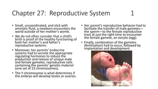
- 1. Chapter 27: Reproductive System 1 • Small, uncoordinated, and slick with amniotic fluid, a newborn encounters the world outside of her mother’s womb. • We do not often consider that a child’s birth is proof of the healthy functioning of both her mother’s and father’s reproductive systems. • Moreover, her parents’ endocrine systems had to secrete the appropriate regulating hormones to induce the production and release of unique male and female gametes, reproductive cells containing the parents’ genetic material (one set of 23 chromosomes). • The Y chromosome is what determines if the embryo will develop testes or ovaries. • Her parent’s reproductive behavior had to facilitate the transfer of male gametes— the sperm—to the female reproductive tract at just the right time to encounter the female gamete, an oocyte (egg). • Finally, combination of the gametes (fertilization) had to occur, followed by implantation and development.
- 2. Male Reproductive System 2 • Scrotum: The testes are located in a skin- covered, highly pigmented, muscular sack called the scrotum that extends from the body behind the penis. • This location is important in sperm production, which occurs within the testes, and proceeds more efficiently when the testes are kept 2 to 4°C below core body temperature. • Unique for its role in human reproduction, a gamete is a specialized sex cell carrying 23 chromosomes—one half the number in body cells. • At fertilization, the chromosomes in one male gamete, called a sperm (or spermatozoon), combine with the chromosomes in one female gamete, called an oocyte and at this point a fertilized egg is produced with all 46 chromosomes. • The function of the male reproductive system is to produce sperm and transfer them to the female reproductive tract.
- 3. Male Reproductive System 3 • Testes: (singular = testis) are the male gonads—that is, the male reproductive organs. They produce both sperm and androgens, such as testosterone, and are active throughout the reproductive lifespan of the male. • The testes are each approximately 4 to 5 cm in length and are housed within the scrotum • During the seventh month of the developmental period of a male fetus, each testis moves through the abdominal musculature to descend into the scrotal cavity. • This is called the “descent of the testis.” • Cryptorchidism is the clinical term used when one or both of the testes fail to descend into the scrotum prior to birth.
- 4. Spermatogenesis Sperm Transport 4 • Spermatogenesis occurs in the seminiferous tubules that form the bulk of each testis. • The process begins at puberty, after which time sperm are produced constantly throughout a man’s life. • One production cycle, from spermatogonia through formed sperm, takes approximately 64 days. A new cycle starts approximately every 16 days, although this timing is not synchronous across the seminiferous tubules. • Sperm counts—the total number of sperm a man produces—slowly decline after age 35, and some studies suggest that smoking can lower sperm counts irrespective of age. • To fertilize an egg, sperm must be moved from the seminiferous tubules in the testes, through the epididymis, and—later during ejaculation—along the length of the penis and out into the female reproductive tract. • From the lumen of the seminiferous tubules, the immotile sperm are surrounded by testicular fluid and moved to the epididymis (plural = epididymides), a coiled tube attached to the testis where newly formed sperm continue to mature. • Though the epididymis does not take up much room in its tightly coiled state, it would be approximately 6 m (20 feet) long if straightened. It takes an average of 12 days for sperm to move through the coils of the epididymis, with the shortest recorded transit time in humans being one day.
- 5. Duct System 5 • During ejaculation, sperm exit the tail of the epididymis and are pushed by smooth muscle contraction to the ductus deferens (also called the vas deferens). • The ductus deferens is a thick, muscular tube that is bundled together inside the scrotum with connective tissue, blood vessels, and nerves into a structure called the spermatic cord • Surgical sterilization to interrupt sperm delivery can be performed by cutting and sealing a small section of the ductus (vas) deferens. • This procedure is called a vasectomy, and it is an effective form of male birth control. • Although it may be possible to reverse a vasectomy, clinicians consider the procedure permanent, and advise men to undergo it only if they are certain they no longer wish to father children.
- 6. SPERM PROSTATE 6 • Sperm make up only 5 percent of the final volume of semen, the thick, milky fluid that the male ejaculates. • The bulk of semen is produced by three critical accessory glands of the male reproductive system: the seminal vesicles, the prostate, and the bulbourethral glands. • As sperm pass through the ampulla of the ductus deferens at ejaculation, they mix with fluid from the associated seminal vesicle. The paired seminal vesicles are glands that contribute approximately 60 percent of the semen volume. • Seminal vesicle fluid contains large amounts of fructose, which is used by the sperm mitochondria to generate ATP to allow movement through the female reproductive tract. • The prostate normally doubles in size during puberty. At approximately age 25, it gradually begins to enlarge again. This enlargement does not usually cause problems; however, abnormal growth of the prostate, or benign prostatic hyperplasia (BPH), can cause constriction of the urethra as it passes through the middle of the prostate gland, leading to a number of lower urinary tract symptoms, such as a frequent and intense urge to urinate, a weak stream, and a sensation that the bladder has not emptied completely. • By age 60, approximately 40 percent of men have some degree of BPH. • By age 80, the number of affected individuals has jumped to as many as 80 percent. Treatments for BPH attempt to relieve the pressure on the urethra so that urine can flow more normally. Mild to moderate symptoms are treated with medication, whereas severe enlargement of the prostate is treated by surgery in which a portion of the prostate tissue is removed.
- 7. Prostate Circumcision 7 • Another common disorder involving the prostate is prostate cancer. According to the Centers for Disease Control and Prevention (CDC), prostate cancer is the second most common cancer in men. However, some forms of prostate cancer grow very slowly and thus may not ever require treatment. • Aggressive forms of prostate cancer, in contrast, involve metastasis to vulnerable organs like the lungs and brain. • There is no link between BPH and prostate cancer, but the symptoms are similar. Prostate cancer is detected by a medical history, a blood test, and a rectal exam that allows physicians to palpate the prostate and check for unusual masses. If a mass is detected, the cancer diagnosis is confirmed by biopsy of the cells. • The skin from the shaft extends down over the glans and forms a collar called the prepuce (or foreskin). • The foreskin also contains a dense concentration of nerve endings, and both lubricate and protect the sensitive skin of the glans penis. • A surgical procedure called circumcision, often performed for religious or social reasons, removes the prepuce, typically within days of birth.
- 8. Testosterone 8 • Testosterone, an androgen, is a steroid hormone produced by Leydig cells. The alternate term for Leydig cells, interstitial cells, reflects their location between the seminiferous tubules in the testes. • In male embryos, testosterone is secreted by Leydig cells by the seventh week of development, with peak concentrations reached in the second trimester. • This early release of testosterone results in the anatomical differentiation of the male sexual organs. In childhood, testosterone concentrations are low. • They increase during puberty, activating characteristic physical changes and initiating spermatogenesis. • The continued presence of testosterone is necessary to keep the male reproductive system working properly, and Leydig cells produce approximately 6 to 7 mg of testosterone per day. • Gonadotropin-release hormone is a hypothalamic hormone that contributes to the regulation of the male reproductive system.
- 9. Erectile Dysfunction 9 • Erectile dysfunction (ED) is a condition in which a man has difficulty either initiating or maintaining an erection. The combined prevalence of minimal, moderate, and complete ED is approximately 40 percent in men at age 40, and reaches nearly 70 percent by 70 years of age. • The possible causes of erectile dysfunction are: In addition to aging, ED is associated with diabetes, vascular disease, • psychiatric disorders, prostate disorders, the use of some drugs such as certain antidepressants, and problems with the testes resulting in low testosterone concentrations. • These physical and emotional conditions can lead to interruptions in the vasodilation pathway and result in an inability to achieve an erection
- 10. Female Reproductive System 10 • The female reproductive system functions to produce gametes and reproductive hormones, just like the male reproductive system; however, it also has the additional task of supporting the developing fetus and delivering it to the outside world. • Unlike its male counterpart, the female reproductive system is located primarily inside the pelvic cavity. • Recall that the ovaries are the female gonads. The gamete they produce is called an oocyte. • The ovaries are the female gonads. Paired ovals, they are each about 2 to 3 cm in length, about the size of an almond. • Oocytes develop within the outer layer of this stroma, each surrounded by supporting cells. This grouping of an oocyte and its supporting cells is called a follicle.
- 11. Ovarian Cycle Ovulation 11 • The ovarian cycle is a set of predictable changes in a female’s oocytes and ovarian follicles. • During a woman’s reproductive years, it is a roughly 28-day cycle that can be correlated with, but is not the same as, the menstrual cycle. • The cycle includes two interrelated processes: oogenesis (the production of female gametes) and folliculogenesis (the growth and development of ovarian follicles). • The initiation of ovulation—the release of an oocyte from the ovary—marks the transition from puberty into reproductive maturity for women. • From then on, throughout a woman’s reproductive years, ovulation occurs approximately once every 28 days. • Just prior to ovulation, a surge of luteinizing hormone triggers the resumption of meiosis in a primary oocyte. This initiates the transition from primary to secondary oocyte.
- 12. Oocyte 12 • Following ovulation, the secondary oocyte surrounded by a few granulosa cells is released into the peritoneal cavity. The nearby uterine tube, either left or right, receives the oocyte. Unlike sperm, oocytes lack flagella, and therefore cannot move on their own. So how do they travel into the uterine tube and toward the uterus? High concentrations of estrogen that occur around the time of ovulation induce contractions of the smooth muscle along the length of the uterine tube. These contractions occur every 4 to 8 seconds, and the result is a coordinated movement that sweeps the surface of the ovary and the pelvic cavity. • Current flowing toward the uterus is generated by coordinated beating of the cilia that line the outside and lumen of the length of the uterine tube. These cilia beat more strongly in response to the high estrogen concentrations that occur around the time of ovulation. • As a result of these mechanisms, the oocyte– granulosa cell complex is pulled into the interior of the tube. Once inside, the muscular contractions and beating cilia move the oocyte slowly toward the uterus. When fertilization does occur, sperm typically meet the egg while it is still moving through the ampulla. • The uterine tubes or fallopian tubes or oviducts. Fertilization of the egg by the sperm typically occurs in the uterine tube. If the egg is fertilized, it will then enter the uterus and implant and continue to grow. • If the egg is not fertilized, it will simply degrade— either in the uterine tube or in the uterus, where it may be shed with the next menstrual period.
- 13. Menstrual Cycle 13 • The menstrual cycle—the series of changes in which the uterine lining is shed, rebuilds, and prepares for implantation. • The timing of the menstrual cycle starts with the first day of menses, referred to as day one of a woman’s period. • Cycle length is determined by counting the days between the onset of bleeding in two subsequent cycles. Because the average length of a woman’s menstrual cycle is 28 days, this is the time period used to identify the timing of events in the cycle. • However, the length of the menstrual cycle varies among women, and even in the same woman from one cycle to the next, typically from 21 to 32 days. • Female fertility (the ability to conceive) peaks when women are in their twenties, and is slowly reduced until a women reaches 35 years of age. • After that time, fertility declines more rapidly, until it ends completely at the end of menopause. • Menopause is the cessation of the menstrual cycle that occurs as a result of the loss of ovarian follicles and the hormones that they produce. A woman is considered to have completed menopause if she has not menstruated in a full year. After that point, she is considered postmenopausal. • The average age for this change is consistent worldwide at between 50 and 52 years of age, but it can normally occur in a woman’s forties, or later in her fifties. Poor health, including smoking, can lead to earlier loss of fertility and earlier menopause.
- 14. Menopause 14 • After menopause, lower amounts of estrogens can lead to other changes. Cardiovascular disease becomes as prevalent in women as in men, possibly because estrogens reduce the amount of cholesterol in the blood vessels. When estrogen is lacking, many women find that they suddenly have problems with high cholesterol and the • cardiovascular issues that accompany it. • Osteoporosis is another problem because bone density decreases rapidly in the first years after menopause. The reduction in bone density leads to a higher incidence of fractures.