SlideShare uma empresa Scribd logo
1 de 50
BIOLOGICAL MEMBRANES
Introduction
• All living cells are surrounded by a highly viscous yet flexible structure called
cell membrane.
• The cell membrane in animal cells is also called as the plasma membrane.
• The cell membrane is the outermost covering of the cell with a thickness of
about 7-10nm which separates a cell from the other.
• The cell membrane also serves as a selective barrier permitting only certain
compounds to enter or leave the cell.
• In addition to the cell membrane, eukaryotic cells also contain internal
membrane systems which form specialized compartments within the cell.
Such intracellular membranes separate many morphologically distinguishable
structures(organelles), e.g. the nuclei, the mitochondria, the endoplasmic
reticulum etc., from each other.
• Cell membrane and other membranes are collectively referred to as biological
membranes that determine which substances are to enter or exit from the
enclosed region.
FUNCTIONS OF BIOLOGICAL MEMBRANE
1. It separates two different microenvironments
• Separates the intracellular compartment from the extracellular matrix or
extracellular fluid.
2. Cell shape
• Maintain shape of the cell and define its boundaries, such as in nerves and
erythrocytes.
3. Cell movement
• Specific arrangement of membrane proteins is critical in controlling the
movements of some cells, e.g. movement of neutrophils from the
intravascular to the extravascular compartment.
4. Enzymes
• Many of the membrane proteins are enzymes and are located either within
or on the cell membrane. The inner mitochondrial membrane is essential for
localization and correct orientation of the respiratory enzymes within it for
their maximum efficiency.
5. Receptor molecules
• Membrane proteins act as recognition sites, such as hormone receptors for
insulin or glucagon.
6. Translocation of substances
• Membrane proteins regulate translocation of molecules, such as amino
acids, glucose and various ions.
7. Signal transduction
• Various membrane proteins help in the transmission of signals, such as for
the transmission of nerve impulses.
COMPOSITION OF A BIOLOGICAL MEMBRANE
Biological membranes are composed of lipids, proteins and
carbohydrates
LIPIDS
• Lipids form more than 50% of the
total membrane constituents.
• Membrane lipids comprise of
both hydrophobic as well as
hydrophilic regions, and thus are
termed as amphipathic
molecules.
• Lipids have a polar head group
and a nonpolar tail.
• Fatty acids may be both saturated
as well as unsaturated.
• Phospholipids form the major
proportion of the lipid
component of cell membranes.
• Besides, free/unesterified
cholesterol, triacylglycerol and
free fatty acids also occur in the
membrane.
In an aqueous solution, amphiphilic
molecules form structurally ordered
aggregates, such as micelles and bilayers.
These aggregates form the structural basis
of biological membranes.
MICELLES
• A micelle is a spheroidal
aggregate where a large
number of amphiphillic
molecules, e.g. soaps and
detergents, are arranged in
such a way that their
hydrophilic groups interact
with the aqueous solvent
while the hydrophobic groups
are associated at the centre,
i.e. away from the solvent.
• Micelles are formed when the
cross-section of the
hydrophilic head group
exceeds that of the
hydrophobic tails.
LIPID BILAYER
• Bilayers are formed when the cross-
section of the hydrophilic head
group of amphiphilic lipids equals
that of the hydrophobic tails.
• A lipid bilayer exists as a sheet, i.e.
an expanded planer-aggregate, in
which hydrophobic regions of
phospholipids are protected from
the aqueous environment while the
hydrophilic regions are immerses in
water.
• These are extremely stable
structures which are held together
by noncovalent interactions of the
hydrocarbon chains and ionic
interactions of the charged head
groups with water.
PROTEINS
• Proteins form another major portion of the membrane.
• Protein concentration varies from about 20% in the myelin sheath to about 80%
in the inner membrane of the mitochondria.
• Membrane proteins are classified, roughly by their mode of interaction with the
membrane, as integral membrane proteins, peripheral membrane proteins and
lipid-linked proteins.
• Proteins that span the cell membrane from one side to the other are called
integral membrane proteins or intrinsic proteins. These are partially or totally
immersed. Glycophorin A is a transmembrane protein which spans the
erythrocyte membrane.
• Proteins which are embedded on any side of the membrane are called
peripheral proteins or extrinsic proteins. These are immersed only partially.
Cytochrome c is a peripheral membrane protein associated with the outer
surface of the inner mitochondrial membrane.
• Some membrane-associated proteins contain covalently attached lipids and are
called as lipid-linked proteins. Removal of the lipid fraction from these
proteolipids leads to denaturation of the membrane proteins and loss of their
biological functions.
• Proteolipids are present in many membranes, e.g. lipophilin present in the
myelin.
CARBOHYDRATES
• Carbohydrate content of
biological membrane may vary
between 3-10%.
• Oligosaccharides (short chains of
carbohydrates) are covalently
attached either to a protein
(glycoprotein) or to a lipid
(glycolipid).
• Oligosaccharide chains are
normally located on the outer
surface of the membrane or the
terminal side of the endoplasmic
reticulum.
STRUCTURE OF A BIOLOGICAL MEMBRANE
• Biological membranes have such a structure where some proteins span the lipid
bilayer whereas others are only immersed partially. This is called ‘fluid mosaic’
model because the membrane consists of a mosaic of proteins and lipids which are
free to drift about in the plane of the membrane.
• In the fluid-mosaic model of biological membrane, the integral proteins are
immersed in the lipid bilayer and have specific domains for the ligand binding, for
catalytic activity and for the attachment of carbohydrates or lipids.
• Peripheral proteins have various modes of attachment. Some apparently, bind to
integral proteins, such as an antigen.
• There also occurs fluidity in the lipid portion of the membrane in which both the
lipids and the proteins move. This is due to the presence of unsaturated fatty acids.
• Since cis-double bonds cause fatty acyl chains to bend (i.e. form kink), the
membrane thus becomes less tightly packed and therefore is more fluid nature.
• The degree of fluidity thus is highly dependent on lipid composition of the
membrane.
• It changes in response to a change in the diet or physiological state of the animal.
• Increased cholesterol and Ca++ decrease membrane fluidity.
• Fluidity of a membrane also affects its functions, e.g. it can control activity of the
membrane bound enzymes, cell growth and other functions such as phagocytosis.
Cholesterol and membrane fluidity
• Cholesterol is made up of three basic
chemical parts – the steroid nucleus
that intercalates between
phospholipid hydrocarbon tails, a long
hydrocarbon chain located in the non-
polar core, and the hydroxyl group
that interacts with the polar head
groups of phospholipids in the
biological membrane.
• At high temperature, cholesterol
prevents lateral movement of
phospholipid hydrocarbon tails
thereby preventing an abnormal rise
in membrane fluidity.
• At low temperature, it prevents the
close packing of the same
hydrocarbon tails thereby preventing
an abnormal fall in membrane fluidity.
• Hence, it is aptly said that cholesterol
‘modulates’ membrane fluidity.
TRANSPORT ACROSS MEMBRANES
• Every living cell must acquire from its surroundings the raw materials for
biosynthesis and for energy production, and must release to its environment the
byproducts of metabolism.
• The lipid bilayer of biological membranes is intrinsically impermeable to ion and
polar molecules, yet certain such species must be able to cross these membranes for
normal cell function.
• A few nonpolar compounds can dissolve in the lipid bilayer and cross the membrane
unassisted, but for polar or charged compounds or ions, a membrane protein is
essential for transmembrane movement.
• In some cases a membrane protein simply facilitates the diffusion of a solute down
its concentration gradient, but transport often occurs against a gradient of
concentration, electrical charge, or both, in which case solutes must be “pumped” in
a process that requires energy.
• The energy may come directly from ATP hydrolysis or may be supplied in the form of
movement of another solute to down its electrochemical gradient with enough
energy to carry another solute up its gradient.
• Ions may also move across membranes via ion-channels formed by proteins, or they
may be carried across by ionophores, small molecules that mask the charge of the
ions and allow them to diffuse through the lipid bilayer.
• With very few exceptions, the traffic of small molecules across the plasma
membrane is mediated by proteins such as transmembrane channels, carriers, or
pumps.
Types of movement
FUNCTIONAL MECHANISMS OF TRANSPORT
• Transport of a substance across the cell membrane can
be described in a functional sense, according to the:
Number of molecules transported, and the direction of
their movement.
 UNIPORT TRANSPORT
• It refers to the process which allows the movement of
one type of molecules in only one direction, e.g.
glucose uptake in erythrocytes.
 COTRANSPORT
• It refers to the process where transfer of one solute
depends upon the simultaneous or sequential transfer
of the other.
• Two types of molecules when move in the same
direction, it is called as symport, e.g. the Na+ - glucose
transporter-1(SGLT1) or the Na+ -amino acid
transporter in the cells lining the small intestine and
proximal renal tubules.
• Two types of molecules when move in the opposite
direction, it is called as antiport, e.g. the Na+ - K+
transporter, the Na+ - Ca++ transporter.
PASSIVE TRANSPORT
• Passive transport is
the movement of
molecules across
the cell membrane
and does not
require energy.
• It is dependent on
the permeability of
the cell membrane.
• There are three
main kinds of
passive transport –
Simple Diffusion,
Facilitated
Diffusion and
Osmosis .
Simple diffusion
• When two aqueous compartments containing unequal concentrations of a soluble
compound or ion are separated by a permeable divider (membrane), the solute
moves by simple diffusion from the region of higher concentration, through the
membrane, to the region of lower concentration, until the two compartments have
equal solute concentrations.
• When ions of opposite charge are separated by a permeable membrane, there is a
transmembrane electrical gradient, a membrane potential, Vm (expressed in volts or
millivolts).
• This membrane potential produces a force opposing ion movements that increase Vm
and driving ion movements that reduce Vm.
• Thus the direction in which a charged solute tends to move spontaneously across a
membrane depends on both the chemical gradient (the difference in solute
concentration) and the electrical gradient (Vm) across the membrane. Together,
these two factors are referred to as the electrochemical gradient or electrochemical
potential.
• Some solutes such as gases (O2, N2, CO2, NO etc.) transport across the cell membrane
by diffusing down an electrochemical gradient and do not require metabolic energy.
• This passive non-mediated transport is called as simple diffusion.
• The direction of flow is always from a higher to a lower concentration and the net
movement of a molecule from one side to the other continues until concentration on
each side is at chemical equilibrium.
• Diffusion of a substance may also occur through transmembrane routes, such as
channels or pores present across the membrane protein.
• Diffusion of a
substance thus
involves three
major steps, i.e.
the solvent must:
1. Leave the
environment on
one side and
enter the
membrane,
2. Transverse the
membrane, and
3. Leave the
membrane so as
to enter, a new
environment, on
the opposite side.
Channels
• In natural membranes, there are transmembrane channels (pore-like structure)
which are composed of proteins.
• These channel permit rapid movement of specific ions or molecules from one
side of membrane to other.
• These channels are formed by integral membrane proteins and selectively
allows substances to pass.
• The permeability of a channel depends upon the size, the extent of hydration,
and the extent of charge density on the ion.
• These channels are very selective and in most cases permit the passage of only
one type of ions.
• Channels are opened transiently, i,e. they are gated.
• The flow of ions or molecules through the channel regulates the opening and
shutting of the passage-way (gate).
• This opening and closing of a membrane channel involves a conformational
change subsequent to a change in the voltage (membrane potential) or to the
binding of a ligand, such as a chemical agent.
Different types of ion channels found in
mammalian cells
Channel Responsive factor
Voltage-gated Change in membrane potential. E.g. Na+, K+ and Ca2+ channels
in the heart
Ligand-gated A specific extracellular molecule, such as acetylcholine for the
aetylcholine receptor channel of the neuromuscular junction.
A specific intracellular molecule, e.g. cAMP for Ca2+ channels
in myocytes.
Ligand gated channels
• These channels are gated in response to the binding of some extra or
intracellular molecule.
• The binding of some extracellular molecules, termed as agonists, controls
opening of a channel, e.g. the nicotineacetylcholine channel, also referred to
as acetylcholine receptor.
• Acetylcholine, a neurotransmitter, is released at the neuromuscular junction
by a neuron when electrically excited.
• It then diffused to the skeletal muscle membrane and interacts with
acetylcholine receptors.
• The binding of acetylcholine with its receptor in turn opens the channel and
allows selective cations to move across the membrane.
• Some channels are regulated by specific intracellular regulatory molecules,
such as cAMP.
• Generation of cAMP in the cell usually activates protein kinase A.
• The liberated catalytic subunit phosphorylates some of the proteins to
produce a cellular effect.
• A number of pharmacologic agents that modulate these channels are used
therapeutically.
Channelopathies
Mutations in genes encoding polypeptide constituents of ion channel may
lead to certain diseases termed as channelopathies, e.g. myasthenia gravis
and cystic fibrosis.
Myasthenia gravis
• It is an acquired autoimmune disease
characterised by muscle weakness due to
decreased neuromuscular signal
transmission.
• Autoantibodies against acetylcholine
receptors accelerate their turnover and
reduce their number.
• Acetylcholineesterase-inhibitor drugs are
given to enhance the stay of acetylcholine
at the neuromuscular junction.
• Ultimately, the patients require the removal
of the culprit antibodies from the plasma at
regular intervals, a process called
‘plasmapheresis’.
Cystic fibrosis
• It is a multiorgan disease
but its gene product is a
cystic fibrosis
transmembrane
conductance regulator
(CFTR), which is a cAMP-
dependent Cl- channel.
• Patients with cystic fibrosis
have reduced permeability
which in turn impairs fluid
and electrolyte secretion
and leads to luminal
dehydration.
Movement of water across biological
membranes
• Water can move rapidly in and out of cells, but the partition coefficient of
water into lipids is low; therefore, the permeability of the membrane lipid
bilayer for water is also low.
• Specific membrane proteins that function as water channels explain the
rapid movement of water across the plasma membrane. These water
channels are small integral membrane proteins known as aquaporins
(AQP).
• Many different forms have been discovered so far; at least six forms are
expressed in cells in the kidneys and seven forms in the gastrointestinal
tract, tissues in which water movement across plasma membranes is
particularly rapid.
Nephrogenic Diabetes Insipidus
• In the kidneys, aquaporin-2 (AQP2) channels are abundant in the collecting
ducts and are the target of the hormone arginine vasopressin, also known as
antidiuretic hormone (ADH).
• This hormone increases water transport in the collecting ducts by stimulating
the insertion of AQP2 proteins into the apical plasma membrane.
• Defects in AQP2 plays a critical role in inherited and acquired disorders of
water reabsorption by the kidney.
• For example, diabetes insipidus is a condition in which the kidney losses its
ability to reabsorb water properly, resulting in excessive loss of water and
excretion of a large volume of dilute urine (polyuria).
• Although inherited forms of diabetes insipidus are relatively rate, it can
develop in patients receiving chronic lithium therapy for psychiatric disorders,
giving rise to the term lithium-induced polyuria.
• Both of these conditions are associated with a decrease in the number of AQP2
proteins in the collecting ducts of the kidney and are therefore called
nephrogenic diabetes insipidus.
Ionophores
• Ionophores are small organic molecules such as antibiotics which are
synthesized by some bacteria and function as shuttles.
• Ionophores increase permeability of the membrane to a particular ion by
binding the ion, diffusing it through the membrane and releasing it on the
other side.
• To ensure the net transport, uncomplexed ionophores return to the original
side of the membrane and are ready to repeat the process.
• Because of their ability to complex specific ions and facilitate their transport,
ionophores contain hydrophilic centres for ion-binding and are surrounded by
pheripheral hydrophobic regions.
• This in turn allows the molecules to dissolve effectively in the membrane and
diffuse through it.
• The net diffusion of a substance thus depends upon:
I. Its concentration gradient across the membrane.
II. The electric potential across the membrane.
III. The permeability coefficient of the substance for the membrane.
IV. The hydrostatic pressure gradient across the membrane, and
V. temperature
• Each ionophore, e.g.
valinomycin or nigericin
has a definite ion
specificity.
• Valinomycin translocates
K+ by an electronegative
import mechanism.
• Nigericin is an electrically
neutral antiporter which
translocates K+ in
exchange for H+ across
the membrane.
• Both ionophores act as
mobile carriers that
diffuse back and forth
across the membrane,
carrying ions from one
side to the other.
Carrier-Mediated Transport
• Molecules that cannot freely diffuse through the lipid bilayer membrane by
themselves, need to be transported in association with specific carrier molecules.
This process is known as mediated transport or carrier-mediated transport.
• The carrier molecules are variously designated as carriers, permeases, porters,
translocases or transporters.
Transport proteins
• Transport proteins, also called as transporters, are proteins that translocate a
molecule or ion across the membrane by binding to and physically moving it.
• These are the integral membrane proteins involved in both passive and active
transport by binding a specific substances on one side of the membrane.
• Most transport proteins have a high degree of structural stereospecificity for the
substance to be transported.
• They demonstrate saturation kinetics, i.e. when binding sites on all the transport
proteins are occupied, the system is saturated and the rate of transport reaches a
plateau.
• They can be inhibited by both competitive and non-competitive inhibitors. The
inhibition can prevent transport by blocking the binding sites or by interacting with
the transport protein and altering its conformation so that it becomes non-
functional.
FACILITATED DIFFUSION
• Facilitated diffusion, leads to the
translocation of solutes through
membrane transport proteins
without the expenditure of
metabolic energy.
• The process can operate either
unidirectionally or bidirectionally
and the net flux across the
membrane occurs down a
concentration gradient, i.e. the
molecules flow from a higher to
the lower concentration.
• A ‘ping-pong’ mechanism explains
facilitated diffusion of molecules
across the biological membrane
with the help of a transport
protein.
MECHANISM OF FACILIATED TRANSPORT
• A ‘ping-pong’ mechanism is put forth to explain the occurrence of facilitated
diffusion.
• According to this mechanism, a transport (carrier) protein exists in two
conformations.
• A transport protein, in the ‘pong’ state in the lipid bilayer, is exposed to high
concentrations of the solute, and the molecules of the solute bind to specific
sites on the carrier protein.
• This results in a conformational change to the ‘ping’ state which ensures that
the solute is released towards the other side of the membrane. This process is
completely reversible.
• The transport of a solute molecule mediated by a transporter protein thus has
four aspects:
1. Recognition
• Transport proteins have receptor sites to which the solute attaches. The
transporter thus recognizes an appropriate solute form the aqueous
environment for its translocation across the membrane.
2. Translocation
• After binding of the solute with the receptor protein, there occurs a
conformational change in the transporter which translocates (moves) the
solute molecule a short distance but into the new environment.
3. Release
• A change in the conformation of the transporter protein decreases affinity
of the solute and releases it to the new environment.
4. Recovery
• After release of the solute, the transport protein reverts to its original
conformation to accept another solute molecule, i.e. the transporter is
recovered in its original conformation.
• Several hormones (such as insulin, glucocorticoids, growth hormone etc.)
regulate facilitated diffusion by changing the number of transport proteins
available.
• Examples of transport proteins which mediate facilitated diffusion, include
glucose transporters, anion transporters, etc.
• A group of transport proteins have been identified in the plasma
membrane of mammalian cells for the transport of D-sugars by a uniport
mechanism.
• Insulin increases glucose transport in muscle and adipose tissue; amino
acid transport in liver and other tissues.
OSMOSIS
• Osmosis (Greek : push) refers to the
movement of solvent (most
frequently water) through a
semipermeable membrane.
• The flow of solvent occurs form a
solution of low concentration (dilute
solution) to a solution of high
concentration (concentrated
solution), when both are separated
by a semipermeable membrane.
• Osmosis is a colligative property i.e.
a character which depends on the
number of solute particles and not
their nature.
• The movement of water in the body
occurs through osmosis, and does
not require energy (ATP).
• Certain medical and health
complications are due to
disturbances in osmosis. E.g. edema,
diarrhoea, cholera etc.
ACTIVE TRANSPORT
• In active transport, the
transport protein moves a
specific molecule against
the concentration
gradient i.e. from a lower
concentration to the
higher concentration.
• It is a process that
requires energy which, in
most cases is coupled to
the hydrolysis of ATP.
• The active transport can
be grouped as
1. Primary active transport
2. Secondary active
transport
PRIMARY ACTIVE TRANSPORT
• This transport system has the same characteristics as the passive transport system
but it is an endergonic process.
• Examples of such transporters include membrane-bound ATPases that translocate
cations.
• They are further classified as:
I. P type transporters
• The transporter protein is phosphorylated and dephosphorylated during the
transport activity.
II. V type transporters
• These are present in the membrane of the lysosomes, the golgi visicles and the
secretory vesicles.
• These are responsible for acidification of the interior of these vesicles.
III. F type transporters
• These are present in mitochondria and are involved in ATP synthesis.
• Primary active transport systems are important in the maintenance of
electrochemical gradient in biological systems and consume nearly one-third of the
total energy expenditure of the cell, with the hydrolysis of intracellular ATP.
• Examples of primary active transport system include Na+/K+ - ATPase, H+/K+-ATPase
and neurotransmitters.
Na+ - K+ PUMP
• The cells have a high intracellular K+
concentration and a low Na+
concentration.
• The Na+ - K+ pump is responsible for the
maintenance of high K+ and low Na+
concentration in the cells.
• This is brought about by an integral
plasma membrane protein, namely the
enzyme Na+ - K+ ATPase.
• Na+ - K+ ATPase pumps 3 Na+ ions from
inside the cell to outside and brings 2 K+
ions from the outside to the inside with a
concomitant hydrolysis of intracellular
ATP.
• Drugs that inhibit Na+ - K+ pump are
1. Ouabain
• It is a steroid derivative and inhibits Na+ -
K+ ATPase .
2. Digoxin
• It is a steroid glycoside and an inhibitor of
Na+ - K+ ATPase.
• It is used in the treatment of cardiac
failure.
Na+ - K+ PUMP and Heart failure
• The cell membrane of cardiomyocytes, i.e.
cells of myocardium, contain many
transport pumps. Two of them are, Na+ - K+
ATPase and Na+/C++ exchanger.
• The Na+ - K+ ATPase serves its usual
function of maintaining low intracellular Na+
concentrations.
• The Na+/C++ exchanger relies on this Na+
gradient to extrude C++ out of the cells.
• Cardiac glycosides such as digoxin and
ouabain abolish this gradient by inhibiting
the Na+ - K+ ATPase .
• High intracellular Na+ concentration slows
the extrusion of C++ by the Na+/C++
exchanger.
• Increased availability of C++ results in
increased force of contraction that is
clinically useful in the management of
cardiac failure.
Ca2+ - ATPase
• Ca2+ is an important intracellular messenger referred to as a second messenger.
• It regulates various cellular processes such as muscle contraction, release of
neurotransmitters and glycogen breakdown.
• It is also important activator of oxidative metabolism.
• In order to maintain low cytosolic Ca2+ concentration, it is actively transported
out of the cell across the plasma membrane, the endoplasmic reticulum or the
sarcoplasmic reticulum.
• The Ca2+ -ATPase (Ca2+ pump) actively pumps two Ca2+ out of the cytosol at the
expense of ATP hydrolysis.
• The mechanism of Ca2+ -ATPase resembles that of the Na+ - K+ ATPase .
• In eukaryotes, the Ca2+ -transporter is regulated by the cytosolic Ca2+ level
through a calcium binding protein termed calmodulin.
H+/K+ - ATPase (proton pump)
• Cells in the gastric mucosa secrete
HCl.
• The secreated protons (H+) are
derived from the intracellular
hydration of CO2 by carbonic
anhydrase.
• The secretion of H+ involves an H+/K+ -
ATPase, also called the proton pump.
• This is an antiport with structure and
properties similar to Na+ - K+ ATPase.
• As H+ is pumped out, the K+ which
enters the cell is subsequently
externalized by its cotransport with
the Cl-.
• The oeverall transported product
therefore is HCL.
• Inhibition of H+/K+ - ATPase is of
clinical importance.
Proton pump and Peptic ulcer
• Excess production of HCL along with the failure of mucosal defence
mechanisms, can damage the gastric mucosa and may lead to peptic ulcer.
• The H+/K+ - ATPase of the gastric mucosa is activated by histamine stimulation
of the cell surface receptor.
• Compounds, such as Cimetidine and its analogs (antihistamines) bind to
histamine receptors.
• These drugs block the process by competing with histamine for its binding to
the receptor an in turn reduce HCl production.
• Histamine analogs are therefore widely used to alleviate the painful and
otherwise fatal symptoms of peptic ulcer.
• Proton pump inhibitors such as Omeprazole, are also used in the treatment of
peptic ulcer.
• They are selective inhibitors of H+/K+ - ATPase and are therefore more powerful
than the antihistamines.
• It is now recognized that many ulcers are caused by infection with the bacteria
Helicobacter pylori and can better be cured by the use of antibiotics besides a
reduction in acidity.
SECONDARY ACTIVE TRANSPORT
• It is also called ion
gradient driven active
transport.
• In this process, free
energy of the
electrochemical
gradient, generated by
an ion-pumping ATPase,
drives the transport of
another substance (a
neutral molecule), such
as a sugar or an amino
acid, against its
concentration gradient.
• e.g. Na+ - glucose
transport system
Fig. Na+ - glucose transport system
Na+ - glucose transport system
• Glucose enters the intestinal epithelial cells by active transport using the
electrogenic Na+ - glucose cotransport system (SGLT1) in the apical membrane.
• This increases the intracellular glucose concentration above the blood glucose
concentration, and the glucose molecules move passively out of the cell and
into the blood via an equilibrating carrier mechanism (GLUT2) in the basolateral
membrane.
• The intestinal GLUT2, like the erythrocyte GLUT1, is a sodium-independent
transporter that moves glucose down its concentration gradient.
• However, unlike GLUT1, the GLUT2 transporter can accept other sugars, such as
galactose and fructose for absorption.
• The Na+/K+ - ATPase that is located in the basolateral membrane pumps out the
sodium ions that enter the cell with the glucose molecules via SGLT1.
• The polarized organization of the epithelial cells and the integrated functions of
the plasma membrane transporters form the basis by which cells accomplish
transcellular movement of both glucose and sodium ions, and is also exploited
clinically.
• In short, the successful uptake of glucose and sodium (symport) is ‘secondarily’
dependent on the Na+ gradient maintained by the primary active Na+/K+ -
ATPase .
ORAL REHYDRATION SOLUTION
• The administration of oral rehydration solution
(ORS) has dramatically reduced the mortality
resulting from cholera and other diseases that
involve extreme losses of water/solutes from the
gastrointestinal tract.
• The main ingredients of ORS are glucose, NaCl or
NaHCO3, KCl and water.
• The glucose and Na+ are reabsorbed by the
sodium-glucose transporter-1 (SGLT1) in the
apical membrane of enterocytes, i.e. epithelial
cells lining the lumen of the small intestine.
• Transfer of solutes on the basolateral aspect of
the enterocytes increases the osmolarity
compared with the luminal osmolarity thereby
favouring the osmotic absorption of water.
• In this manner, the absorption of glucose
accompanied by the obligatory increase in
absorption of NaCl and water, help to
compensate for the diarrhoeal losses of
water/solutes, i.e. dehydration.
Comparison between facilitated diffusion and active transport
Parameter Facilitated diffusion Active transport
Specific binding site Present Present
Saturation kinetics Yes Yes
Inhibition by structural
analogs
Yes Yes
Direction of operation Uni or bidirectional Unidirectional
Mode of operation Along electrical/chemical
gradient
Against electrical/chemical
gradient
Energy dependent No yes
TRANSPORT OF MACROMOLECULES (vesicular translocation)
The transport of macromolecules such as proteins, polysaccharides and
polynucleotides across the membrane is brought about by two independent
mechanisms namely endocytosis and exocytosis.
ENDOCYTOSIS
• It is a mechanism for the uptake of macromolecules by the cells (e.g. uptake of
LDL by cells).
• In this process, a region of the plasma membrane invaginates, enclosing a small
volume of the extracellular fluid and its contents within a bud, and generates
endocytotic vesicles.
• The vesicle then pinches-off, as fusion of the plasma membrane seals the neak
of the vesicle at the original site of invagination.
• The resulting small vesicle is called an endosome.
• It moves into the interior of the cell and delivers its contents to some other
organelle, bound by a single membrane, e.g. a lysosome, by fusion of the two
membranes.
• The ‘hybrid vesicle’ is called a secondary lysosome.
• Due to the
presence of
hydrolytic enzymes
, the
macromolecular
contents are
digested to their
monomers, such as
amino acids,
simple sugars or
nucleotides, which
then diffuses out
of the vesicle in
the cytoplasm.
• There are two
general types of
processes referred
to as endocytosis,
i.e. phagocytosis
and pinocytosis.
Phagocytosis
• Phagocytosis
(or cell
eating)
occurs only in
specialized
cells like
macrophages
and
granulocytes
for the
ingestion of
large
particles,
such as
bacteria,
viruses etc.
Pinocytosis
• Pinocytosis (or cell drinking) leads to cellular uptake of fluid
and its contents as a result of invagination of the plasma
membrane.
• The receptor mediated pinocytosis is a very selective type
of pinocytosis that occurs in coated pits, lined with the
protein clathrin, resulting in the formation of the clathrin-
coated vesicles.
• The high affinity receptors permit selective concentration
of the ligand from the medium, e.g. LDL, transferrin etc.
and the receptors are subsequently internalized by means
of the coated pits containing the receptors.
• The coated vesicle may fuse with lysosomes, the contents
are digested and clathrin is recycled back to the
membranes.
• Sometimes, in case of some hormones, clathrin is not
required for receptor-mediated pinocytosis. The
internalized vesicle fuses with another organelle such as
golgi complex, i.e. no secondary lysosomes are formed. The
process is known as adsorptive pinocytosis.
EXOCYTOSIS
• Exocytosis is the reverse of endocytosis.
• It involves contact of two inside surface monolayers from the cytosolic side and
release of macromolecules to the exterior of a cell.
• A secretory vesicle in the cytoplasm, originating in the golgi complex or the
endoplasmic reticulum, moves to the inner surface of the plasma membrane
and fuses with it, releasing the vesicular contents outside the membrane.
• The secreated/exocytosed molecules may have either of three possible fates:
1. They become a part of the cell membrane surface, e.g. antigens.
2. They become a part of the extracellular matrix, e.g. collagens.
3. They enter the blood and carried to distant sites, e.g. hormones like insulin.
• In some diseases characterized by uncontrolled cell division, vesicles may be
thrown out to the cell exterior, and contain molecules actually meant for
intracellular use only.
• The process is not a true exocytosis and such vesicles are not true secretory
vesicles. They are called exosomes.
• The exosomes can be
isolated from the
blood and the
transcriptome
subjected to reverse
transcription
polymerase chain
reaction for
detecting DNA
mutations.
• Such blood-based
detection of
mutations is very
valuable in
diagnosing tumors
where tissue
availability is limited,
as in case of cancers
of lungs, pancreas
and ovaries.

Mais conteúdo relacionado

Mais procurados

Mais procurados (20)

Biomembranes (lipids, proteins, carbohydrates)
Biomembranes (lipids, proteins, carbohydrates)Biomembranes (lipids, proteins, carbohydrates)
Biomembranes (lipids, proteins, carbohydrates)
 
Active transport
Active transport Active transport
Active transport
 
Cell adhesion molecules
Cell adhesion moleculesCell adhesion molecules
Cell adhesion molecules
 
Structure and composition of Biomembranes
Structure and composition of BiomembranesStructure and composition of Biomembranes
Structure and composition of Biomembranes
 
Membrane Dynamics:Properties of biological membrane (plasma membrane)
Membrane Dynamics:Properties of biological membrane (plasma membrane)Membrane Dynamics:Properties of biological membrane (plasma membrane)
Membrane Dynamics:Properties of biological membrane (plasma membrane)
 
active and passive transport of plasma membrane
active and passive transport of plasma membraneactive and passive transport of plasma membrane
active and passive transport of plasma membrane
 
Cell membrane
Cell membraneCell membrane
Cell membrane
 
Cell membrane (Plasma Membrane) & Cellular Junctions
Cell membrane (Plasma Membrane) & Cellular JunctionsCell membrane (Plasma Membrane) & Cellular Junctions
Cell membrane (Plasma Membrane) & Cellular Junctions
 
Structure and function of plasma membrane 2
Structure and function of plasma membrane 2Structure and function of plasma membrane 2
Structure and function of plasma membrane 2
 
Membrane proteins
Membrane proteinsMembrane proteins
Membrane proteins
 
chemical composition of plasma membrane
chemical composition of plasma membranechemical composition of plasma membrane
chemical composition of plasma membrane
 
The cell membrane function
The cell membrane functionThe cell membrane function
The cell membrane function
 
TRANSPORT ACROSS CELL MEMBRANE
TRANSPORT ACROSS CELL MEMBRANETRANSPORT ACROSS CELL MEMBRANE
TRANSPORT ACROSS CELL MEMBRANE
 
Biomembranes
BiomembranesBiomembranes
Biomembranes
 
Membrane transport
Membrane transportMembrane transport
Membrane transport
 
Endoplasmic reticulum- cell Organelle
Endoplasmic reticulum- cell OrganelleEndoplasmic reticulum- cell Organelle
Endoplasmic reticulum- cell Organelle
 
5. Cell Membrane and Membrane Transport
5. Cell Membrane and Membrane Transport5. Cell Membrane and Membrane Transport
5. Cell Membrane and Membrane Transport
 
Structure and functions of endoplasmic reticulum
Structure and functions of endoplasmic reticulumStructure and functions of endoplasmic reticulum
Structure and functions of endoplasmic reticulum
 
Structure of model membrane
Structure of model membrane Structure of model membrane
Structure of model membrane
 
Golgi complex structure and functions
Golgi complex structure and functionsGolgi complex structure and functions
Golgi complex structure and functions
 

Semelhante a Transport across membrane

Lecture 4 Membrane Structure and Membrane Transport of Small Molecules PT_7.pdf
Lecture 4 Membrane Structure and Membrane Transport of Small Molecules PT_7.pdfLecture 4 Membrane Structure and Membrane Transport of Small Molecules PT_7.pdf
Lecture 4 Membrane Structure and Membrane Transport of Small Molecules PT_7.pdf
RajveerChoudhary28
 
cell and its organelles
cell and its organellescell and its organelles
cell and its organelles
saqib khan
 

Semelhante a Transport across membrane (20)

BIOLOGICAL CELL MEMBRANE OF THE CELLS PRESENT IN LIFE
BIOLOGICAL CELL MEMBRANE OF THE CELLS PRESENT IN LIFEBIOLOGICAL CELL MEMBRANE OF THE CELLS PRESENT IN LIFE
BIOLOGICAL CELL MEMBRANE OF THE CELLS PRESENT IN LIFE
 
2membrane
2membrane2membrane
2membrane
 
Plasma membrane.pptx
Plasma membrane.pptxPlasma membrane.pptx
Plasma membrane.pptx
 
Ultra-structure and functions of plasma membrane.pptx
Ultra-structure and functions of plasma membrane.pptxUltra-structure and functions of plasma membrane.pptx
Ultra-structure and functions of plasma membrane.pptx
 
Ultra-structure and functions of plasma membrane.pptx
Ultra-structure and functions of plasma membrane.pptxUltra-structure and functions of plasma membrane.pptx
Ultra-structure and functions of plasma membrane.pptx
 
1.plasma membrane
1.plasma membrane1.plasma membrane
1.plasma membrane
 
Cell Membrane.pptx
Cell Membrane.pptxCell Membrane.pptx
Cell Membrane.pptx
 
cell,subcellular organelles,and transport
cell,subcellular organelles,and transportcell,subcellular organelles,and transport
cell,subcellular organelles,and transport
 
CELL STRUCTURE.pptx
CELL STRUCTURE.pptxCELL STRUCTURE.pptx
CELL STRUCTURE.pptx
 
3. Structure and function of cell membrane (Biochemistry)
3. Structure and function of cell membrane (Biochemistry)3. Structure and function of cell membrane (Biochemistry)
3. Structure and function of cell membrane (Biochemistry)
 
Lecture 4 Membrane Structure and Membrane Transport of Small Molecules PT_7.pdf
Lecture 4 Membrane Structure and Membrane Transport of Small Molecules PT_7.pdfLecture 4 Membrane Structure and Membrane Transport of Small Molecules PT_7.pdf
Lecture 4 Membrane Structure and Membrane Transport of Small Molecules PT_7.pdf
 
Cell organelles
Cell organellesCell organelles
Cell organelles
 
Membrane structure and membrane chemistry.pptx
Membrane structure and membrane chemistry.pptxMembrane structure and membrane chemistry.pptx
Membrane structure and membrane chemistry.pptx
 
cell membrane.pptx
cell membrane.pptxcell membrane.pptx
cell membrane.pptx
 
CELL Biochemistry.ppt
CELL Biochemistry.pptCELL Biochemistry.ppt
CELL Biochemistry.ppt
 
Introduction to Anatomy (Lec 3) 2021-2022-1.pdf
Introduction to Anatomy (Lec 3) 2021-2022-1.pdfIntroduction to Anatomy (Lec 3) 2021-2022-1.pdf
Introduction to Anatomy (Lec 3) 2021-2022-1.pdf
 
cell and its organelles
cell and its organellescell and its organelles
cell and its organelles
 
Cell and their function.pptx
Cell and their function.pptxCell and their function.pptx
Cell and their function.pptx
 
CELL Biochemistry - new.ppt
CELL Biochemistry - new.pptCELL Biochemistry - new.ppt
CELL Biochemistry - new.ppt
 
the cell and its functions
the cell and its functionsthe cell and its functions
the cell and its functions
 

Mais de Kamlesh Yadav

Mais de Kamlesh Yadav (15)

Translation b.pharm
Translation b.pharmTranslation b.pharm
Translation b.pharm
 
Transcription b.pharm
Transcription b.pharmTranscription b.pharm
Transcription b.pharm
 
Tca cycle b.pharm
Tca cycle b.pharmTca cycle b.pharm
Tca cycle b.pharm
 
Regulation of gene expression b,pharm
Regulation of gene expression b,pharmRegulation of gene expression b,pharm
Regulation of gene expression b,pharm
 
Mutation, repair, recombination
Mutation, repair, recombinationMutation, repair, recombination
Mutation, repair, recombination
 
Ketone bodies
Ketone bodiesKetone bodies
Ketone bodies
 
Introduction to metabolism b.pharm
Introduction to metabolism b.pharmIntroduction to metabolism b.pharm
Introduction to metabolism b.pharm
 
Enzymes b.pharm
Enzymes b.pharmEnzymes b.pharm
Enzymes b.pharm
 
Dna replication b.pharm
Dna replication b.pharmDna replication b.pharm
Dna replication b.pharm
 
Carbohydrate metabolism b.pharm
Carbohydrate metabolism b.pharmCarbohydrate metabolism b.pharm
Carbohydrate metabolism b.pharm
 
Biochemical organization of the cell
Biochemical organization of the cellBiochemical organization of the cell
Biochemical organization of the cell
 
Electrophoresis
ElectrophoresisElectrophoresis
Electrophoresis
 
Electrophoresis
ElectrophoresisElectrophoresis
Electrophoresis
 
Radioisotopes jps
Radioisotopes jpsRadioisotopes jps
Radioisotopes jps
 
Chromatography
ChromatographyChromatography
Chromatography
 

Último

Call Girl In Indore 📞9235973566📞 Just📲 Call Inaaya Indore Call Girls Service ...
Call Girl In Indore 📞9235973566📞 Just📲 Call Inaaya Indore Call Girls Service ...Call Girl In Indore 📞9235973566📞 Just📲 Call Inaaya Indore Call Girls Service ...
Call Girl In Indore 📞9235973566📞 Just📲 Call Inaaya Indore Call Girls Service ...
Sheetaleventcompany
 
Dehradun Call Girls Service {8854095900} ❤️VVIP ROCKY Call Girl in Dehradun U...
Dehradun Call Girls Service {8854095900} ❤️VVIP ROCKY Call Girl in Dehradun U...Dehradun Call Girls Service {8854095900} ❤️VVIP ROCKY Call Girl in Dehradun U...
Dehradun Call Girls Service {8854095900} ❤️VVIP ROCKY Call Girl in Dehradun U...
Sheetaleventcompany
 
Kolkata Call Girls Service ❤️🍑 9xx000xx09 👄🫦 Independent Escort Service Kolka...
Kolkata Call Girls Service ❤️🍑 9xx000xx09 👄🫦 Independent Escort Service Kolka...Kolkata Call Girls Service ❤️🍑 9xx000xx09 👄🫦 Independent Escort Service Kolka...
Kolkata Call Girls Service ❤️🍑 9xx000xx09 👄🫦 Independent Escort Service Kolka...
Sheetaleventcompany
 
💚Chandigarh Call Girls Service 💯Piya 📲🔝8868886958🔝Call Girls In Chandigarh No...
💚Chandigarh Call Girls Service 💯Piya 📲🔝8868886958🔝Call Girls In Chandigarh No...💚Chandigarh Call Girls Service 💯Piya 📲🔝8868886958🔝Call Girls In Chandigarh No...
💚Chandigarh Call Girls Service 💯Piya 📲🔝8868886958🔝Call Girls In Chandigarh No...
Sheetaleventcompany
 

Último (20)

Call Girls in Lucknow Just Call 👉👉 8875999948 Top Class Call Girl Service Ava...
Call Girls in Lucknow Just Call 👉👉 8875999948 Top Class Call Girl Service Ava...Call Girls in Lucknow Just Call 👉👉 8875999948 Top Class Call Girl Service Ava...
Call Girls in Lucknow Just Call 👉👉 8875999948 Top Class Call Girl Service Ava...
 
💰Call Girl In Bangalore☎️63788-78445💰 Call Girl service in Bangalore☎️Bangalo...
💰Call Girl In Bangalore☎️63788-78445💰 Call Girl service in Bangalore☎️Bangalo...💰Call Girl In Bangalore☎️63788-78445💰 Call Girl service in Bangalore☎️Bangalo...
💰Call Girl In Bangalore☎️63788-78445💰 Call Girl service in Bangalore☎️Bangalo...
 
ANATOMY AND PHYSIOLOGY OF RESPIRATORY SYSTEM.pptx
ANATOMY AND PHYSIOLOGY OF RESPIRATORY SYSTEM.pptxANATOMY AND PHYSIOLOGY OF RESPIRATORY SYSTEM.pptx
ANATOMY AND PHYSIOLOGY OF RESPIRATORY SYSTEM.pptx
 
Independent Bangalore Call Girls (Adult Only) 💯Call Us 🔝 7304373326 🔝 💃 Escor...
Independent Bangalore Call Girls (Adult Only) 💯Call Us 🔝 7304373326 🔝 💃 Escor...Independent Bangalore Call Girls (Adult Only) 💯Call Us 🔝 7304373326 🔝 💃 Escor...
Independent Bangalore Call Girls (Adult Only) 💯Call Us 🔝 7304373326 🔝 💃 Escor...
 
❤️Call Girl Service In Chandigarh☎️9814379184☎️ Call Girl in Chandigarh☎️ Cha...
❤️Call Girl Service In Chandigarh☎️9814379184☎️ Call Girl in Chandigarh☎️ Cha...❤️Call Girl Service In Chandigarh☎️9814379184☎️ Call Girl in Chandigarh☎️ Cha...
❤️Call Girl Service In Chandigarh☎️9814379184☎️ Call Girl in Chandigarh☎️ Cha...
 
Call Girls in Lucknow Just Call 👉👉8630512678 Top Class Call Girl Service Avai...
Call Girls in Lucknow Just Call 👉👉8630512678 Top Class Call Girl Service Avai...Call Girls in Lucknow Just Call 👉👉8630512678 Top Class Call Girl Service Avai...
Call Girls in Lucknow Just Call 👉👉8630512678 Top Class Call Girl Service Avai...
 
Low Cost Call Girls Bangalore {9179660964} ❤️VVIP NISHA Call Girls in Bangalo...
Low Cost Call Girls Bangalore {9179660964} ❤️VVIP NISHA Call Girls in Bangalo...Low Cost Call Girls Bangalore {9179660964} ❤️VVIP NISHA Call Girls in Bangalo...
Low Cost Call Girls Bangalore {9179660964} ❤️VVIP NISHA Call Girls in Bangalo...
 
Race Course Road } Book Call Girls in Bangalore | Whatsapp No 6378878445 VIP ...
Race Course Road } Book Call Girls in Bangalore | Whatsapp No 6378878445 VIP ...Race Course Road } Book Call Girls in Bangalore | Whatsapp No 6378878445 VIP ...
Race Course Road } Book Call Girls in Bangalore | Whatsapp No 6378878445 VIP ...
 
❤️Chandigarh Escorts Service☎️9814379184☎️ Call Girl service in Chandigarh☎️ ...
❤️Chandigarh Escorts Service☎️9814379184☎️ Call Girl service in Chandigarh☎️ ...❤️Chandigarh Escorts Service☎️9814379184☎️ Call Girl service in Chandigarh☎️ ...
❤️Chandigarh Escorts Service☎️9814379184☎️ Call Girl service in Chandigarh☎️ ...
 
Call Girl In Indore 📞9235973566📞 Just📲 Call Inaaya Indore Call Girls Service ...
Call Girl In Indore 📞9235973566📞 Just📲 Call Inaaya Indore Call Girls Service ...Call Girl In Indore 📞9235973566📞 Just📲 Call Inaaya Indore Call Girls Service ...
Call Girl In Indore 📞9235973566📞 Just📲 Call Inaaya Indore Call Girls Service ...
 
Dehradun Call Girls Service {8854095900} ❤️VVIP ROCKY Call Girl in Dehradun U...
Dehradun Call Girls Service {8854095900} ❤️VVIP ROCKY Call Girl in Dehradun U...Dehradun Call Girls Service {8854095900} ❤️VVIP ROCKY Call Girl in Dehradun U...
Dehradun Call Girls Service {8854095900} ❤️VVIP ROCKY Call Girl in Dehradun U...
 
Call Girls Shahdol Just Call 8250077686 Top Class Call Girl Service Available
Call Girls Shahdol Just Call 8250077686 Top Class Call Girl Service AvailableCall Girls Shahdol Just Call 8250077686 Top Class Call Girl Service Available
Call Girls Shahdol Just Call 8250077686 Top Class Call Girl Service Available
 
Kolkata Call Girls Service ❤️🍑 9xx000xx09 👄🫦 Independent Escort Service Kolka...
Kolkata Call Girls Service ❤️🍑 9xx000xx09 👄🫦 Independent Escort Service Kolka...Kolkata Call Girls Service ❤️🍑 9xx000xx09 👄🫦 Independent Escort Service Kolka...
Kolkata Call Girls Service ❤️🍑 9xx000xx09 👄🫦 Independent Escort Service Kolka...
 
Call Girls Kathua Just Call 8250077686 Top Class Call Girl Service Available
Call Girls Kathua Just Call 8250077686 Top Class Call Girl Service AvailableCall Girls Kathua Just Call 8250077686 Top Class Call Girl Service Available
Call Girls Kathua Just Call 8250077686 Top Class Call Girl Service Available
 
💚Reliable Call Girls Chandigarh 💯Niamh 📲🔝8868886958🔝Call Girl In Chandigarh N...
💚Reliable Call Girls Chandigarh 💯Niamh 📲🔝8868886958🔝Call Girl In Chandigarh N...💚Reliable Call Girls Chandigarh 💯Niamh 📲🔝8868886958🔝Call Girl In Chandigarh N...
💚Reliable Call Girls Chandigarh 💯Niamh 📲🔝8868886958🔝Call Girl In Chandigarh N...
 
💰Call Girl In Bangalore☎️7304373326💰 Call Girl service in Bangalore☎️Bangalor...
💰Call Girl In Bangalore☎️7304373326💰 Call Girl service in Bangalore☎️Bangalor...💰Call Girl In Bangalore☎️7304373326💰 Call Girl service in Bangalore☎️Bangalor...
💰Call Girl In Bangalore☎️7304373326💰 Call Girl service in Bangalore☎️Bangalor...
 
Kolkata Call Girls Shobhabazar 💯Call Us 🔝 8005736733 🔝 💃 Top Class Call Gir...
Kolkata Call Girls Shobhabazar  💯Call Us 🔝 8005736733 🔝 💃  Top Class Call Gir...Kolkata Call Girls Shobhabazar  💯Call Us 🔝 8005736733 🔝 💃  Top Class Call Gir...
Kolkata Call Girls Shobhabazar 💯Call Us 🔝 8005736733 🔝 💃 Top Class Call Gir...
 
💚Chandigarh Call Girls Service 💯Piya 📲🔝8868886958🔝Call Girls In Chandigarh No...
💚Chandigarh Call Girls Service 💯Piya 📲🔝8868886958🔝Call Girls In Chandigarh No...💚Chandigarh Call Girls Service 💯Piya 📲🔝8868886958🔝Call Girls In Chandigarh No...
💚Chandigarh Call Girls Service 💯Piya 📲🔝8868886958🔝Call Girls In Chandigarh No...
 
Call Girls Bangalore - 450+ Call Girl Cash Payment 💯Call Us 🔝 6378878445 🔝 💃 ...
Call Girls Bangalore - 450+ Call Girl Cash Payment 💯Call Us 🔝 6378878445 🔝 💃 ...Call Girls Bangalore - 450+ Call Girl Cash Payment 💯Call Us 🔝 6378878445 🔝 💃 ...
Call Girls Bangalore - 450+ Call Girl Cash Payment 💯Call Us 🔝 6378878445 🔝 💃 ...
 
Ahmedabad Call Girls Book Now 9630942363 Top Class Ahmedabad Escort Service A...
Ahmedabad Call Girls Book Now 9630942363 Top Class Ahmedabad Escort Service A...Ahmedabad Call Girls Book Now 9630942363 Top Class Ahmedabad Escort Service A...
Ahmedabad Call Girls Book Now 9630942363 Top Class Ahmedabad Escort Service A...
 

Transport across membrane

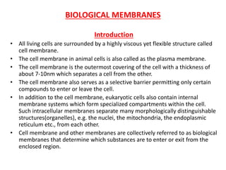
  • 1. BIOLOGICAL MEMBRANES Introduction • All living cells are surrounded by a highly viscous yet flexible structure called cell membrane. • The cell membrane in animal cells is also called as the plasma membrane. • The cell membrane is the outermost covering of the cell with a thickness of about 7-10nm which separates a cell from the other. • The cell membrane also serves as a selective barrier permitting only certain compounds to enter or leave the cell. • In addition to the cell membrane, eukaryotic cells also contain internal membrane systems which form specialized compartments within the cell. Such intracellular membranes separate many morphologically distinguishable structures(organelles), e.g. the nuclei, the mitochondria, the endoplasmic reticulum etc., from each other. • Cell membrane and other membranes are collectively referred to as biological membranes that determine which substances are to enter or exit from the enclosed region.
  • 2. FUNCTIONS OF BIOLOGICAL MEMBRANE 1. It separates two different microenvironments • Separates the intracellular compartment from the extracellular matrix or extracellular fluid. 2. Cell shape • Maintain shape of the cell and define its boundaries, such as in nerves and erythrocytes. 3. Cell movement • Specific arrangement of membrane proteins is critical in controlling the movements of some cells, e.g. movement of neutrophils from the intravascular to the extravascular compartment. 4. Enzymes • Many of the membrane proteins are enzymes and are located either within or on the cell membrane. The inner mitochondrial membrane is essential for localization and correct orientation of the respiratory enzymes within it for their maximum efficiency.
  • 3. 5. Receptor molecules • Membrane proteins act as recognition sites, such as hormone receptors for insulin or glucagon. 6. Translocation of substances • Membrane proteins regulate translocation of molecules, such as amino acids, glucose and various ions. 7. Signal transduction • Various membrane proteins help in the transmission of signals, such as for the transmission of nerve impulses.
  • 4. COMPOSITION OF A BIOLOGICAL MEMBRANE Biological membranes are composed of lipids, proteins and carbohydrates
  • 5. LIPIDS • Lipids form more than 50% of the total membrane constituents. • Membrane lipids comprise of both hydrophobic as well as hydrophilic regions, and thus are termed as amphipathic molecules. • Lipids have a polar head group and a nonpolar tail. • Fatty acids may be both saturated as well as unsaturated. • Phospholipids form the major proportion of the lipid component of cell membranes. • Besides, free/unesterified cholesterol, triacylglycerol and free fatty acids also occur in the membrane. In an aqueous solution, amphiphilic molecules form structurally ordered aggregates, such as micelles and bilayers. These aggregates form the structural basis of biological membranes.
  • 6. MICELLES • A micelle is a spheroidal aggregate where a large number of amphiphillic molecules, e.g. soaps and detergents, are arranged in such a way that their hydrophilic groups interact with the aqueous solvent while the hydrophobic groups are associated at the centre, i.e. away from the solvent. • Micelles are formed when the cross-section of the hydrophilic head group exceeds that of the hydrophobic tails.
  • 7. LIPID BILAYER • Bilayers are formed when the cross- section of the hydrophilic head group of amphiphilic lipids equals that of the hydrophobic tails. • A lipid bilayer exists as a sheet, i.e. an expanded planer-aggregate, in which hydrophobic regions of phospholipids are protected from the aqueous environment while the hydrophilic regions are immerses in water. • These are extremely stable structures which are held together by noncovalent interactions of the hydrocarbon chains and ionic interactions of the charged head groups with water.
  • 8. PROTEINS • Proteins form another major portion of the membrane. • Protein concentration varies from about 20% in the myelin sheath to about 80% in the inner membrane of the mitochondria. • Membrane proteins are classified, roughly by their mode of interaction with the membrane, as integral membrane proteins, peripheral membrane proteins and lipid-linked proteins. • Proteins that span the cell membrane from one side to the other are called integral membrane proteins or intrinsic proteins. These are partially or totally immersed. Glycophorin A is a transmembrane protein which spans the erythrocyte membrane. • Proteins which are embedded on any side of the membrane are called peripheral proteins or extrinsic proteins. These are immersed only partially. Cytochrome c is a peripheral membrane protein associated with the outer surface of the inner mitochondrial membrane. • Some membrane-associated proteins contain covalently attached lipids and are called as lipid-linked proteins. Removal of the lipid fraction from these proteolipids leads to denaturation of the membrane proteins and loss of their biological functions. • Proteolipids are present in many membranes, e.g. lipophilin present in the myelin.
  • 9. CARBOHYDRATES • Carbohydrate content of biological membrane may vary between 3-10%. • Oligosaccharides (short chains of carbohydrates) are covalently attached either to a protein (glycoprotein) or to a lipid (glycolipid). • Oligosaccharide chains are normally located on the outer surface of the membrane or the terminal side of the endoplasmic reticulum.
  • 10. STRUCTURE OF A BIOLOGICAL MEMBRANE • Biological membranes have such a structure where some proteins span the lipid bilayer whereas others are only immersed partially. This is called ‘fluid mosaic’ model because the membrane consists of a mosaic of proteins and lipids which are free to drift about in the plane of the membrane. • In the fluid-mosaic model of biological membrane, the integral proteins are immersed in the lipid bilayer and have specific domains for the ligand binding, for catalytic activity and for the attachment of carbohydrates or lipids. • Peripheral proteins have various modes of attachment. Some apparently, bind to integral proteins, such as an antigen. • There also occurs fluidity in the lipid portion of the membrane in which both the lipids and the proteins move. This is due to the presence of unsaturated fatty acids. • Since cis-double bonds cause fatty acyl chains to bend (i.e. form kink), the membrane thus becomes less tightly packed and therefore is more fluid nature. • The degree of fluidity thus is highly dependent on lipid composition of the membrane. • It changes in response to a change in the diet or physiological state of the animal. • Increased cholesterol and Ca++ decrease membrane fluidity. • Fluidity of a membrane also affects its functions, e.g. it can control activity of the membrane bound enzymes, cell growth and other functions such as phagocytosis.
  • 11. Cholesterol and membrane fluidity • Cholesterol is made up of three basic chemical parts – the steroid nucleus that intercalates between phospholipid hydrocarbon tails, a long hydrocarbon chain located in the non- polar core, and the hydroxyl group that interacts with the polar head groups of phospholipids in the biological membrane. • At high temperature, cholesterol prevents lateral movement of phospholipid hydrocarbon tails thereby preventing an abnormal rise in membrane fluidity. • At low temperature, it prevents the close packing of the same hydrocarbon tails thereby preventing an abnormal fall in membrane fluidity. • Hence, it is aptly said that cholesterol ‘modulates’ membrane fluidity.
  • 12. TRANSPORT ACROSS MEMBRANES • Every living cell must acquire from its surroundings the raw materials for biosynthesis and for energy production, and must release to its environment the byproducts of metabolism. • The lipid bilayer of biological membranes is intrinsically impermeable to ion and polar molecules, yet certain such species must be able to cross these membranes for normal cell function. • A few nonpolar compounds can dissolve in the lipid bilayer and cross the membrane unassisted, but for polar or charged compounds or ions, a membrane protein is essential for transmembrane movement. • In some cases a membrane protein simply facilitates the diffusion of a solute down its concentration gradient, but transport often occurs against a gradient of concentration, electrical charge, or both, in which case solutes must be “pumped” in a process that requires energy. • The energy may come directly from ATP hydrolysis or may be supplied in the form of movement of another solute to down its electrochemical gradient with enough energy to carry another solute up its gradient. • Ions may also move across membranes via ion-channels formed by proteins, or they may be carried across by ionophores, small molecules that mask the charge of the ions and allow them to diffuse through the lipid bilayer. • With very few exceptions, the traffic of small molecules across the plasma membrane is mediated by proteins such as transmembrane channels, carriers, or pumps.
  • 14.
  • 15. FUNCTIONAL MECHANISMS OF TRANSPORT • Transport of a substance across the cell membrane can be described in a functional sense, according to the: Number of molecules transported, and the direction of their movement.  UNIPORT TRANSPORT • It refers to the process which allows the movement of one type of molecules in only one direction, e.g. glucose uptake in erythrocytes.  COTRANSPORT • It refers to the process where transfer of one solute depends upon the simultaneous or sequential transfer of the other. • Two types of molecules when move in the same direction, it is called as symport, e.g. the Na+ - glucose transporter-1(SGLT1) or the Na+ -amino acid transporter in the cells lining the small intestine and proximal renal tubules. • Two types of molecules when move in the opposite direction, it is called as antiport, e.g. the Na+ - K+ transporter, the Na+ - Ca++ transporter.
  • 16. PASSIVE TRANSPORT • Passive transport is the movement of molecules across the cell membrane and does not require energy. • It is dependent on the permeability of the cell membrane. • There are three main kinds of passive transport – Simple Diffusion, Facilitated Diffusion and Osmosis .
  • 17. Simple diffusion • When two aqueous compartments containing unequal concentrations of a soluble compound or ion are separated by a permeable divider (membrane), the solute moves by simple diffusion from the region of higher concentration, through the membrane, to the region of lower concentration, until the two compartments have equal solute concentrations. • When ions of opposite charge are separated by a permeable membrane, there is a transmembrane electrical gradient, a membrane potential, Vm (expressed in volts or millivolts). • This membrane potential produces a force opposing ion movements that increase Vm and driving ion movements that reduce Vm. • Thus the direction in which a charged solute tends to move spontaneously across a membrane depends on both the chemical gradient (the difference in solute concentration) and the electrical gradient (Vm) across the membrane. Together, these two factors are referred to as the electrochemical gradient or electrochemical potential. • Some solutes such as gases (O2, N2, CO2, NO etc.) transport across the cell membrane by diffusing down an electrochemical gradient and do not require metabolic energy. • This passive non-mediated transport is called as simple diffusion. • The direction of flow is always from a higher to a lower concentration and the net movement of a molecule from one side to the other continues until concentration on each side is at chemical equilibrium. • Diffusion of a substance may also occur through transmembrane routes, such as channels or pores present across the membrane protein.
  • 18. • Diffusion of a substance thus involves three major steps, i.e. the solvent must: 1. Leave the environment on one side and enter the membrane, 2. Transverse the membrane, and 3. Leave the membrane so as to enter, a new environment, on the opposite side.
  • 19. Channels • In natural membranes, there are transmembrane channels (pore-like structure) which are composed of proteins. • These channel permit rapid movement of specific ions or molecules from one side of membrane to other. • These channels are formed by integral membrane proteins and selectively allows substances to pass. • The permeability of a channel depends upon the size, the extent of hydration, and the extent of charge density on the ion. • These channels are very selective and in most cases permit the passage of only one type of ions. • Channels are opened transiently, i,e. they are gated. • The flow of ions or molecules through the channel regulates the opening and shutting of the passage-way (gate). • This opening and closing of a membrane channel involves a conformational change subsequent to a change in the voltage (membrane potential) or to the binding of a ligand, such as a chemical agent.
  • 20. Different types of ion channels found in mammalian cells Channel Responsive factor Voltage-gated Change in membrane potential. E.g. Na+, K+ and Ca2+ channels in the heart Ligand-gated A specific extracellular molecule, such as acetylcholine for the aetylcholine receptor channel of the neuromuscular junction. A specific intracellular molecule, e.g. cAMP for Ca2+ channels in myocytes.
  • 21.
  • 22. Ligand gated channels • These channels are gated in response to the binding of some extra or intracellular molecule. • The binding of some extracellular molecules, termed as agonists, controls opening of a channel, e.g. the nicotineacetylcholine channel, also referred to as acetylcholine receptor. • Acetylcholine, a neurotransmitter, is released at the neuromuscular junction by a neuron when electrically excited. • It then diffused to the skeletal muscle membrane and interacts with acetylcholine receptors. • The binding of acetylcholine with its receptor in turn opens the channel and allows selective cations to move across the membrane. • Some channels are regulated by specific intracellular regulatory molecules, such as cAMP. • Generation of cAMP in the cell usually activates protein kinase A. • The liberated catalytic subunit phosphorylates some of the proteins to produce a cellular effect. • A number of pharmacologic agents that modulate these channels are used therapeutically.
  • 23. Channelopathies Mutations in genes encoding polypeptide constituents of ion channel may lead to certain diseases termed as channelopathies, e.g. myasthenia gravis and cystic fibrosis. Myasthenia gravis • It is an acquired autoimmune disease characterised by muscle weakness due to decreased neuromuscular signal transmission. • Autoantibodies against acetylcholine receptors accelerate their turnover and reduce their number. • Acetylcholineesterase-inhibitor drugs are given to enhance the stay of acetylcholine at the neuromuscular junction. • Ultimately, the patients require the removal of the culprit antibodies from the plasma at regular intervals, a process called ‘plasmapheresis’. Cystic fibrosis • It is a multiorgan disease but its gene product is a cystic fibrosis transmembrane conductance regulator (CFTR), which is a cAMP- dependent Cl- channel. • Patients with cystic fibrosis have reduced permeability which in turn impairs fluid and electrolyte secretion and leads to luminal dehydration.
  • 24. Movement of water across biological membranes • Water can move rapidly in and out of cells, but the partition coefficient of water into lipids is low; therefore, the permeability of the membrane lipid bilayer for water is also low. • Specific membrane proteins that function as water channels explain the rapid movement of water across the plasma membrane. These water channels are small integral membrane proteins known as aquaporins (AQP). • Many different forms have been discovered so far; at least six forms are expressed in cells in the kidneys and seven forms in the gastrointestinal tract, tissues in which water movement across plasma membranes is particularly rapid.
  • 25. Nephrogenic Diabetes Insipidus • In the kidneys, aquaporin-2 (AQP2) channels are abundant in the collecting ducts and are the target of the hormone arginine vasopressin, also known as antidiuretic hormone (ADH). • This hormone increases water transport in the collecting ducts by stimulating the insertion of AQP2 proteins into the apical plasma membrane. • Defects in AQP2 plays a critical role in inherited and acquired disorders of water reabsorption by the kidney. • For example, diabetes insipidus is a condition in which the kidney losses its ability to reabsorb water properly, resulting in excessive loss of water and excretion of a large volume of dilute urine (polyuria). • Although inherited forms of diabetes insipidus are relatively rate, it can develop in patients receiving chronic lithium therapy for psychiatric disorders, giving rise to the term lithium-induced polyuria. • Both of these conditions are associated with a decrease in the number of AQP2 proteins in the collecting ducts of the kidney and are therefore called nephrogenic diabetes insipidus.
  • 26. Ionophores • Ionophores are small organic molecules such as antibiotics which are synthesized by some bacteria and function as shuttles. • Ionophores increase permeability of the membrane to a particular ion by binding the ion, diffusing it through the membrane and releasing it on the other side. • To ensure the net transport, uncomplexed ionophores return to the original side of the membrane and are ready to repeat the process. • Because of their ability to complex specific ions and facilitate their transport, ionophores contain hydrophilic centres for ion-binding and are surrounded by pheripheral hydrophobic regions. • This in turn allows the molecules to dissolve effectively in the membrane and diffuse through it. • The net diffusion of a substance thus depends upon: I. Its concentration gradient across the membrane. II. The electric potential across the membrane. III. The permeability coefficient of the substance for the membrane. IV. The hydrostatic pressure gradient across the membrane, and V. temperature
  • 27. • Each ionophore, e.g. valinomycin or nigericin has a definite ion specificity. • Valinomycin translocates K+ by an electronegative import mechanism. • Nigericin is an electrically neutral antiporter which translocates K+ in exchange for H+ across the membrane. • Both ionophores act as mobile carriers that diffuse back and forth across the membrane, carrying ions from one side to the other.
  • 28. Carrier-Mediated Transport • Molecules that cannot freely diffuse through the lipid bilayer membrane by themselves, need to be transported in association with specific carrier molecules. This process is known as mediated transport or carrier-mediated transport. • The carrier molecules are variously designated as carriers, permeases, porters, translocases or transporters. Transport proteins • Transport proteins, also called as transporters, are proteins that translocate a molecule or ion across the membrane by binding to and physically moving it. • These are the integral membrane proteins involved in both passive and active transport by binding a specific substances on one side of the membrane. • Most transport proteins have a high degree of structural stereospecificity for the substance to be transported. • They demonstrate saturation kinetics, i.e. when binding sites on all the transport proteins are occupied, the system is saturated and the rate of transport reaches a plateau. • They can be inhibited by both competitive and non-competitive inhibitors. The inhibition can prevent transport by blocking the binding sites or by interacting with the transport protein and altering its conformation so that it becomes non- functional.
  • 29. FACILITATED DIFFUSION • Facilitated diffusion, leads to the translocation of solutes through membrane transport proteins without the expenditure of metabolic energy. • The process can operate either unidirectionally or bidirectionally and the net flux across the membrane occurs down a concentration gradient, i.e. the molecules flow from a higher to the lower concentration. • A ‘ping-pong’ mechanism explains facilitated diffusion of molecules across the biological membrane with the help of a transport protein.
  • 31. • A ‘ping-pong’ mechanism is put forth to explain the occurrence of facilitated diffusion. • According to this mechanism, a transport (carrier) protein exists in two conformations. • A transport protein, in the ‘pong’ state in the lipid bilayer, is exposed to high concentrations of the solute, and the molecules of the solute bind to specific sites on the carrier protein. • This results in a conformational change to the ‘ping’ state which ensures that the solute is released towards the other side of the membrane. This process is completely reversible. • The transport of a solute molecule mediated by a transporter protein thus has four aspects: 1. Recognition • Transport proteins have receptor sites to which the solute attaches. The transporter thus recognizes an appropriate solute form the aqueous environment for its translocation across the membrane. 2. Translocation • After binding of the solute with the receptor protein, there occurs a conformational change in the transporter which translocates (moves) the solute molecule a short distance but into the new environment.
  • 32. 3. Release • A change in the conformation of the transporter protein decreases affinity of the solute and releases it to the new environment. 4. Recovery • After release of the solute, the transport protein reverts to its original conformation to accept another solute molecule, i.e. the transporter is recovered in its original conformation. • Several hormones (such as insulin, glucocorticoids, growth hormone etc.) regulate facilitated diffusion by changing the number of transport proteins available. • Examples of transport proteins which mediate facilitated diffusion, include glucose transporters, anion transporters, etc. • A group of transport proteins have been identified in the plasma membrane of mammalian cells for the transport of D-sugars by a uniport mechanism. • Insulin increases glucose transport in muscle and adipose tissue; amino acid transport in liver and other tissues.
  • 33. OSMOSIS • Osmosis (Greek : push) refers to the movement of solvent (most frequently water) through a semipermeable membrane. • The flow of solvent occurs form a solution of low concentration (dilute solution) to a solution of high concentration (concentrated solution), when both are separated by a semipermeable membrane. • Osmosis is a colligative property i.e. a character which depends on the number of solute particles and not their nature. • The movement of water in the body occurs through osmosis, and does not require energy (ATP). • Certain medical and health complications are due to disturbances in osmosis. E.g. edema, diarrhoea, cholera etc.
  • 34. ACTIVE TRANSPORT • In active transport, the transport protein moves a specific molecule against the concentration gradient i.e. from a lower concentration to the higher concentration. • It is a process that requires energy which, in most cases is coupled to the hydrolysis of ATP. • The active transport can be grouped as 1. Primary active transport 2. Secondary active transport
  • 35. PRIMARY ACTIVE TRANSPORT • This transport system has the same characteristics as the passive transport system but it is an endergonic process. • Examples of such transporters include membrane-bound ATPases that translocate cations. • They are further classified as: I. P type transporters • The transporter protein is phosphorylated and dephosphorylated during the transport activity. II. V type transporters • These are present in the membrane of the lysosomes, the golgi visicles and the secretory vesicles. • These are responsible for acidification of the interior of these vesicles. III. F type transporters • These are present in mitochondria and are involved in ATP synthesis. • Primary active transport systems are important in the maintenance of electrochemical gradient in biological systems and consume nearly one-third of the total energy expenditure of the cell, with the hydrolysis of intracellular ATP. • Examples of primary active transport system include Na+/K+ - ATPase, H+/K+-ATPase and neurotransmitters.
  • 36. Na+ - K+ PUMP • The cells have a high intracellular K+ concentration and a low Na+ concentration. • The Na+ - K+ pump is responsible for the maintenance of high K+ and low Na+ concentration in the cells. • This is brought about by an integral plasma membrane protein, namely the enzyme Na+ - K+ ATPase. • Na+ - K+ ATPase pumps 3 Na+ ions from inside the cell to outside and brings 2 K+ ions from the outside to the inside with a concomitant hydrolysis of intracellular ATP. • Drugs that inhibit Na+ - K+ pump are 1. Ouabain • It is a steroid derivative and inhibits Na+ - K+ ATPase . 2. Digoxin • It is a steroid glycoside and an inhibitor of Na+ - K+ ATPase. • It is used in the treatment of cardiac failure.
  • 37. Na+ - K+ PUMP and Heart failure • The cell membrane of cardiomyocytes, i.e. cells of myocardium, contain many transport pumps. Two of them are, Na+ - K+ ATPase and Na+/C++ exchanger. • The Na+ - K+ ATPase serves its usual function of maintaining low intracellular Na+ concentrations. • The Na+/C++ exchanger relies on this Na+ gradient to extrude C++ out of the cells. • Cardiac glycosides such as digoxin and ouabain abolish this gradient by inhibiting the Na+ - K+ ATPase . • High intracellular Na+ concentration slows the extrusion of C++ by the Na+/C++ exchanger. • Increased availability of C++ results in increased force of contraction that is clinically useful in the management of cardiac failure.
  • 38. Ca2+ - ATPase • Ca2+ is an important intracellular messenger referred to as a second messenger. • It regulates various cellular processes such as muscle contraction, release of neurotransmitters and glycogen breakdown. • It is also important activator of oxidative metabolism. • In order to maintain low cytosolic Ca2+ concentration, it is actively transported out of the cell across the plasma membrane, the endoplasmic reticulum or the sarcoplasmic reticulum. • The Ca2+ -ATPase (Ca2+ pump) actively pumps two Ca2+ out of the cytosol at the expense of ATP hydrolysis. • The mechanism of Ca2+ -ATPase resembles that of the Na+ - K+ ATPase . • In eukaryotes, the Ca2+ -transporter is regulated by the cytosolic Ca2+ level through a calcium binding protein termed calmodulin.
  • 39. H+/K+ - ATPase (proton pump) • Cells in the gastric mucosa secrete HCl. • The secreated protons (H+) are derived from the intracellular hydration of CO2 by carbonic anhydrase. • The secretion of H+ involves an H+/K+ - ATPase, also called the proton pump. • This is an antiport with structure and properties similar to Na+ - K+ ATPase. • As H+ is pumped out, the K+ which enters the cell is subsequently externalized by its cotransport with the Cl-. • The oeverall transported product therefore is HCL. • Inhibition of H+/K+ - ATPase is of clinical importance.
  • 40. Proton pump and Peptic ulcer • Excess production of HCL along with the failure of mucosal defence mechanisms, can damage the gastric mucosa and may lead to peptic ulcer. • The H+/K+ - ATPase of the gastric mucosa is activated by histamine stimulation of the cell surface receptor. • Compounds, such as Cimetidine and its analogs (antihistamines) bind to histamine receptors. • These drugs block the process by competing with histamine for its binding to the receptor an in turn reduce HCl production. • Histamine analogs are therefore widely used to alleviate the painful and otherwise fatal symptoms of peptic ulcer. • Proton pump inhibitors such as Omeprazole, are also used in the treatment of peptic ulcer. • They are selective inhibitors of H+/K+ - ATPase and are therefore more powerful than the antihistamines. • It is now recognized that many ulcers are caused by infection with the bacteria Helicobacter pylori and can better be cured by the use of antibiotics besides a reduction in acidity.
  • 41. SECONDARY ACTIVE TRANSPORT • It is also called ion gradient driven active transport. • In this process, free energy of the electrochemical gradient, generated by an ion-pumping ATPase, drives the transport of another substance (a neutral molecule), such as a sugar or an amino acid, against its concentration gradient. • e.g. Na+ - glucose transport system Fig. Na+ - glucose transport system
  • 42. Na+ - glucose transport system • Glucose enters the intestinal epithelial cells by active transport using the electrogenic Na+ - glucose cotransport system (SGLT1) in the apical membrane. • This increases the intracellular glucose concentration above the blood glucose concentration, and the glucose molecules move passively out of the cell and into the blood via an equilibrating carrier mechanism (GLUT2) in the basolateral membrane. • The intestinal GLUT2, like the erythrocyte GLUT1, is a sodium-independent transporter that moves glucose down its concentration gradient. • However, unlike GLUT1, the GLUT2 transporter can accept other sugars, such as galactose and fructose for absorption. • The Na+/K+ - ATPase that is located in the basolateral membrane pumps out the sodium ions that enter the cell with the glucose molecules via SGLT1. • The polarized organization of the epithelial cells and the integrated functions of the plasma membrane transporters form the basis by which cells accomplish transcellular movement of both glucose and sodium ions, and is also exploited clinically. • In short, the successful uptake of glucose and sodium (symport) is ‘secondarily’ dependent on the Na+ gradient maintained by the primary active Na+/K+ - ATPase .
  • 43. ORAL REHYDRATION SOLUTION • The administration of oral rehydration solution (ORS) has dramatically reduced the mortality resulting from cholera and other diseases that involve extreme losses of water/solutes from the gastrointestinal tract. • The main ingredients of ORS are glucose, NaCl or NaHCO3, KCl and water. • The glucose and Na+ are reabsorbed by the sodium-glucose transporter-1 (SGLT1) in the apical membrane of enterocytes, i.e. epithelial cells lining the lumen of the small intestine. • Transfer of solutes on the basolateral aspect of the enterocytes increases the osmolarity compared with the luminal osmolarity thereby favouring the osmotic absorption of water. • In this manner, the absorption of glucose accompanied by the obligatory increase in absorption of NaCl and water, help to compensate for the diarrhoeal losses of water/solutes, i.e. dehydration.
  • 44. Comparison between facilitated diffusion and active transport Parameter Facilitated diffusion Active transport Specific binding site Present Present Saturation kinetics Yes Yes Inhibition by structural analogs Yes Yes Direction of operation Uni or bidirectional Unidirectional Mode of operation Along electrical/chemical gradient Against electrical/chemical gradient Energy dependent No yes
  • 45.
  • 46. TRANSPORT OF MACROMOLECULES (vesicular translocation) The transport of macromolecules such as proteins, polysaccharides and polynucleotides across the membrane is brought about by two independent mechanisms namely endocytosis and exocytosis. ENDOCYTOSIS • It is a mechanism for the uptake of macromolecules by the cells (e.g. uptake of LDL by cells). • In this process, a region of the plasma membrane invaginates, enclosing a small volume of the extracellular fluid and its contents within a bud, and generates endocytotic vesicles. • The vesicle then pinches-off, as fusion of the plasma membrane seals the neak of the vesicle at the original site of invagination. • The resulting small vesicle is called an endosome. • It moves into the interior of the cell and delivers its contents to some other organelle, bound by a single membrane, e.g. a lysosome, by fusion of the two membranes. • The ‘hybrid vesicle’ is called a secondary lysosome.
  • 47. • Due to the presence of hydrolytic enzymes , the macromolecular contents are digested to their monomers, such as amino acids, simple sugars or nucleotides, which then diffuses out of the vesicle in the cytoplasm. • There are two general types of processes referred to as endocytosis, i.e. phagocytosis and pinocytosis.
  • 48. Phagocytosis • Phagocytosis (or cell eating) occurs only in specialized cells like macrophages and granulocytes for the ingestion of large particles, such as bacteria, viruses etc. Pinocytosis • Pinocytosis (or cell drinking) leads to cellular uptake of fluid and its contents as a result of invagination of the plasma membrane. • The receptor mediated pinocytosis is a very selective type of pinocytosis that occurs in coated pits, lined with the protein clathrin, resulting in the formation of the clathrin- coated vesicles. • The high affinity receptors permit selective concentration of the ligand from the medium, e.g. LDL, transferrin etc. and the receptors are subsequently internalized by means of the coated pits containing the receptors. • The coated vesicle may fuse with lysosomes, the contents are digested and clathrin is recycled back to the membranes. • Sometimes, in case of some hormones, clathrin is not required for receptor-mediated pinocytosis. The internalized vesicle fuses with another organelle such as golgi complex, i.e. no secondary lysosomes are formed. The process is known as adsorptive pinocytosis.
  • 49. EXOCYTOSIS • Exocytosis is the reverse of endocytosis. • It involves contact of two inside surface monolayers from the cytosolic side and release of macromolecules to the exterior of a cell. • A secretory vesicle in the cytoplasm, originating in the golgi complex or the endoplasmic reticulum, moves to the inner surface of the plasma membrane and fuses with it, releasing the vesicular contents outside the membrane. • The secreated/exocytosed molecules may have either of three possible fates: 1. They become a part of the cell membrane surface, e.g. antigens. 2. They become a part of the extracellular matrix, e.g. collagens. 3. They enter the blood and carried to distant sites, e.g. hormones like insulin. • In some diseases characterized by uncontrolled cell division, vesicles may be thrown out to the cell exterior, and contain molecules actually meant for intracellular use only. • The process is not a true exocytosis and such vesicles are not true secretory vesicles. They are called exosomes.
  • 50. • The exosomes can be isolated from the blood and the transcriptome subjected to reverse transcription polymerase chain reaction for detecting DNA mutations. • Such blood-based detection of mutations is very valuable in diagnosing tumors where tissue availability is limited, as in case of cancers of lungs, pancreas and ovaries.

Notas do Editor

  1. Cross section : a surface or shape exposed by making a straight cut through something, especially at right angles to an axis.
  2. Ligand : an ion or molecule attached to a metal atom by coordinate bonding.
  3. Acetylcholinesterase (HGNC symbol ACHE), also known as AChE or acetylhydrolase, is the primary cholinesterase in the body. It is an enzyme that catalyzes the breakdown of acetylcholine and of some other choline esters that function as neurotransmitters.
  4. Mucosal defence mechanism : In the stomach several mucosal defence mechanisms protect the stomach against hydrochloric acid and noxious agents. The pre-epithelial protection is made up by the mucus-bicarbonate barrier. Peptic ulcer : An ulcer in the lining of the duodenum, the lower end of the esophagus, or the stomach (usually along the lesser curvature). Peptic ulcer disease is a common illness, affecting about 10% of men and 5% of women during their lifetimes. Common causes of peptic ulcer are factors that increase gastric acid priduction or impair mucosal barrier protection, such use of salicylates and nonsteroidal anti-inflammatory drugs (NSAIDS), tobacco smoking, Helicobacter pylori infection or the upper gastrointestinal tract, pathologic hypersecretory disorders, consumption of alcohol and coffee and severe physiological stress. Alleviate : To lessen the effect of.
  5. Enterocyte : A nutrient absorbing cell located on the surface of the small intestinal villus. Its free surface cell membrane is folded into microvilli that increase the surface area available for absorption. Osmolarity : The concentration of a solution expressed as the total number of solute particles per litre.
  6. Pinches off : to press (something, esp. flesh) tightly between two surfaces, esp. between a finger and the thumb (see nip1) 2 to confine, squeeze, or painfully press (toes, fingers, etc.)
  7. Transcriptome : The sum total of all the messenger RNA molecules expressed from the genes of an organism.