SlideShare uma empresa Scribd logo
1 de 16
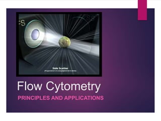
Flow Cytometry
PRINCIPLES AND APPLICATIONS
What is Flow Cytometry?
‘Flow Cytometry’ as the name suggests is a technique for cell
counting and measurement of different properties of the cell
(‘cyto’= cell; ‘metry’=count/measurement).
It is a laser based technology that measures and analyses
different physical and chemical properties of the cells/particles
flowing in a stream of fluid through a beam of light.
References:
http://www.d.umn.edu/~biomed/flowcytometry/introflowcytometry.pdf
A Flow Cytometer
Place your
sample here
Historical Perspective: Evolution of Flow
Cytometry
17th
Century
1934
1947 to
1949
1953 19651879 1968
1970s
onwards…
Development of
light microscope
by
Leeuwenhoek.
Principles of Droplet
formation by Lord
Rayleigh.
Counting of RBCs
by Moldavan by
forcing a
suspension of cells
through capillary
tube.
Development of
Coulter Principle by
Wallace Coulter and
counting of RBCs
using the first Coulter
Counter.
Optical counting of
RBCs by Crosland-
Taylor by use of
laminar flow
principles
Development of
electrostatic
inkjet droplet
deflection by
Richard Sweet
Application of
Sweet’s principle
and Coulter principle
to develop the first
cell sorter by M.
Fulwyler
Development of
fluorescence
based cell sorter
by Wolfgang
Gohde
Development of
FACS and other
advances.
Principles of working of Flow
Cytometer
Coulter
Principle
Principle
s of
Laminar
Flow
Electro
statics
Optics &
Light
Scatteri
ng
Flow
Cytome
try
Components of a Flow Cytometer
A flow cytometer is made up of three main systems: fluidics, optics, and
electronics.
 The fluidics system transports particles in a stream to the laser beam for
interrogation.
 The optics system consists of lasers to illuminate the particles in the
sample stream and optical filters to direct the resulting light signals to the
appropriate detectors.
 The electronics system converts the detected light signals into electronic
signals that can be processed by the computer. For some instruments
equipped with a sorting feature, the electronics system is also capable of
initiating sorting decisions to charge and deflect particles.
References:
http://www.d.umn.edu/~biomed/flowcytometry/introflowcytometry.pdf
Working of a Flow Cytometer
 In the flow cytometer, particles are carried to the laser intercept in a fluid
stream. Any suspended particle or cell from 0.2–150 micrometers in size is
suitable for analysis.
 The portion of the fluid stream where particles are located is called the sample
core.
 When particles pass through the laser intercept, they scatter laser light. Any
fluorescent molecules present on the particle fluoresce.
 The scattered and fluorescent light is collected by appropriately positioned
lenses.
 A combination of beam splitters and filters steers the scattered and fluorescent
light to the appropriate detectors.
 The detectors produce electronic signals proportional to the optical signals
striking them.
References:
http://www.d.umn.edu/~biomed/flowcytometry/introflowcytometry.pdf
Applications of Flow
Cytometry
Flow cytometry is the sine qua non (without which, nothing)of the modern
researcher’s toolbox.
Flow cytometry measures multiple characteristics of individual particles
flowing in single file in a stream of fluid. Light scattering at different angles can
distinguish differences in size and internal complexity, whereas light emitted
from fluorescently labeled antibodies can identify a wide array of cell surface
and cytoplasmic antigens. This approach makes flow cytometry a powerful
tool for detailed analysis of complex populations in a short period of time.
References:
Kuby Immunology, 7th Edition
http://www.clinchem.org/content/46/8/1221.full
Applications
Immunophenotyping
Cell subsets are measured by labeling
population-specific proteins with a fluorescent
tag on the cell surface. In clinical labs,
immunophenotyping is useful in diagnosing
hematological malignancies such as
lymphomas and leukemia.
Cell Sorting
The cell sorter is a specialized flow
cytometer with the ability to physically
isolate cells of interest into separate
collection tubes. The sorter uses
sophisticated electronics and fluidics to
identify and "kick" the cells of interest out of
the fluidic stream into a test tube.
DNA Content Analysis
The measurement of cellular DNA content by
flow cytometry uses fluorescent dyes, such as
propidium iodide, that intercalate into the DNA
helical structure. The fluorescent signal is directly
proportional to the amount of DNA in the nucleus
and can identify gross gains or losses in DNA.
Cell Cycle Analysis
Flow cytometry can analyze replication
states using fluorescent dyes to measure
the four distinct phases of the cell cycle.
Along with determining cell cycle replication
states, the assay can measure cell
aneuploidy associated with chromosomal
abnormalities.
Apoptosis
The two distinct types of cell death,
apoptosis and necrosis, can be
distinguished by flow cytometry on the basis
of differences in morphological, biochemical
and molecular changes occurring in the
dying cells.
Cell Proliferation Assays
The flow cytometer can measure proliferation by
labeling resting cells with a cell membrane
fluorescent dye, carboxyfluorescein succinimidyl
ester (CFSE). When the cells are activated, they
begin to proliferate and undergo mitosis. As the
cells divide, half of the original dye is passed on
to each daughter cell. By measuring the
reduction of the fluorescence signal, researchers
can calculate cellular activation and proliferation.
References:
http://www.clinchem.org/content/46/8/1221.full
http://www.seattlechildrens.org/research/cores/flow-cytometry/applications-of-flow-cytometry/
Fluorescence Activated Cell
Sorting (FACS)
 Consider a group of lymphocytes from a mouse that have been stained with
green fluorescent antibodies specific for CD4 (e.g., fluorescein isothiocyanate,
or FITC anti-CD4) and red fluorescent antibodies specific for CD8 (e.g.,
phycoerythrin, or PE anti-CD8).
 Both the labeled cells generate SSC and FSC as they pass through the laser
beam creating voltage pulses that are recorded by the computer.
 However, each labeled cell will also emit light of specific wavelength as a
result of the fluorescent label. For instance, CD4 cells will emit green
fluorescent light of wavelength 525-530 nm while CD8 cells emit orange light
of wavelength 560 nm. These fluorescent signals pass through the
Photomultiplier tubes and generate voltage pulses.
 The software integrates all the information for a particular cell allowing
characterization of individual cells.
References:
Kuby Immunology, 7th Edition
Clinical Applications: DNA
Content Analysis
 Investigators are currently using techniques of DNA flow cytometry to measure ploidy status
(DNA content) and proliferative potential (S phase fraction) in a wide variety of solid tumors.
These measurements have shown relevance for diagnosis, prognosis, and treatment for
patients with cancer.
 The measurement of cellular DNA content by flow cytometry uses fluorescent dyes, such as
propidium iodide, that intercalate into the DNA helical structure. The fluorescent signal is
directly proportional to the amount of DNA in the nucleus and can identify gross gains or losses
in DNA.
 Abnormal DNA content, also known as “DNA content aneuploidy”, can be determined in a tumor
cell population. DNA aneuploidy generally is associated with malignancy; however, certain
benign conditions may appear aneuploid.
 Cell Cycle Analysis: This technique is based on the premise that cells in G0 or G1 phases of
the cell cycle possess a normal diploid chromosomal, and hence DNA content (2n) whereas
cells in G2 and just prior to mitosis (M) contain exactly twice this amount (4n). As DNA is
synthesized during S-phase, cells are found with a DNA content ranging between 2n and 4n. A
histogram plot of DNA content against cell numbers gives the classical DNA profile for a
proliferating cell culture.
References:
http://europepmc.org/abstract/med/2645625
http://www.clinchem.org/content/46/8/1221.full
http://www.icms.qmul.ac.uk/flowcytometry/uses/cellcycleanalysis/cellcycle/index.html
Flow Cytometry and Ecology
 Assessments of diversity, abundance, and activity of water column
microorganisms are fundamental to studies in aquatic microbiology.
 Currently, most applications of flow cytometry to environmental samples make
use of various morphological and physiological characteristics of the cells (e.g.,
size and pigment content of photosynthetic organisms).
 These criteria generally are not sufficient for identification at the genus or
species level. Staining with DNA-specific fluorochromes offers information
about numbers of bacterial cells but not about their identity.
 The combined use of dyes that bind preferentially to G- C or A. T base pairs
has been used to distinguish organisms of different G+C content
References:
Appl.%20Environ.%20Microbiol.-1990-Amann-1919-25.pdf
Flow Cytometry and Cancer
Research
 The prognosis of patients with cancer is largely determined by the specific histological diagnosis,
tumor mass stage, and host performance status.
 Quantitative cytology in the form of flow cytometry has greatly advanced the objective elucidation of
tumor cell heterogeneity by using probes that discriminate tumor and normal cells and assess
differentiate as well as proliferative tumor cell properties.
 Both DNA content analysis and FACS can be utilised in cancer research.
 Abnormal nuclear DMA content is a conclusive marker of malignancy and is found with increasing
frequency in leukemia (23% among 793 patients), in lymphoma (53% among 360 patients), and in
myeloma (76% among 177 patients), as well as in solid tumors (75% among 3611 patients), for an
overall incidence of 67% in 4941 patients.
 Flow cytometric immunophenotyping (FCI) aids in the differentiation of chronic lymphocytic leukemia
(CLL) from mantle cell lymphoma (MCL); however, overlapping phenotypes may occur. CD11c
expression has been reported in up to 90% of CLL cases but has rarely been reported in MCL.
Whether CD11c can be used to exclude MCL has not been directly addressed. FCI reports were
reviewed for 90 MCL cases (44 patients) and 355 CLL/small lymphocytic lymphoma (SLL) cases (158
patients).
References:
http://ajcp.ascpjournals.org/content/134/2/271.full.pdf+html
http://cancerres.aacrjournals.org/content/43/9/3982.full.pdf+html
References
 http://flowcytometry.berkeley.edu/pdfs/Basic%20Flow%20Cytometry.pdf
 http://www.azom.com/article.aspx?ArticleID=6020
 https://www.beckmancoulter.com/wsrportal/wsr/industrial/particle-technologies/coulter-principle/index.htm
 http://www.cyto.purdue.edu/cdroms/cyto2/6/coulter/ss000103.htm
 http://ajcp.ascpjournals.org/content/134/2/271.full.pdf+html
 http://cancerres.aacrjournals.org/content/43/9/3982.full.pdf+html
 Appl.%20Environ.%20Microbiol.-1990-Amann-1919-25.pdf
 http://europepmc.org/abstract/med/2645625
 http://www.clinchem.org/content/46/8/1221.full
 http://www.icms.qmul.ac.uk/flowcytometry/uses/cellcycleanalysis/cellcycle/index.html
 Kuby Immunology, 7th Edition
 http://www.clinchem.org/content/46/8/1221.full
 http://www.seattlechildrens.org/research/cores/flow-cytometry/applications-of-flow-cytometry
 http://www.d.umn.edu/~biomed/flowcytometry/introflowcytometry.pdf
Thank You.

Mais conteúdo relacionado

Mais procurados

Flow cytometry and fluorescence activated cell sorting (FACS)
Flow cytometry and fluorescence activated cell sorting (FACS)Flow cytometry and fluorescence activated cell sorting (FACS)
Flow cytometry and fluorescence activated cell sorting (FACS)Abu Sufiyan Chhipa
 
fish- Fluorescence in situ hybridization
fish- Fluorescence in situ hybridization fish- Fluorescence in situ hybridization
fish- Fluorescence in situ hybridization gaurav raja
 
Flow Cytometry - basics, principles and applications
Flow Cytometry - basics, principles and applicationsFlow Cytometry - basics, principles and applications
Flow Cytometry - basics, principles and applicationsAnkit Raiyani
 
Agarose Gel Electrophoresis
Agarose Gel ElectrophoresisAgarose Gel Electrophoresis
Agarose Gel ElectrophoresisHarshit Jadav
 
Flow cytometry definition, principle, parts, steps, types, uses
Flow cytometry definition, principle, parts, steps, types, usesFlow cytometry definition, principle, parts, steps, types, uses
Flow cytometry definition, principle, parts, steps, types, usesGayathri Devi S
 
Flow cytometry
Flow cytometryFlow cytometry
Flow cytometryprachann
 
Radio immunoassay (RIA)
Radio immunoassay (RIA)Radio immunoassay (RIA)
Radio immunoassay (RIA)MehulJain143
 
Immunodiffusion principles and application
Immunodiffusion principles and  applicationImmunodiffusion principles and  application
Immunodiffusion principles and applicationsuniu
 
Mtt Assay for cell viability
Mtt Assay for cell viabilityMtt Assay for cell viability
Mtt Assay for cell viabilitysakeena gilani
 
Characterization of Cell Line
Characterization of Cell LineCharacterization of Cell Line
Characterization of Cell LineVarij Nayan
 
Insuite hybridization
Insuite hybridizationInsuite hybridization
Insuite hybridizationNoman Ch
 
Pcr and its types
Pcr and its typesPcr and its types
Pcr and its typesnirvarna gr
 
Presentation on Fluorescence in-Situ Hybridization (FISH)
Presentation on Fluorescence in-Situ Hybridization (FISH)Presentation on Fluorescence in-Situ Hybridization (FISH)
Presentation on Fluorescence in-Situ Hybridization (FISH)Dr. Kaushik Kumar Panigrahi
 

Mais procurados (20)

Flow cytometry and fluorescence activated cell sorting (FACS)
Flow cytometry and fluorescence activated cell sorting (FACS)Flow cytometry and fluorescence activated cell sorting (FACS)
Flow cytometry and fluorescence activated cell sorting (FACS)
 
fish- Fluorescence in situ hybridization
fish- Fluorescence in situ hybridization fish- Fluorescence in situ hybridization
fish- Fluorescence in situ hybridization
 
Flow Cytometry - basics, principles and applications
Flow Cytometry - basics, principles and applicationsFlow Cytometry - basics, principles and applications
Flow Cytometry - basics, principles and applications
 
Agarose Gel Electrophoresis
Agarose Gel ElectrophoresisAgarose Gel Electrophoresis
Agarose Gel Electrophoresis
 
Flow cytometry definition, principle, parts, steps, types, uses
Flow cytometry definition, principle, parts, steps, types, usesFlow cytometry definition, principle, parts, steps, types, uses
Flow cytometry definition, principle, parts, steps, types, uses
 
Flow Cytometry (FCM)
Flow Cytometry (FCM) Flow Cytometry (FCM)
Flow Cytometry (FCM)
 
Flow cytometry
Flow cytometryFlow cytometry
Flow cytometry
 
Immunoelectrophoresis
ImmunoelectrophoresisImmunoelectrophoresis
Immunoelectrophoresis
 
Flow cytometry
Flow cytometryFlow cytometry
Flow cytometry
 
Radio immunoassay (RIA)
Radio immunoassay (RIA)Radio immunoassay (RIA)
Radio immunoassay (RIA)
 
Immunodiffusion principles and application
Immunodiffusion principles and  applicationImmunodiffusion principles and  application
Immunodiffusion principles and application
 
Mtt Assay for cell viability
Mtt Assay for cell viabilityMtt Assay for cell viability
Mtt Assay for cell viability
 
Characterization of Cell Line
Characterization of Cell LineCharacterization of Cell Line
Characterization of Cell Line
 
Cell culture media
Cell culture mediaCell culture media
Cell culture media
 
Immunofluorescence
ImmunofluorescenceImmunofluorescence
Immunofluorescence
 
Flow cytometry
Flow cytometry Flow cytometry
Flow cytometry
 
Insuite hybridization
Insuite hybridizationInsuite hybridization
Insuite hybridization
 
Pcr and its types
Pcr and its typesPcr and its types
Pcr and its types
 
Presentation on Fluorescence in-Situ Hybridization (FISH)
Presentation on Fluorescence in-Situ Hybridization (FISH)Presentation on Fluorescence in-Situ Hybridization (FISH)
Presentation on Fluorescence in-Situ Hybridization (FISH)
 
Western blotting
Western blottingWestern blotting
Western blotting
 

Semelhante a Flow cytometry: Principles and Applications

Rohan gupta 2015 b1ab651p facs
Rohan gupta 2015 b1ab651p   facsRohan gupta 2015 b1ab651p   facs
Rohan gupta 2015 b1ab651p facsRohan Gupta
 
Flow Cytometry- Presentation
Flow Cytometry- PresentationFlow Cytometry- Presentation
Flow Cytometry- PresentationBIDISHA MANDAL
 
Flow cytometry in diagnostics
Flow cytometry in diagnosticsFlow cytometry in diagnostics
Flow cytometry in diagnosticsKaberi Nath
 
Presentation on flow cytometry1
Presentation on flow cytometry1Presentation on flow cytometry1
Presentation on flow cytometry1Nagendra sharma
 
Flow cytometry ready
Flow cytometry readyFlow cytometry ready
Flow cytometry readyYra Yunus
 
Applications of Flow Cytometry | Cell Analysis
Applications of Flow Cytometry | Cell AnalysisApplications of Flow Cytometry | Cell Analysis
Applications of Flow Cytometry | Cell AnalysisUniversity of The Punjab
 
Flow Cytometry: Guide to Understanding Applications and Benefits | The Lifesc...
Flow Cytometry: Guide to Understanding Applications and Benefits | The Lifesc...Flow Cytometry: Guide to Understanding Applications and Benefits | The Lifesc...
Flow Cytometry: Guide to Understanding Applications and Benefits | The Lifesc...The Lifesciences Magazine
 
An introduction to flow cytometry- Ashwini.R
An introduction to flow cytometry- Ashwini.RAn introduction to flow cytometry- Ashwini.R
An introduction to flow cytometry- Ashwini.RAshwini R
 
Analytical chemistry 2013 qian liu
Analytical chemistry 2013 qian liuAnalytical chemistry 2013 qian liu
Analytical chemistry 2013 qian liuQian Liu, phD
 
Medcrave Group - Microfluidic technologies
Medcrave Group - Microfluidic technologiesMedcrave Group - Microfluidic technologies
Medcrave Group - Microfluidic technologiesMedCrave
 
Microbiological techniques
Microbiological techniquesMicrobiological techniques
Microbiological techniquesRemshaHussain
 
Laboratory diagnosis of neoplasm
Laboratory diagnosis of neoplasmLaboratory diagnosis of neoplasm
Laboratory diagnosis of neoplasmSunita Patil
 

Semelhante a Flow cytometry: Principles and Applications (20)

Rohan gupta 2015 b1ab651p facs
Rohan gupta 2015 b1ab651p   facsRohan gupta 2015 b1ab651p   facs
Rohan gupta 2015 b1ab651p facs
 
Flow cytometry
Flow cytometryFlow cytometry
Flow cytometry
 
Flow Cytometry- Presentation
Flow Cytometry- PresentationFlow Cytometry- Presentation
Flow Cytometry- Presentation
 
Flow Cytometry technique
Flow Cytometry technique Flow Cytometry technique
Flow Cytometry technique
 
Flow cytometry in diagnostics
Flow cytometry in diagnosticsFlow cytometry in diagnostics
Flow cytometry in diagnostics
 
Flowcytometry 1
Flowcytometry 1Flowcytometry 1
Flowcytometry 1
 
Presentation on flow cytometry1
Presentation on flow cytometry1Presentation on flow cytometry1
Presentation on flow cytometry1
 
Flow cytometry
Flow cytometryFlow cytometry
Flow cytometry
 
Flow cytometry ready
Flow cytometry readyFlow cytometry ready
Flow cytometry ready
 
Applications of Flow Cytometry | Cell Analysis
Applications of Flow Cytometry | Cell AnalysisApplications of Flow Cytometry | Cell Analysis
Applications of Flow Cytometry | Cell Analysis
 
Flow Cytometry: Guide to Understanding Applications and Benefits | The Lifesc...
Flow Cytometry: Guide to Understanding Applications and Benefits | The Lifesc...Flow Cytometry: Guide to Understanding Applications and Benefits | The Lifesc...
Flow Cytometry: Guide to Understanding Applications and Benefits | The Lifesc...
 
Flow cytometry
Flow cytometryFlow cytometry
Flow cytometry
 
An introduction to flow cytometry- Ashwini.R
An introduction to flow cytometry- Ashwini.RAn introduction to flow cytometry- Ashwini.R
An introduction to flow cytometry- Ashwini.R
 
Analytical chemistry 2013 qian liu
Analytical chemistry 2013 qian liuAnalytical chemistry 2013 qian liu
Analytical chemistry 2013 qian liu
 
Medcrave Group - Microfluidic technologies
Medcrave Group - Microfluidic technologiesMedcrave Group - Microfluidic technologies
Medcrave Group - Microfluidic technologies
 
Flow cytometry
Flow cytometryFlow cytometry
Flow cytometry
 
Haematology Analyzer
Haematology Analyzer  Haematology Analyzer
Haematology Analyzer
 
Preclinical Studies
Preclinical StudiesPreclinical Studies
Preclinical Studies
 
Microbiological techniques
Microbiological techniquesMicrobiological techniques
Microbiological techniques
 
Laboratory diagnosis of neoplasm
Laboratory diagnosis of neoplasmLaboratory diagnosis of neoplasm
Laboratory diagnosis of neoplasm
 

Último

Natural Polymer Based Nanomaterials
Natural Polymer Based NanomaterialsNatural Polymer Based Nanomaterials
Natural Polymer Based NanomaterialsAArockiyaNisha
 
TEST BANK For Radiologic Science for Technologists, 12th Edition by Stewart C...
TEST BANK For Radiologic Science for Technologists, 12th Edition by Stewart C...TEST BANK For Radiologic Science for Technologists, 12th Edition by Stewart C...
TEST BANK For Radiologic Science for Technologists, 12th Edition by Stewart C...ssifa0344
 
Nightside clouds and disequilibrium chemistry on the hot Jupiter WASP-43b
Nightside clouds and disequilibrium chemistry on the hot Jupiter WASP-43bNightside clouds and disequilibrium chemistry on the hot Jupiter WASP-43b
Nightside clouds and disequilibrium chemistry on the hot Jupiter WASP-43bSérgio Sacani
 
VIRUSES structure and classification ppt by Dr.Prince C P
VIRUSES structure and classification ppt by Dr.Prince C PVIRUSES structure and classification ppt by Dr.Prince C P
VIRUSES structure and classification ppt by Dr.Prince C PPRINCE C P
 
Disentangling the origin of chemical differences using GHOST
Disentangling the origin of chemical differences using GHOSTDisentangling the origin of chemical differences using GHOST
Disentangling the origin of chemical differences using GHOSTSérgio Sacani
 
Labelling Requirements and Label Claims for Dietary Supplements and Recommend...
Labelling Requirements and Label Claims for Dietary Supplements and Recommend...Labelling Requirements and Label Claims for Dietary Supplements and Recommend...
Labelling Requirements and Label Claims for Dietary Supplements and Recommend...Lokesh Kothari
 
Stunning ➥8448380779▻ Call Girls In Panchshil Enclave Delhi NCR
Stunning ➥8448380779▻ Call Girls In Panchshil Enclave Delhi NCRStunning ➥8448380779▻ Call Girls In Panchshil Enclave Delhi NCR
Stunning ➥8448380779▻ Call Girls In Panchshil Enclave Delhi NCRDelhi Call girls
 
All-domain Anomaly Resolution Office U.S. Department of Defense (U) Case: “Eg...
All-domain Anomaly Resolution Office U.S. Department of Defense (U) Case: “Eg...All-domain Anomaly Resolution Office U.S. Department of Defense (U) Case: “Eg...
All-domain Anomaly Resolution Office U.S. Department of Defense (U) Case: “Eg...Sérgio Sacani
 
GBSN - Microbiology (Unit 1)
GBSN - Microbiology (Unit 1)GBSN - Microbiology (Unit 1)
GBSN - Microbiology (Unit 1)Areesha Ahmad
 
DIFFERENCE IN BACK CROSS AND TEST CROSS
DIFFERENCE IN  BACK CROSS AND TEST CROSSDIFFERENCE IN  BACK CROSS AND TEST CROSS
DIFFERENCE IN BACK CROSS AND TEST CROSSLeenakshiTyagi
 
STERILITY TESTING OF PHARMACEUTICALS ppt by DR.C.P.PRINCE
STERILITY TESTING OF PHARMACEUTICALS ppt by DR.C.P.PRINCESTERILITY TESTING OF PHARMACEUTICALS ppt by DR.C.P.PRINCE
STERILITY TESTING OF PHARMACEUTICALS ppt by DR.C.P.PRINCEPRINCE C P
 
Chemistry 4th semester series (krishna).pdf
Chemistry 4th semester series (krishna).pdfChemistry 4th semester series (krishna).pdf
Chemistry 4th semester series (krishna).pdfSumit Kumar yadav
 
SOLUBLE PATTERN RECOGNITION RECEPTORS.pptx
SOLUBLE PATTERN RECOGNITION RECEPTORS.pptxSOLUBLE PATTERN RECOGNITION RECEPTORS.pptx
SOLUBLE PATTERN RECOGNITION RECEPTORS.pptxkessiyaTpeter
 
Formation of low mass protostars and their circumstellar disks
Formation of low mass protostars and their circumstellar disksFormation of low mass protostars and their circumstellar disks
Formation of low mass protostars and their circumstellar disksSérgio Sacani
 
Botany krishna series 2nd semester Only Mcq type questions
Botany krishna series 2nd semester Only Mcq type questionsBotany krishna series 2nd semester Only Mcq type questions
Botany krishna series 2nd semester Only Mcq type questionsSumit Kumar yadav
 
Recombination DNA Technology (Nucleic Acid Hybridization )
Recombination DNA Technology (Nucleic Acid Hybridization )Recombination DNA Technology (Nucleic Acid Hybridization )
Recombination DNA Technology (Nucleic Acid Hybridization )aarthirajkumar25
 
9654467111 Call Girls In Raj Nagar Delhi Short 1500 Night 6000
9654467111 Call Girls In Raj Nagar Delhi Short 1500 Night 60009654467111 Call Girls In Raj Nagar Delhi Short 1500 Night 6000
9654467111 Call Girls In Raj Nagar Delhi Short 1500 Night 6000Sapana Sha
 
Broad bean, Lima Bean, Jack bean, Ullucus.pptx
Broad bean, Lima Bean, Jack bean, Ullucus.pptxBroad bean, Lima Bean, Jack bean, Ullucus.pptx
Broad bean, Lima Bean, Jack bean, Ullucus.pptxjana861314
 
Hire 💕 9907093804 Hooghly Call Girls Service Call Girls Agency
Hire 💕 9907093804 Hooghly Call Girls Service Call Girls AgencyHire 💕 9907093804 Hooghly Call Girls Service Call Girls Agency
Hire 💕 9907093804 Hooghly Call Girls Service Call Girls AgencySheetal Arora
 

Último (20)

Natural Polymer Based Nanomaterials
Natural Polymer Based NanomaterialsNatural Polymer Based Nanomaterials
Natural Polymer Based Nanomaterials
 
TEST BANK For Radiologic Science for Technologists, 12th Edition by Stewart C...
TEST BANK For Radiologic Science for Technologists, 12th Edition by Stewart C...TEST BANK For Radiologic Science for Technologists, 12th Edition by Stewart C...
TEST BANK For Radiologic Science for Technologists, 12th Edition by Stewart C...
 
Nightside clouds and disequilibrium chemistry on the hot Jupiter WASP-43b
Nightside clouds and disequilibrium chemistry on the hot Jupiter WASP-43bNightside clouds and disequilibrium chemistry on the hot Jupiter WASP-43b
Nightside clouds and disequilibrium chemistry on the hot Jupiter WASP-43b
 
CELL -Structural and Functional unit of life.pdf
CELL -Structural and Functional unit of life.pdfCELL -Structural and Functional unit of life.pdf
CELL -Structural and Functional unit of life.pdf
 
VIRUSES structure and classification ppt by Dr.Prince C P
VIRUSES structure and classification ppt by Dr.Prince C PVIRUSES structure and classification ppt by Dr.Prince C P
VIRUSES structure and classification ppt by Dr.Prince C P
 
Disentangling the origin of chemical differences using GHOST
Disentangling the origin of chemical differences using GHOSTDisentangling the origin of chemical differences using GHOST
Disentangling the origin of chemical differences using GHOST
 
Labelling Requirements and Label Claims for Dietary Supplements and Recommend...
Labelling Requirements and Label Claims for Dietary Supplements and Recommend...Labelling Requirements and Label Claims for Dietary Supplements and Recommend...
Labelling Requirements and Label Claims for Dietary Supplements and Recommend...
 
Stunning ➥8448380779▻ Call Girls In Panchshil Enclave Delhi NCR
Stunning ➥8448380779▻ Call Girls In Panchshil Enclave Delhi NCRStunning ➥8448380779▻ Call Girls In Panchshil Enclave Delhi NCR
Stunning ➥8448380779▻ Call Girls In Panchshil Enclave Delhi NCR
 
All-domain Anomaly Resolution Office U.S. Department of Defense (U) Case: “Eg...
All-domain Anomaly Resolution Office U.S. Department of Defense (U) Case: “Eg...All-domain Anomaly Resolution Office U.S. Department of Defense (U) Case: “Eg...
All-domain Anomaly Resolution Office U.S. Department of Defense (U) Case: “Eg...
 
GBSN - Microbiology (Unit 1)
GBSN - Microbiology (Unit 1)GBSN - Microbiology (Unit 1)
GBSN - Microbiology (Unit 1)
 
DIFFERENCE IN BACK CROSS AND TEST CROSS
DIFFERENCE IN  BACK CROSS AND TEST CROSSDIFFERENCE IN  BACK CROSS AND TEST CROSS
DIFFERENCE IN BACK CROSS AND TEST CROSS
 
STERILITY TESTING OF PHARMACEUTICALS ppt by DR.C.P.PRINCE
STERILITY TESTING OF PHARMACEUTICALS ppt by DR.C.P.PRINCESTERILITY TESTING OF PHARMACEUTICALS ppt by DR.C.P.PRINCE
STERILITY TESTING OF PHARMACEUTICALS ppt by DR.C.P.PRINCE
 
Chemistry 4th semester series (krishna).pdf
Chemistry 4th semester series (krishna).pdfChemistry 4th semester series (krishna).pdf
Chemistry 4th semester series (krishna).pdf
 
SOLUBLE PATTERN RECOGNITION RECEPTORS.pptx
SOLUBLE PATTERN RECOGNITION RECEPTORS.pptxSOLUBLE PATTERN RECOGNITION RECEPTORS.pptx
SOLUBLE PATTERN RECOGNITION RECEPTORS.pptx
 
Formation of low mass protostars and their circumstellar disks
Formation of low mass protostars and their circumstellar disksFormation of low mass protostars and their circumstellar disks
Formation of low mass protostars and their circumstellar disks
 
Botany krishna series 2nd semester Only Mcq type questions
Botany krishna series 2nd semester Only Mcq type questionsBotany krishna series 2nd semester Only Mcq type questions
Botany krishna series 2nd semester Only Mcq type questions
 
Recombination DNA Technology (Nucleic Acid Hybridization )
Recombination DNA Technology (Nucleic Acid Hybridization )Recombination DNA Technology (Nucleic Acid Hybridization )
Recombination DNA Technology (Nucleic Acid Hybridization )
 
9654467111 Call Girls In Raj Nagar Delhi Short 1500 Night 6000
9654467111 Call Girls In Raj Nagar Delhi Short 1500 Night 60009654467111 Call Girls In Raj Nagar Delhi Short 1500 Night 6000
9654467111 Call Girls In Raj Nagar Delhi Short 1500 Night 6000
 
Broad bean, Lima Bean, Jack bean, Ullucus.pptx
Broad bean, Lima Bean, Jack bean, Ullucus.pptxBroad bean, Lima Bean, Jack bean, Ullucus.pptx
Broad bean, Lima Bean, Jack bean, Ullucus.pptx
 
Hire 💕 9907093804 Hooghly Call Girls Service Call Girls Agency
Hire 💕 9907093804 Hooghly Call Girls Service Call Girls AgencyHire 💕 9907093804 Hooghly Call Girls Service Call Girls Agency
Hire 💕 9907093804 Hooghly Call Girls Service Call Girls Agency
 

Flow cytometry: Principles and Applications

  • 2. What is Flow Cytometry? ‘Flow Cytometry’ as the name suggests is a technique for cell counting and measurement of different properties of the cell (‘cyto’= cell; ‘metry’=count/measurement). It is a laser based technology that measures and analyses different physical and chemical properties of the cells/particles flowing in a stream of fluid through a beam of light. References: http://www.d.umn.edu/~biomed/flowcytometry/introflowcytometry.pdf
  • 3. A Flow Cytometer Place your sample here
  • 4. Historical Perspective: Evolution of Flow Cytometry 17th Century 1934 1947 to 1949 1953 19651879 1968 1970s onwards… Development of light microscope by Leeuwenhoek. Principles of Droplet formation by Lord Rayleigh. Counting of RBCs by Moldavan by forcing a suspension of cells through capillary tube. Development of Coulter Principle by Wallace Coulter and counting of RBCs using the first Coulter Counter. Optical counting of RBCs by Crosland- Taylor by use of laminar flow principles Development of electrostatic inkjet droplet deflection by Richard Sweet Application of Sweet’s principle and Coulter principle to develop the first cell sorter by M. Fulwyler Development of fluorescence based cell sorter by Wolfgang Gohde Development of FACS and other advances.
  • 5. Principles of working of Flow Cytometer Coulter Principle Principle s of Laminar Flow Electro statics Optics & Light Scatteri ng Flow Cytome try
  • 6. Components of a Flow Cytometer A flow cytometer is made up of three main systems: fluidics, optics, and electronics.  The fluidics system transports particles in a stream to the laser beam for interrogation.  The optics system consists of lasers to illuminate the particles in the sample stream and optical filters to direct the resulting light signals to the appropriate detectors.  The electronics system converts the detected light signals into electronic signals that can be processed by the computer. For some instruments equipped with a sorting feature, the electronics system is also capable of initiating sorting decisions to charge and deflect particles. References: http://www.d.umn.edu/~biomed/flowcytometry/introflowcytometry.pdf
  • 7. Working of a Flow Cytometer  In the flow cytometer, particles are carried to the laser intercept in a fluid stream. Any suspended particle or cell from 0.2–150 micrometers in size is suitable for analysis.  The portion of the fluid stream where particles are located is called the sample core.  When particles pass through the laser intercept, they scatter laser light. Any fluorescent molecules present on the particle fluoresce.  The scattered and fluorescent light is collected by appropriately positioned lenses.  A combination of beam splitters and filters steers the scattered and fluorescent light to the appropriate detectors.  The detectors produce electronic signals proportional to the optical signals striking them. References: http://www.d.umn.edu/~biomed/flowcytometry/introflowcytometry.pdf
  • 8. Applications of Flow Cytometry Flow cytometry is the sine qua non (without which, nothing)of the modern researcher’s toolbox. Flow cytometry measures multiple characteristics of individual particles flowing in single file in a stream of fluid. Light scattering at different angles can distinguish differences in size and internal complexity, whereas light emitted from fluorescently labeled antibodies can identify a wide array of cell surface and cytoplasmic antigens. This approach makes flow cytometry a powerful tool for detailed analysis of complex populations in a short period of time. References: Kuby Immunology, 7th Edition http://www.clinchem.org/content/46/8/1221.full
  • 9. Applications Immunophenotyping Cell subsets are measured by labeling population-specific proteins with a fluorescent tag on the cell surface. In clinical labs, immunophenotyping is useful in diagnosing hematological malignancies such as lymphomas and leukemia. Cell Sorting The cell sorter is a specialized flow cytometer with the ability to physically isolate cells of interest into separate collection tubes. The sorter uses sophisticated electronics and fluidics to identify and "kick" the cells of interest out of the fluidic stream into a test tube. DNA Content Analysis The measurement of cellular DNA content by flow cytometry uses fluorescent dyes, such as propidium iodide, that intercalate into the DNA helical structure. The fluorescent signal is directly proportional to the amount of DNA in the nucleus and can identify gross gains or losses in DNA. Cell Cycle Analysis Flow cytometry can analyze replication states using fluorescent dyes to measure the four distinct phases of the cell cycle. Along with determining cell cycle replication states, the assay can measure cell aneuploidy associated with chromosomal abnormalities. Apoptosis The two distinct types of cell death, apoptosis and necrosis, can be distinguished by flow cytometry on the basis of differences in morphological, biochemical and molecular changes occurring in the dying cells. Cell Proliferation Assays The flow cytometer can measure proliferation by labeling resting cells with a cell membrane fluorescent dye, carboxyfluorescein succinimidyl ester (CFSE). When the cells are activated, they begin to proliferate and undergo mitosis. As the cells divide, half of the original dye is passed on to each daughter cell. By measuring the reduction of the fluorescence signal, researchers can calculate cellular activation and proliferation. References: http://www.clinchem.org/content/46/8/1221.full http://www.seattlechildrens.org/research/cores/flow-cytometry/applications-of-flow-cytometry/
  • 10. Fluorescence Activated Cell Sorting (FACS)  Consider a group of lymphocytes from a mouse that have been stained with green fluorescent antibodies specific for CD4 (e.g., fluorescein isothiocyanate, or FITC anti-CD4) and red fluorescent antibodies specific for CD8 (e.g., phycoerythrin, or PE anti-CD8).  Both the labeled cells generate SSC and FSC as they pass through the laser beam creating voltage pulses that are recorded by the computer.  However, each labeled cell will also emit light of specific wavelength as a result of the fluorescent label. For instance, CD4 cells will emit green fluorescent light of wavelength 525-530 nm while CD8 cells emit orange light of wavelength 560 nm. These fluorescent signals pass through the Photomultiplier tubes and generate voltage pulses.  The software integrates all the information for a particular cell allowing characterization of individual cells. References: Kuby Immunology, 7th Edition
  • 11.
  • 12. Clinical Applications: DNA Content Analysis  Investigators are currently using techniques of DNA flow cytometry to measure ploidy status (DNA content) and proliferative potential (S phase fraction) in a wide variety of solid tumors. These measurements have shown relevance for diagnosis, prognosis, and treatment for patients with cancer.  The measurement of cellular DNA content by flow cytometry uses fluorescent dyes, such as propidium iodide, that intercalate into the DNA helical structure. The fluorescent signal is directly proportional to the amount of DNA in the nucleus and can identify gross gains or losses in DNA.  Abnormal DNA content, also known as “DNA content aneuploidy”, can be determined in a tumor cell population. DNA aneuploidy generally is associated with malignancy; however, certain benign conditions may appear aneuploid.  Cell Cycle Analysis: This technique is based on the premise that cells in G0 or G1 phases of the cell cycle possess a normal diploid chromosomal, and hence DNA content (2n) whereas cells in G2 and just prior to mitosis (M) contain exactly twice this amount (4n). As DNA is synthesized during S-phase, cells are found with a DNA content ranging between 2n and 4n. A histogram plot of DNA content against cell numbers gives the classical DNA profile for a proliferating cell culture. References: http://europepmc.org/abstract/med/2645625 http://www.clinchem.org/content/46/8/1221.full http://www.icms.qmul.ac.uk/flowcytometry/uses/cellcycleanalysis/cellcycle/index.html
  • 13. Flow Cytometry and Ecology  Assessments of diversity, abundance, and activity of water column microorganisms are fundamental to studies in aquatic microbiology.  Currently, most applications of flow cytometry to environmental samples make use of various morphological and physiological characteristics of the cells (e.g., size and pigment content of photosynthetic organisms).  These criteria generally are not sufficient for identification at the genus or species level. Staining with DNA-specific fluorochromes offers information about numbers of bacterial cells but not about their identity.  The combined use of dyes that bind preferentially to G- C or A. T base pairs has been used to distinguish organisms of different G+C content References: Appl.%20Environ.%20Microbiol.-1990-Amann-1919-25.pdf
  • 14. Flow Cytometry and Cancer Research  The prognosis of patients with cancer is largely determined by the specific histological diagnosis, tumor mass stage, and host performance status.  Quantitative cytology in the form of flow cytometry has greatly advanced the objective elucidation of tumor cell heterogeneity by using probes that discriminate tumor and normal cells and assess differentiate as well as proliferative tumor cell properties.  Both DNA content analysis and FACS can be utilised in cancer research.  Abnormal nuclear DMA content is a conclusive marker of malignancy and is found with increasing frequency in leukemia (23% among 793 patients), in lymphoma (53% among 360 patients), and in myeloma (76% among 177 patients), as well as in solid tumors (75% among 3611 patients), for an overall incidence of 67% in 4941 patients.  Flow cytometric immunophenotyping (FCI) aids in the differentiation of chronic lymphocytic leukemia (CLL) from mantle cell lymphoma (MCL); however, overlapping phenotypes may occur. CD11c expression has been reported in up to 90% of CLL cases but has rarely been reported in MCL. Whether CD11c can be used to exclude MCL has not been directly addressed. FCI reports were reviewed for 90 MCL cases (44 patients) and 355 CLL/small lymphocytic lymphoma (SLL) cases (158 patients). References: http://ajcp.ascpjournals.org/content/134/2/271.full.pdf+html http://cancerres.aacrjournals.org/content/43/9/3982.full.pdf+html
  • 15. References  http://flowcytometry.berkeley.edu/pdfs/Basic%20Flow%20Cytometry.pdf  http://www.azom.com/article.aspx?ArticleID=6020  https://www.beckmancoulter.com/wsrportal/wsr/industrial/particle-technologies/coulter-principle/index.htm  http://www.cyto.purdue.edu/cdroms/cyto2/6/coulter/ss000103.htm  http://ajcp.ascpjournals.org/content/134/2/271.full.pdf+html  http://cancerres.aacrjournals.org/content/43/9/3982.full.pdf+html  Appl.%20Environ.%20Microbiol.-1990-Amann-1919-25.pdf  http://europepmc.org/abstract/med/2645625  http://www.clinchem.org/content/46/8/1221.full  http://www.icms.qmul.ac.uk/flowcytometry/uses/cellcycleanalysis/cellcycle/index.html  Kuby Immunology, 7th Edition  http://www.clinchem.org/content/46/8/1221.full  http://www.seattlechildrens.org/research/cores/flow-cytometry/applications-of-flow-cytometry  http://www.d.umn.edu/~biomed/flowcytometry/introflowcytometry.pdf