The reproductive system
•Transferir como PPTX, PDF•
2 gostaram•422 visualizações

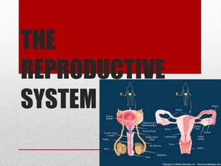
the Male Reproductive System

Recomendados
Mais conteúdo relacionado
Mais procurados
Mais procurados (20)
Destaque
Destaque (20)
Semelhante a The reproductive system
Semelhante a The reproductive system (20)
Último
Último (20)
The reproductive system
- 2. THE MALE REPRODUCTIVE SYSTEM • FUNCTION The male reproductive system functions in the production of sperm cells, the sustaining and transferring of sperm cells to the female, and the production of male sex hormones which are important in the normal functioning and development of the reproductive system behavior.
- 3. Anatomy of the Male Reproductive System
- 4. SCROTUM A sac-like structures that protects the primary male reproductive organs –testes. Beneath the skin of the scrotum is a sheet of loose connective tissue and another sheet of smooth muscle called dartos muscle –functions in the regulation of testicular temperatures by either contracting or relaxing on the environmental temperature
- 5. • Cold temperature –the muscle, together with the cremaster muscles, contract to pull the skin of the scrotum together giving it a wrinkled appearance and drawing it nearer to the body. • Warm temperature –the cremaster and the dartos muscle relax to give the scrotum a loose and thin appearance and extending the testes away from the body. • Extreme temperature –sperm production and development may not occur.
- 6. TESTES • A tough, white fibrous connective tissue capsule, the tunica albunigea, surrounds each testes and extends inward to form septa that partition the organ into lobules. • Each testis is an oval structure about 5 centimetres long and 3 centimetres in diameter. About 250 lobules in each testis. Each lobule contains 1 to 4 highly coiled seminiferous tubules that converge to form a single straight tubule, which leads into the rete testes. Short efferent ducts exit the testes Interstitial cells (cells of Leydig), which produce male sex hormones, are located between the seminiferous tubules within a lobule.
- 7. EPIDIDYMIS • Sperm leave the testes through a series of ducts that enter the epididymis. o A long (about 6 meters) tube that is tightly coiled to form a comma-shaped organ located along the superior and posterior margins of the testes. o The sperm leave the testes as immature and incapable of fertilizing ova. o Sperm complete their maturation process and become fertile as they move through the epididymis where they are stored in the lower portion, or tail of the epididymis.
- 8. DUCTUS DEFERENS • The ductus deferens, also called “vas deferens”, is a fibromuscular tube that is continuous (or contiguous) with the epididymis. • Begins at the bottom (tail) of the epididymis then turns sharply upward along the posterior margin of the testes. • Enters the abdominopelvic cavity through the inguinal canal and passes along the lateral pelvic wall. • Crosses over the ureter and posterior portion of the urinary bladder, and then descends along the posterior wall of the bladder toward the prostate gland. • Before reaching prostate gland, each ductus deferens enlarges to form an ampulla
- 9. DUCTUS DEFERENS • Sperm are stored in the proximal portion of the ductus deferens, near the epididymis, and peristaltic movements propel the sperm through the tube. • The proximal portion of the ducts deferens is a component of the spermatic cord, which contains vascular and neural structures that supply the testes. • The spermatic cord contains the ductus deferens, testicular artery and veins, lymph vessels, testicular nerve, cremaster muscle that elevates the testes for warmth and at times of sexual stimulation, and a connective covering.
- 10. EJACULATORY DUCT • Each ducts deferens, at the ampulla, joins the duct from the adjacent seminal vesicle (one of the accessory glands) to from a short ejaculatory duct. Each ejaculatory duct passes through the prostrate gland and empties into the urethra.
- 11. URETHRA • The urethra extends from the urinary bladder to the external urethral orifice at the tip of the penis. It is a passageway for sperm and fluids from the reproductive system and urine from the urinary system. While reproductive fluids are passing through the urethra, sphincters contract tightly to keep urine from entering the urethra. The male urethra is divided into three regions. o Prostatic urethra o Membranous urethra o Penile urethra
- 12. URETHRA • Prostatic urethra is the proximal portion that passes through the prostate gland. • Receives the ejaculatory duct, which contains sperm and secretions from the seminal vesicle. • Receives numerous ducts from the prostrate gland • Membranous urethra, is a short region that passes through the pelvic floor. • Penile urethra is the longest portion (also called spongy urethra or cavernous urethra), which extends the length of the penis and opens to the outside at the external urethral orifice. *The ducts from the bulbourethral glands open into the urethra of the penis.
- 13. SEMINAL VESICLES • The paired seminal vesicles are saccular glands posterior to the urinary bladder. Each gland has a short duct that joins with the deferens at the ampulla to form an ejaculatory duct, with then empties into the urethra. The fluid from the seminal vesicles is viscous and contains the following; o Fructose –which provides an energy source for the sperm o Prostaglandins –which contribute the mobility and viability of the sperm. o Proteins –that causes slight coagulation reactions in the semen after ejaculation.
- 14. PROSTATE • The prostate gland is a firm, dense structure that is located just inferior to the urinary bladder. It is about the size of a walnut and encircles the urethra as it leaves the urinary bladder. Numerous short ducts from the substance of the prostate gland empty into the prostatic urethra. The secretions of the prostrate are thin, milky–coloured and, alkaline. They function to enhance the motility of the sperm.
- 15. BULBOURETHRAL GLANDS • The paired bulbourethral (Cowper’s) glands re small, about the size and located near the base of the penis. A short duct from each gland enters the proximal end of the penile urethra. In response to sexual stimulation, the bulbourethral glands secrete an alkaline mucus-like fluid. This fluid neutralizes the acidity of the urine residue in the urethra, helps to neutralize the acidity of the vagina, and provides some lubrication for the tipp of the penis during intercourse.
- 16. SEMINAL FLUID • Seminal fluid, or semen, is alkaline mixture of sperm cells and secretions from the accessory glands, o Secretions from the seminal vesicles make up about 60% of the volume of the semen, with most of the remainder coming from the prostrate gland. o The sperm and secretions from the bulbourethral gland contribute only a small volume. The volume of semen in a single ejaculation may vary from 1.5 to 6.0 ml. o There are usually between 50 and 150 million sperm per millilitre of semen. o Sperm counts below 10 to 20 million per millilitre usually present fertility problems. o Although only one sperm actually penetrates and fertilizes the ovum, it takes several million sperm in an ejaculation to ensure that fertilization will take place.
- 17. PENIS • The penis, the male copulatory organ, is a cylindrical pendant organ located anterior to the scrotum and functions to transfer sperm to the vagina. The penis consist of three in connective tissue that wrapped in connective tissue and covered with skin. o The two dorsal columns are the corpora cavernosa o The single, midline ventral column surrounds the urethra and is called the corpus spongiosum. o The penis has a root, body (shaft), and glans penis. o The root of the penis attaches it to the public arch and the body is the visible, pendant portion. o The corpus spongiosum expands at the distal end to form the glans penis. o The urethra, which extends throughout the length of the corpus spongiosum, opens through the external urethral office at the tip of the glans penis. o A loose fold skin, called the prepuce, of foreskin, covers the glans penis.
- 18. SPERMATOGENESIS • Sperm are produced by spermatogenesis within the seminiferous tubules. A transverse section of a seminiferous tubule shoes that is packed with cells in various stages of development. Interspersed with these cells, there are large cells that extend from the periphery of the tubule to the lumen. These large cells are the supporting, or sustentacular cells (Sertoli’s cells), which supports and nourish the other cells.
- 19. SPERMATOGENESIS 1. Each primary spermatocytes goes through the first meiotic division, meiosis I, to produce two secondary spermatocytes, each with 23 chromosomes (haploid). 2. One chromosomes, consisting of two chromatids, goes to each secondary spermatocyte. 3. In the second meiotic division, meiosis II, each secondary spermatocyte divides to produce two spermatids. 4. Centromere divides so that a single-stranded chromatid goes to each cell.
- 20. SPERMATOGENESIS • As a result of the two meiotic divisions, each primary spermatocyte produces four spermatids. During spermatogenesis there are two cellular divisions, but only one replication of DNA so that each spermatid has 23 chromosomes, one from each pair in the original primary spermatocyte. Each successive stage in spermatogenesis is pushed toward the center of the tubule so that the more immature cells are at the periphery and more differentiated cells are nearer the center.
- 22. EARLY DEVELOPMENT • Early in embryonic development. Primordial germ cells enter the testes and differentiate into spermatogonia, immature cells that remain dormant until puberty. o Spermatogonia are diploid cells, each 46 chromosomes (23 pairs) located around the periphery of the seminiferous tubules. o At puberty, hormones stimulate these cells to begin dividing by mitosis. o Some of the daughter cells produced by mitosis remain at the periphery as spermatogonia. o Others are pushed toward the lumen, undergo some changes and become primary spermatocytes. o Because they are produced by mitosis, primary spermatocytes, like spermatogonia, are diploid and have 46 chromosomes.
- 23. SPERMIOGENESIS • The final step in the development of sperm is called spermiogenesis. In this process, the spermatids formed from spermatogenesis become mature spermatozoa, or sperm. The mature sperm cell has a head, midpiece and tail. o The head, also called the nuclear region, contains the 23 chromosomes surrounded by a nuclear membrane. o The tip of the head is covered b an acrosome, which contains enzymes that help the sperm penetrate the female gamete. o The midpiece, metabolic region, contains mitochondria that provide adenosine triphosphate (ATP). o The tail, locomotors region, uses as a typical flagellum for locomotion.
- 24. SPERMIOGENESIS • The sperm are released into the lumen of the seminiferous tubule and leave the testes. They then enter the epididymis where they undergo their final maturation and become capable of fertilizing a female gamete. o Sperm production begins at puberty and continues throughout the life of a male. o The entire process, beginning with primary spermatocyte, takes about 74 days. o After ejaculation, the sperm can live for about 48 hours in the female reproductive tract.
- 25. ERECTION AND EJACULATION • The male sexual response includes erection and orgasm accompanied by ejaculation. Orgasm followed a variable time period during which it is not possible to achieve another erection.
- 26. ERECTION • Erection occurs because of parasympathetic action potentials coming from the sacral portion of the spinal cord. o Arteries of the erectile tissues dilate which increases blood supply to the erectile tissues. o Blood also fills venous sinuses known as sinusoids which compress the veins that reduce blood flow the penis. o The increased blood pressure in the sinusoids causes the erectile tissue to inflate and become rigid. o The mucous glands within the urethra and the bulbourethral bladder glands also start secreting mucus.
- 27. EJACULATION • Prior to ejaculation, sperm cells and testicular fluid are propelled from the epididymis and into the ductus deferens. The sperm cells, testicular secretions, and seminal fluid move into the urethra in order to mix secretions from the prostate gland. Sympathetic nervous stimulation, primarily from the lumbar section of the spinal cord, results to emission or discharge. This results to contractions of the reproductive tracts and stimulate the seminal vesicles and prostate gland to release secretions. Thus, semen accumulates in the urethra.
- 28. EJACULATION • Ejaculation is then brought about by the contraction of smooth muscles in the urethral wall and the skeletal muscles that surround the base of the penis. Immediately before ejaculation, nerve impulses are sent to the skeletal muscles at the base of the penis. The semen is forced out the urethra when rhythmic contractions are produce. Also, muscle tension increase all throughout the body.
- 29. FIN….