Recomendados

Recomendados
Mais conteúdo relacionado
Mais procurados
Mais procurados (20)
Destaque
Destaque (20)
Semelhante a JL Poster
Semelhante a JL Poster (20)
JL Poster
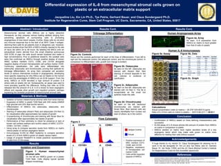
- 1. Mesenchymal stromal cells (MSCs) are a highly attractive therapeutic as they possess various healing abilities ranging from preventing apoptosis of surrounding cells to promoting angiogenesis of new blood vessels. Further, these cells do not elicit any immune response due to the lack of an MHC Class II antigen, deeming them safe for all patients even in allogeneic use. However, previous studies show that 90% of MSCs directly injected to the site of injury die within 24 to 48 hours. By seeding the MSCs onto an extracellular matrix derived from the small intestinal submucosa (SIS ECM) of pigs, the cells would remain localized and viable in high concentrations. Prior to additional research, the isolated cells were first confirmed as hMSCs through positive display of known hMSC surface markers CD73, CD90, and CD105 alongside negative display of known hematopoietic cell surface marker CD45. Continuing classification, the cells were differentiated into osteocytes, adipocytes, and chondrocytes, proving their aptitude for trilineage differentiation. An array then compared cell secretion levels of various chemokines involved in angiogenesis, developing more specific reasoning for why MSCs are so helpful to the human body and the advantages of hMSCs on an ECM support. From the array, hMSCs on ECM secreted a high amount of protein IL-8 where cells on plastic did not. Subsequently, an assay was used to more precisely quantify the difference, finding that cells on ECM released 40x the amount of IL-8. IL-8 is known to have angiogenic effects and regulate other growth and migration proteins, perhaps explaining the remedial properties of mesenchymal stromal cells. Figure 2a. Controls Above are the controls performed for each of the lines of differentiation. From left to right are the osteocyte control, the adipocyte control, and the chondrocyte control. In comparison to differentiated cells, growth and change is evident. Differential expression of IL-8 from mesenchymal stromal cells grown on plastic or an extracellular matrix support Jacqueline Liu, Xin Lin Ph.D., Tye Petrie, Gerhard Bauer, and Claus Sondergaard Ph.D. Institute for Regenerative Cures, Stem Cell Program, UC Davis, Sacramento, CA, United States, 95817 Abstract / Introduction Materials & Methods Conclusion Results Results Cont. Acknowledgements References • Isolation of human mesenchymal stromal cells from bone marrow • Expansion of hMSC in plastic T225 flask with D20 media (DMEM high glucose and 20% fetal bovine serum) • Differentiation of hMSCs into osteocytes, adipocytes, and chondrocytes • Staining of osteocytes (Alizarin Red) and adipocytes (Oil Red O) for visualization after approximately two weeks of growth • Cryosectioning of chondrocytes and staining with Alcian Blue for visualization after approximately two weeks of growth • Performance of flow cytometry to check for presence or lack of known cell surface markers and confirm status of hMSC (CD73+, CD90+, CD105+, CD45-) • Using array to compare secretion levels from hMSCs on matrix versus plastic of various angiogenic factors • Immunoassay ELISA kit (R&D Systems) to compare secretion levels of IL-8 from hMSCs grown on plastic versus matrix • Usage of program ReaderFit to create graphical representation of data from ELISA assay Isolation and Expansion Results Cont. Figure 1. Human mesenchymal stromal cells On the left are hMSCs grown on a plastic T225 flask. Cells display typical spindle shaped morphology. Trilineage Differentiation Figure 2b. Osteocytes As seen on the left, osteocytes are stained with Alizarin Red. Red staining of mineral deposits in the cell cultures is evidence of calcification. Figure 2c. Adipocytes As seen on the left, adipocytes are stained with Oil Red O. The fat is recognizable as the small red spheres inside the cells. Flow Cytometry Human IL-8 Immunoassay • Confirmation of hMSCs based on three defining characteristics was successful • ELISA assay of IL-8 shows almost 40x amount of protein from hMSCs on matrix in comparison to plastic • hMSCs seeded on matrix have higher secretion levels of a key angiogenic factor which may make cells grown on matrix more effective for healing than cells without support A huge thanks to my mentor Dr. Claus Sondergaard for allowing me to work in his lab alongside Dr. Xin Lin and Tye Petrie, and Dr. Gerhard Bauer, Dr. Jan Nolta, CIRM, and the UC Davis Medical Center’s Institute for Regenerative Cures for this incredible opportunity. Id Name Response Independent Calculated 1 Std1 1.8425 2000 2003.635 2 Std2 1.127 1000 986.6755 3 Std3 0.6895 500 518.1953 4 Std4 0.3755 250 242.7631 5 Std5 0.217 125 123.5553 6 Std6 0.1265 62.5 62.7066 7 Std7 0.0715 31.2 29.2378 8 Std8 (0) 0.014 0 0 9 Matrix (diluted 1:100) 0.0665 26.376 10 Plastic 0.132 66.2211 Calculations Actual concentration (cells on matrix)— 26.376*100=2637.6 pg/mL Ratio of IL-8 from cells on matrix to plastic— 2637.6:66.2211 = 39.83:1 Osteocyte Control Well 1 Well 2 Adipocyte Control Well 1 Well 2 Standard hMSC on matrix (diluted 1:100) hMSC on plastic Figure 5b. DataFigure 5a. Assay Figure 5c. Graph b. CD90+ d. CD45-c. CD105+ a. CD73+ Isotype Control Actual Control for CD45 is not to scale. • Dominici M, Le Blanc K, Mueller I, Slaper-Cortenbach I, Marini F, Krause D, et al. Minimal criteria for defining multipotent mesenchymal stromal cells. The International Society for Cellular Therapy position statement. Cytotherapy. 2006;8:315–317. • Keating A. Mesenchymal stromal cells. Curr Opin Hematol. 2006;13:419–425. • AE Koch, PJ Polverini, SL Kunkel, LA Harlow, LA DiPietro, VM Elner, SG Elner, and RM Strieter. Interleukin-8 as a macrophage-derived mediator of angiogenesis. Science 11 December 1992: 258 (5089), 1798-1801. Human Angiogenesis Array Figure 4a. Array As seen, IL-8 expression from hMSCs on matrix is much greater than that of cells on plastic.Cells on matrix Cells on plastic IL-8 Expression IL-8 Expression 200 µm 200 µm 200 µm 200 µm 200 µm 200 µm 200 µm • Positive expression of MSC markers CD73, CD90, and CD105 • Negative expression of hematopoietic cell marker CD45 • Cells exhibit expected aspects of hMSCs Figure 2d. Chondrocytes As seen on the left, sectioned chondrocytes are stained with Alcian Blue. Blue stained cells are clearly identifiable from nuclear red stain of others, as in the control. Chondrocyte Control 200 µm 200 µm 200 µm Slide 1 Slide 2 Figure 3