SlideShare uma empresa Scribd logo
1 de 11
Preliminary Analyses of Xanthomonas citri Avirulence Proteins, AvrGf1 and AvrGf2
Harrison Toney
Introduction:
Citrus canker disease is caused by the bacteria Xanthomonas citri which affects many citrus species
world-wide1
. A strain of X. citri has recently been found in a Florida orchard that infects Key limes. A
genetic analysis of this organism identified two genes crucial for infection, avrGf1 and avrGf2. These
genes were transferred to a bacterium that was normally non-pathogenic to citrus, Xanthomonas
perforans, and the resulting recombinant organism was able to infect grapefruit2
, thereby proving their
importance as virulence factors for the bacterium. There are no homologues of known function in the
protein sequence or structural databases, which has prevented explanation of the mechanism of action
of these proteins, despite their demonstrated importance in the infectious process. An analysis of the
amino acid sequence of the proteins encoded by each gene using the MEME software suite
(http://www.mendeley.com/catalog/meme-suite-tools-motif-discovery-searching-1/) has identified
three important sequence motifs that have revealed some key features of the proteins. Each protein has
a chloroplast localization sequence which spans the first 87 amino acids of AvrGf1 and the first 69 amino
acids of AvrGf2; each protein has a cyclophilin recognition sequence containing the amino acid sequence
GPLL (amino acids 381-384 in AvrGf1 and amino acids 356-359 in AvrGf2). A cyclophilin is a peptidyl-
prolyl isomerase that converts the peptide bond between proline and another amino acid from the cis-
conformation to the trans- conformation3
. Each protein also contains two C-terminal domains that are
shared by proteins that are secreted by the Type III Secretion System (T3SS), (AvrGf1: Domain 1: amino
acids 419 to 468 and Domain 2: 473 to 523; AvrGf2: Domain 1: amino acids 393 to 441 and Domain 2:
amino acids 447 to 497) which underscores their role as effector proteins since the T3SS is used by
pathogenic bacteria to directly inject proteins into the host and bypass defense systems4
. Based upon
the amino acid sequence analysis, it is our belief that AvrGf1 and AvrGf2 are injected into the plant cell
where they are imported into the chloroplast. Once in the chloroplast, citrus cyclophilin (CsCyp) flips
the peptide bond between glycine and proline in the GPLL motifs of the proteins, thereby activating the
protein. Recently, the structure of a citrus CsCyp from the chloroplast of an orange (Citrus sinesis) was
determined by x-ray crystallography5
and with the use of both molecular docking and isothermal
titration calorimetry experiments it was determined that CsCyp showed a preference for substrates with
proline-rich amino acid sequences such as YSPSAP. Even though AvrGf1 and AvrGf2 don’t have such a
sequence, we speculate they will still interact with CsCyp. By mutating the Glycine in the GPLL sequence
to a serine (SPLL) or alanine (AASL), it has been shown that no hypersensitive response is triggered
inside the citrus plant (Alberto Gochez unpublished data). This suggests that the structural change
induced by flipping the bond is important for activation. In order to gain a better understanding of the
exact function of AvrGf1 and AvrGf2, the ultimate goal for this research is to express and purify large
quantitiesof eachtostudyhowAvrGf1and 2 interactwithCsCyp.
AvrGf1: 532 amino acids
MAPSMHSAASPVSVLHLRDTSMRTKAQLPLTAIQRFLAHDAASTQAPSASASTSLHKNETAGLLAALPARN
ARQGAQRKSGEKEGARQNNGGRGGQWASRAAKYALGIAGAGYVADNFVLSTTSLVDGKGGFTSNDRLDKA
CAKAETYYARYHSATEDERASHSRPFVPIRTCGSNQFATMTDYRAATKVHVGHLFDSQAARESLVTNLACL
KGERIKQECIIRYAPAQVPADPDLSKSELYDRKNKYSLVGMPNAQTGASGYTSRSITQPFINRGMEHFRQAS
QSDKALSLRQCMQSLERALQDTDKLGKQAQHAAGQAILNFRQVYAADEHWGHPEKVIMKTLIANGLLSQE
QTDRIDATLMFEDPSISVLKRNTSIAGPLLQKLETKIQSKRLQDQPETLADFMEMAKQKNMEGLPIAHFKL
NAEGTGFEDCSGLGDSFTSANAVACINHARLMSGEPRLSKEDVGVVVACLNAVYDDASSIRHSLHEIARGCF
VGAGYTTEDADAFYEQICKDAARAFYAGKSMTSSD
AvrGf2:508 amino acids
MRVAKHNVKPVVSLLVNEASKNSPTSAQTIGGANSLNEILSGLPSFSKNANRTSSSRKTPRRLASLAKTAMK
YVAGTAGAAYVYDNTANRFFLSTTSLHDGKQGFTSDARLQEAEKKAEAIYAEYHAHECPDKIEVKRTSLWP
KLVGENAFVTMLDFRSATKVHLKELINTKEARDSIALNISCILGERIKPALLTEHGVVQAPVAFDITKQDDFE
LKNKYSLLGVPNNDTGSYGYASRSILNPFIEKGEKHHAQAIASDQALAPRDCVAALQPMLKNSQSLIPEAQF
RAGQALLILRPLYCGPKTWGDAHKVLMPFLESKGLASTRENQRLGETRPFTPSDMEKGVARRNTSVAGPL
LHELNMLIQKSIYKKDEEGMSDLRSKNLREMKYIPISHFRMNDDCTGFEDSSGLADSFTGYNVAAYINHARL
LSGEDRLSKQDVVAVVGCLNAVYDNASSERHTLREIAHGCFVGAGYTVEDAEAFYKDVCKDSAREFYGGEA
LRAAKSN
Box:GPLL domain,Chloroplastlocalizationsequence,PutativeC-Terminal Domain1,
Putative C-Terminal Domain2
Figure 1: Amino acidsequencemotifs identifiedin Xanthomonas citriby MEMEanalysis.
Materialsand Methods:
Expression of recombinant Proteins: Bacterial expression plasmids containing the structural gene for
AvrGf1, AvrGf2 or CsCyp ligated into a pET21a vector backbone were obtained from our collaborators in
the Department of Plant Pathology at the University of Florida. Aliquots (2µL) of each plasmid were used
to transform calcium competent samples of Escherichia coli NiCo BL21(DE3). Aliquots (50 µL) were
placed on ice and the plasmid DNA was added to them. The cells were incubated on ice for 10 minutes,
removed, and then placed in a 42°C water bath for 2 minutes at which point the cells were placed back
on ice for 10 minutes. After the ice incubation, 500µL of Luria-Bertani (LB) broth was added to the cell
aliquots which were then incubated for 45 minutes at 37°C with shaking. After the 37°C incubation, the
cells were plated onto LB Agar plates containing 100 µg/mL ampicillin and the agar plates were
incubated in the 37°C incubator overnight. A colony from each plate was selected and added to 100 mL
of Luria-Bertani (LB) medium containing 100μg/mL ampicillin which was then incubated overnight at
37°C with shaking (150 rpm). After 18 hours of growth, each 100 mL culture flask was used to inoculate
1L of LB medium containing 100μg/mL ampicillin and the resulting culture was incubated at 37°C with
shaking (150 rpm) until the optical density at 600nm reached 0.6. Once this optical density was reached,
protein expression was induced by the addition of IPTG to a final concentration of 0.1 mM in each flask.
The flasks were then incubated under the previously given conditions for another 3hrs. The cell cultures
were harvested by centrifugation for 30 minutes at 7500 rpm and 4°C. The pellet was collected, and
weighed. The cell pellet was suspended in lysis buffer (50 mM NaH2PO4 and 500 mM NaCl pH 7.2) with
5 mL of buffer used to suspend each 1g of cell pellet and 100µL of protease inhibitor cocktail (Amresco
Inc). The resulting suspension was placed in a stainless steel beaker on ice and sonicated using a
protocol that exposed the sample to 5 seconds of sonication at 30% power and 15 seconds off. This
cycle was repeated for 8 minutes and typically resulted in 20 – 30 kJ of energy being transferred. After
lysis, the sample was centrifuged for 20 minutes at 15000 rpm and 4°C. The supernatant was collected
for purificationandthe pelletwasdiscarded.
Purification: The post-lysis supernatants were purifed by Ni2+
-Metal Chelating Affinity Chromatography
(MCAC) using a 5 mL HisTrap FF column (GE Healthcare, Inc.). Once the sample was loaded onto the
column, the column was washed for 20 column volumes with 50mM Na2HPO4, 500mM NaCl, 10mM
imidazole, pH 7.2 (Buffer A) at a flowrate of 2.5 mL/min. Upon completion of the wash step, the
imidazole concentration in the buffer was increased to 50 mM by increasing the Buffer B concentration
in the mobile phase to 10% (Buffer B: 50mM Na2HPO4, 500mM NaCl, 500mM imidazole, pH 7.2). The
column was washed with 10% buffer B for 10 column volumes then a linear gradient ranging from
50mM imidazole to 500mM(10-100% Buffer B) imidazole was applied to the column to elute the bound
proteins. The absorbance of the column eluate was monitored at 280 nm and the eluate was collected in
fractions (7 mL fraction volumes for the column wash and 2 mL for all gradient steps). Peak fractions
determined to contain the recombinant protein of interest by Sodium Dodecyl-Sulfide Polyacrylamide
Gel Electrophoresis (SDS-PAGE) and -hexahistidine Western blot were combined and concentrated to
2mL using 10 kDa molecular weight cut off (MWCO) spin concentrators (Centriprep, Amicon, Inc.).
Concentrators were loaded with 5 mL of sample and centrifuged for 15 minutes at 4°C and 5000 rpm.
This was repeated until the final volume of solution was 2mL. Once concentrated, eluate was further
purified by gel filtration chromatography (GFC) using a Superdex 200 column (GE Healthcare, Inc.)
equilibrated in 25 mMNa2HPO4, 150 mMNaCl, pH 7.2. The protein was eluted using this same buffer at
a flowrate of 0.5 mL/min. The eluate fractions were collected and analyzed by sodium dodecyl sulfate
polyacrylamidegel electrophoresis(SDS-PAGE).
SDS-PAGE analysis of chromatographic fractions: SDS-PAGE gels were employed to analyze the purity
and relative masses of the proteins in the fractions that eluted from the chromatography columns. The
1X SDS-PAGE buffer consisted 16.5mM Tris, 3.5mM SDS and 193mM Glycine. An SDS-PAGE gel was
prepared with a final resolving gel percentage of 12%. The resolving gel was prepared by combining
3.75mL of 1M Tris pH 8.8, 50μL of 20% SDS, 3mL of 40% acrylamide, 3.2mL of distilled-deionized H2O,
100μL of 10% ammonium persulfate (APS) and 10μL of Tetramethylethylenediamine (TEMED). The
resolving gel was allowed 30-60 minutes for polymerization. Once polymerized, the stacking gel was
prepared by combining: 630μL 1M Tris pH 6.8, 50μL 20% SDS, 830μL of 40% Acrylamide, 3.4mL distilled-
deionized H2O, 50μL of 10% APS and 5μL of (TEMED). A comb with 10 wells was chosen and added to
gel. The stacking gel was allowed 30-60 minutes to polymerize as well. Samples for the SDS-PAGE gel
were preparedwith 20μL of protein samples and 5μL of 5x standard SDS dye. Each gel was charged with
160 volts until the dye front reached the buffer line. Upon completion of electrophoresis, the gels were
stained using the Gelcode Blue staining reagent. They were stained overnight, and then destained using
deionized water. Gels were imaged on a ChemiDoc XRS system (BioRad, Inc.) using the Quantity One 1-D
AnalysisSoftware.
Expression Test of recombinant AvrGf1 and AvrGf2: Bacterial expression plasmids containing the
structural gene for either AvrGf1 or AvrGf2 ligated into a pET21a vector backbone were obtained from
our collaborators in the Department of Plant Pathology at the University of Florida. Aliquots (2µL) of
each plasmid were used to transform calcium competent samples of Escherichia coli NiCo BL21(DE3) and
Escherichia coli Rosetta 2. Aliquots (50 µL) were placed on ice and the plasmid DNA was added to them.
The cells were incubated on ice for 10 minutes, removed, and then placed in a 42°C water bath for 2
minutes at which point the cells were placed back on ice for 10 minutes. After the ice incubation, 500µL
of Luria-Bertani (LB) broth was added to the cell aliquots which were then incubated for 45 minutes at
37°C with shaking. After the 37°C incubation, the cells were plated onto LB Agar plates containing 100
µg/mL ampicillin and the agar plates were incubated in the 37°C incubator overnight. A colony from
each plate was selected and added to 25 mL of Luria-Bertani (LB) medium containing 100μg/mL
ampicillin which was then incubated overnight at 37°C with shaking (150 rpm). After 18hrs of incubation
2.5 mL from each culture tube was used to inoculate 25 mL of LB medium containing 100μg/mL
ampicillin and the resulting cultures were incubated at 37°C with shaking (150 rpm) until the optical
density at 600nm reached 0.6. Once this optical density was reached, protein expression was induced by
the addition of IPTG to a final concentration of 0.1 mM in each flask. Aliquots (500 μL) were collected at
the time of induction, 1hr, 2hrs and 3hrs post induction. The samples were micro-centrifuged for 1.5
minutes at 14000rpm and 4°C. Following centrifugation, the supernatant was discarded and 200 μL of
lysis Buffer (50 mM NaH2PO4 and 500 mM NaCl pH 7.2) and 50 μL of 5x standard SDS dye was added to
sample inpreparationforSDS-PAGEanalysis.
WesternBlot analysisof SDS-PAGEgel:A westernblotanalysisof the 30°C expressiontestof AvrGf2
containingampicillinandchloramphenicol resistance wasdone todetermineif anyof the expressed
proteinsbore the carboxy-terminalhexahistidinetagthatwas engineeredintothe expressed
recombinantproteins.The primaryantibodyusedwasraisedagainstahexahistidinetagandan alkaline
phosphatase conjugatedsecondaryantibodyraisedagainstthe Fcfragmentof thatsame organism
producedinthe primaryantibody.Once the gel wasremovedfromthe electrophoresistank,itwas
washedwithddH2Oand soakedinTransferBuffer(48mMTris, 39mM glycine,20% methanol).A
polyvinyldifluoride(PVDF) membrane(Millipore)wascutto match the size of the SDS-PAGEgel.The
membrane wassoakedin100% methanol towetitand itwas thentransferredtotransferbuffer.A
sandwichconsistingof cellulose saturationpads(GelmanInstrumentCompany,Cat#51334), filterpaper
(Whatman150 mmcircles,Cat# 1450150), PVDF,SDS-PAGEgel,filterpaperandcellulose saturationpad
was assembledonthe bedof the Trans-Blotunit.A 10mL pipette wasusedtoroll outany bubbles.The
Trans-blotSDwas connectedtothe powersupplyandthe transferwasperformedatroom temperature
at 20 voltsfor 15 minutes.Aftertransfer,the PVDFmembrane wasplacedina5000x dilute solutionof
primaryantibodyinTris-bufferedsalineand0.01% Tween-20(TBST) (20mM Tris,150mM Nacl,0.01%
(v/v) Tween-20,pH7.5) with20g of powderedmilk andwasthenincubatedinthe coldroomwith
shakingovernight.Afterincubation,the primaryantibodysolutionwasdiscardedandthe blotwas
washedfourtimesinTBST for15 minuteseachtime inthe coldroom.A 5000x folddilutionof secondary
antibody,whichwaschickenanti-mouse,inTBSTwas addedtothe membrane andthe membrane was
incubatedfor1 hour withshakinginthe coldroom. Once the hour wascomplete,the secondary
antibodysolutionwasdiscardedandthe membrane waswashedwithTBSTfourtimesfor15 minutes
each time.Visualizationof the proteinswasperformedbywashingthe blotwithdeionizedwater,adding
BCIP/NBTreagent(Sigma-Aldrich) andincubatingatroomtemperature until bandsappeared. The color
developmentreactionwasterminatedbywashingthe blotindeionizedwaterafterwhichthe
membrane wasdriedona papertowel.
Results
Purificationofrecombinantproteins
CsCyp
CsCyp was purified using nickel affinity chromatography. The absorbance by protein at 280 nm was
recorded and the representative chromatogram is shown in Fig. 1A. The flow-through peak containing
non-bound proteins is seen spanning fractions 2-18. Loosely bound proteins are shown eluting during
the second peak which spans fractions 27-37. A final predominant peak is seen eluting at approximately
20% buffer B which spans fractions 37-45. Those fractions were collected and concentrated for GFC. A
representative chromatogram of the elution process can be seen in Figure 2. A major peak is seen
eluting between fractions 17 and 18. Those fractions from the GFC of CsCyp were collected and analyzed
by SDS-PAGE. The chromatogram can be seen in Figure 2 accompanied by the SDS-PAGE gel. Protein
bands can be seen for both fractions at approximately 25kDa which is expected due to an amino acid
sequence analysiswhichdeterminedthe molecularweighttobe 25,080 Da
AvrGf1
Purification of AvrGf1 was attempted using nickel affinity chromatography. The representative
chromatogram is shown in Fig. 1B. The flow-through peak containing non-bound proteins is seen
spanning fractions 2-12. A predominant second and final peak that spans fractions 28-32 were collected
and analyzed by SDS-PAGE which can be seen in Figure 3. It was determined due to a lack of any
prominent bands at the calculated mass (57kDa) and the impurity of samples collected that no protein
had beenexpressedduringthisprocedure.
1 3 5
A B
Figure 1. Purification of Recombinant Proteins. (A) Chromatogram illustrating purification of recombinant CsCyp.
(B) Chromatogram illustrating purification of recombinant AvrGf1. The Y-axis is the absorbance of protein at 280
nm and the X-axis is time(min). The lightgreen linedepicts a linear gradientof 10-500mM imidazole.
25kDa
37kDa
50kDa
75kDa
Figure 2. GFC Purification of CsCyp. Representative chromatogram illustrating GFC purification of CsCyp with the
corresponding SDS-PAGE gel. The Y-axis depicts protein absorbance at 280 nm vs. X-axis which is time. The light
green line depicts the gel filtration buffer containing 25 mM NaPO4, 150 mM NaCl, pH 7.2. Lane 1 contains fraction
17 and lane3 contains fraction 18.Molecular weightmarker is shown in lane5.
Expressiontest of recombinantproteins
AvrGf1
1 2 3 4 6
25kDa
37kDa
50kDa
a
75kDa
150kDa
Figure 3. SDS-PAGE of AvrGf1 Purification Attempt. Lane 6 contains molecular weight marker. Lane 1=Fraction 28;
Lane 2=Fraction 29,Lane 3=Fraction 30, Lane 4=Fraction 31.
A B
1 2 3 4 5 6 7 8 9 1 2 3 4 5 6 7 8 9
C
Figure 4: Expression Test of AvrGf1. (A) AvrGf1 in E. coli NiCo BL21 (DE3) 25°C, (B) AvrGf1 in E. coli NiCo BL21
(DE3) 37°C, (C) AvrGf1 in E. coli Rosetta 2 37°C. Each gel: Lanes 1&2 0hr-(uninduced, induced); Lanes 3&4 1hr-
(uninduced, induced); Lanes 5&6 2hr-(uninduced, induced); Lanes 7&8 3hr-(uninduced,induced); Lane 9
(Ladder)
1 2 3 4 5 6 7 8 9
25kDa
37kDa
50kDa
a
75kDa
150kDa
25kDa
37kDa
50kDa
a
75kDa
100kDa
150kDa
100kDa
AvrGf2
A B
1 2 3 4 5 6 7 8 9 1 2 3 4 5 6 7 8 9
A B
1 2 3 4 5 6 7 8 9 1 2 3 4 5 6 7 8 9
Figure 5: Expression Test of AvrGf2. (A) AvrGf2 in E. coli NiCo BL21 (DE3) @ 30°C (B) AvrGf2 in E. coli
NiCo BL21 (DE3) @ 37°C. Each gel: Lanes 1&2 0hr-(uninduced,induced); Lanes 3&4 1hr-(uninduced,
induced); Lanes 5&6 2hr-(uninduced,induced); Lanes 7&8 3hr-(uninduced, induced); Lane 9 (Ladder)
Figure 6: Expression test of AvrGf2 double antibiotic. (A) AvrGf2 in E. Coli Rosetta 2 @ 30°C (B)
AvrGf2 in E. coli Rosetta 2 @ 37°C. Each gel: Lanes 1&2 0hr-(uninduced,induced); Lanes 3&4 1hr-
(uninduced, induced); Lanes 5&6 2hr-(uninduced, induced); Lanes 7&8 3hr-(uninduced,induced);
Lane 9 (Ladder)
25kDa
37kDa
50kDa
a
75kDa
150kDa
25kDa
37kDa
50kDa
a
75kDa
150kDa
100kDa
100kDa
Discussion
Expression of CsCyp was done using the E. coli NiCo BL21(DE3) expression strain. Purification was done
using nickel affinity chromatography and gel filtration chromatography. The expression and purification
procedures for CsCyp produced high quantities of pure protein which was indicated by an SDS-PAGE
analysis after the purification process. The SDS-PAGE gel highlighted prominent protein bands at the
expectedmolecularweightof CsCyp(25kDaexpectedmolecularweight).
Analysis by SDS-PAGE leads us believe CsCyp has been successfully purified and structural analysis can
now be done. Molecular docking experiments between CsCyp and AvrGf1 can also be done to determine
how they interact. We can use that information to compare it to previous studies that determined the
bindingbetweenCsCypandthe preferential sequence YSPASP5
.
AvrGf1 was purified using the same protocol as that used to purify CsCyp, however, the results were not
similar. The SDS-PAGE analysis after the MCAC column step revealed many impurities and no protein
was seenatthe expectedmolecular weight(57kDaexpectedmolecularweight).
I have concluded from the lack of expressed AvrGf1 protein that a problem occurred during either the
expression or purification step of the procedure. It is believed that an error occurred during addition of
the protease inhibitor during the lysis procedure. The protease inhibitor used contained EDTA which
may have interfered with purification by stripping the nickel from the chelating resin. However, it is also
possible thatAvrGf1was not expressedin culture in the firstplace.
To determine if recombinant AvrGf1 is expressed in culture, tests using small scale cultures of
recombinant E. coli NiCo BL21 (DE3) and Rosetta2 expression strains harboring the plasmids encoding
AvrGf1 and AvrGf2 in pET21a were performed at 25°C, 30°C, and 37°C. The expression tests compared
induced cultures to non-induced cultures at time intervals of 0hr, 1hr, 2hr, and 3hr post-induction. SDS-
PAGE analysis of the cultures revealed no detectable expression of AvrGf1 in either strain regardless of
growth temperature. Tests on expression of AvrGf2 yielded the same results; a lack of expression
regardless of strain or temperature. An anti-hexahistidine western blot of the AvrGf2 was performed to
Figure 7: Western Blot of AvrGf2. Molecular weight ladder inlane 1.
100kDa
75kDa
50kDa
37kDa
1
determine if the protein was expressed at low levels. The western blot proved that expression was
unsuccessful,withnobanddetectedinanyof the samples .
We were able to express and purify large quantities of citrus cyclophilin. Unfortunately, we were unable
to detect any expression of X. citri AvrGf1 or AvrGf2 in the strains and conditions used. Future work will
test the role of IPTG concentration and media type in expression of these novel proteins. Production of
recombinant protein is a crucial step in the structure determination of AvrGf1 and AvrGf2. Given the
lack of homologous protein sequences in the public databases, the three dimensional structures of
AvrGf1 and AvrGf2 and may be the only source of insight into the roles they play during the
phytopathogenicprocess.
References
1. Almeida, N.; Digiampietri, L.; Potnus, N.; Adi, S.; Bortolossi, J.; Siva de, A.; Moreira, L.;
Nalvo, A.F.; Ferraz, A.; Moraes, F.; Oliviera, J.; Souza, R.; Facincani, A.; Ferro, M.; Furlan,
L.; Gimenez, D.; Jones, J.; Kitajima, E.; Laia, M.; Leite, R.; Nishiyama, M.; Rodrigues, N.J.;
Letícia, N.A.; Norman, D.J.; Ostroski, E.H.; Pereira, H.A.; Staskawicz, B.; Tezza, R.; Ferro, J.;
Vinatzer, B.; Setubal, J. Novel Insights into the Genomic Basis of Citrus Canker Based on
the Genome Sequences of Two Strains of Xanthomonas fuscans subsp. aurantifolii. BMC
Genomics, 2010, 11, 238.
2. Rybak, M. Genetic Determinants of Host Range Specificity Of the Wellington Strain
Xanthomonas axonopodis pv. citri. 2005. Doctoral dissertation submitted to the
University of Florida.
3. Campos, B.M.; Ambrosio, A.L.; Domingues, M.N.; Brasil de Souza, A.; Barbosa, J.A.; Paes
Leme, A.F.; Perez, C.A.; Whittaker, S.B.; Murakami, M.T.; Zeri, A.C.; Benedetti, C.E. A
Redox 2-Cys Mechanism Regulates the Catalytic Activity of Divergent Cyclophilins. Plant
Physiol., 2013, 162, 1311-1323.
4. Buttner, D.; Sheng Y.; Type III Protein Secretion in Plant Pathogenic Bacteria. Plant
Physiol., 2009, 4, 1656-1664.
5. Brunings, A.M; Gabriel, D.W. Xanthomonas citri: Breaking the Surface. Physiol Mol. Plant
Path., 2003, 4, 141-157.
6. Bernstein, G.; Davis, T.; Eisenmesser, E.; Finerty Jr, P.; Lee, T.; Mackenzie, F.; Ouyang, H.;
Paramanathan, R.; Wolfram, C.S.; Walker, J.; Wen Hwa Lee, S. Structural and Biochemical
Characterization of the Human Cyclophilin Family of Peptidyl-Prolyl Isomerases. PLoS
Biol., 2010, 8(7): e1000439.
7. Balague, C.; Pontier D.; Roby, D. The Hypersensitive Response: A Programmed Cell Death
Associated with Plant Resistance. Comptes Rendus de l'Académie des Sciences, 1998,
321, 721–734.
8. Danal, J.; Morel, J.B. The Hypersensitive response and the Induction of Cell Death in
Plants. Cell Death Differ., 1997, 4, 671-683.
9. Gochez, A. 2012. Personal Communication
10. Gottig, N.; Garavaglia, B.S.; Garofalo, C.G.; Zimaro, T.; Sgro, G.G.; Ficarra, F.A.; Dunger, G.;
Daurelio, L.D.; Thomas, L.; Gehring, C; Orellano, E.G.; Ottado, J. Mechanism of Infection
used by Xanthomonas axonopodis pv. citri in Citrus Canker Disease. Tech. and Educ.
Topics in App. Micro. and Micro. Biotech., 2010, 196-204.
11. Howard, B.; Hill, C.; Li, S.; Sundquist, W. Structural Insights into the Catalytic Mechanism
of Cyclophilin A. Struct. Biol., 2003, 6, 475-481.
12. Nilsson, J.; Stahl, S.; Lundeberg, J.; Nygren, P.A.; Uhlen, M. Affinity Fusion Stategies for
Detection, Purification, and Immobilization of Recombinant Proteins. Protein Expres.
Purif., 1997, 11, 1-16.
13. O’Fagain, C.; Cummins, P.; O’Connor, B. Gel-Filtration Chromatography. http://doras.dcu.ie
(accessed August 6, 2013).

Mais conteúdo relacionado

Mais procurados

Biocatalytic properties of a recombinant aldo keto reductase with broad subst...
Biocatalytic properties of a recombinant aldo keto reductase with broad subst...Biocatalytic properties of a recombinant aldo keto reductase with broad subst...
Biocatalytic properties of a recombinant aldo keto reductase with broad subst...Ailen Ramos
 
Molecular_bilogy_lab_report_1
Molecular_bilogy_lab_report_1Molecular_bilogy_lab_report_1
Molecular_bilogy_lab_report_1Elijah Willie
 
Effect of estradiol -17 β on arachidonic acid metabolism in sheep uterus: in ...
Effect of estradiol -17 β on arachidonic acid metabolism in sheep uterus: in ...Effect of estradiol -17 β on arachidonic acid metabolism in sheep uterus: in ...
Effect of estradiol -17 β on arachidonic acid metabolism in sheep uterus: in ...iosrjce
 
[Final] Purification Of B-Gal Formal Report
[Final] Purification Of B-Gal Formal Report[Final] Purification Of B-Gal Formal Report
[Final] Purification Of B-Gal Formal ReportAndy Chand
 
Expression of Genetically Engineered Chitinase Gene of Pyrococcus furiosus
Expression of Genetically Engineered Chitinase Gene of Pyrococcus furiosusExpression of Genetically Engineered Chitinase Gene of Pyrococcus furiosus
Expression of Genetically Engineered Chitinase Gene of Pyrococcus furiosusIJERDJOURNAL
 
Protein Protein Interactions Of Glycine Oxidase (Thi O)
Protein Protein Interactions Of Glycine Oxidase (Thi O)Protein Protein Interactions Of Glycine Oxidase (Thi O)
Protein Protein Interactions Of Glycine Oxidase (Thi O)bturne
 
2014_BKCS_기생충
2014_BKCS_기생충2014_BKCS_기생충
2014_BKCS_기생충Je-Hyun Baek
 
Dorobantu Adina BMS2 - Molecular Biology FLR.pdf
Dorobantu Adina BMS2 - Molecular Biology FLR.pdfDorobantu Adina BMS2 - Molecular Biology FLR.pdf
Dorobantu Adina BMS2 - Molecular Biology FLR.pdfAdinaGeorgiana7
 
Temp-Sensitive Inhibition of Development in Dictyostelium - Dev Bio 251 18-26...
Temp-Sensitive Inhibition of Development in Dictyostelium - Dev Bio 251 18-26...Temp-Sensitive Inhibition of Development in Dictyostelium - Dev Bio 251 18-26...
Temp-Sensitive Inhibition of Development in Dictyostelium - Dev Bio 251 18-26...James Silverman
 
A visual chip-based coimmunoprecipitation technique for analysis of protein–p...
A visual chip-based coimmunoprecipitation technique for analysis of protein–p...A visual chip-based coimmunoprecipitation technique for analysis of protein–p...
A visual chip-based coimmunoprecipitation technique for analysis of protein–p...Qing Chen
 
Poyton and Clarkson J Biol Chem 1989
Poyton and Clarkson J Biol Chem 1989Poyton and Clarkson J Biol Chem 1989
Poyton and Clarkson J Biol Chem 1989George Clarkson
 
Appl Microbiol Biotechnol
Appl Microbiol Biotechnol Appl Microbiol Biotechnol
Appl Microbiol Biotechnol Charles Zhang
 
Optimization of experimental protocols for cellular lysis
Optimization of experimental protocols for cellular lysisOptimization of experimental protocols for cellular lysis
Optimization of experimental protocols for cellular lysisExpedeon
 
35.Isolation and purification of endopolygalacturonase produced by Alternaria...
35.Isolation and purification of endopolygalacturonase produced by Alternaria...35.Isolation and purification of endopolygalacturonase produced by Alternaria...
35.Isolation and purification of endopolygalacturonase produced by Alternaria...Annadurai B
 

Mais procurados (20)

Biocatalytic properties of a recombinant aldo keto reductase with broad subst...
Biocatalytic properties of a recombinant aldo keto reductase with broad subst...Biocatalytic properties of a recombinant aldo keto reductase with broad subst...
Biocatalytic properties of a recombinant aldo keto reductase with broad subst...
 
Molecular_bilogy_lab_report_1
Molecular_bilogy_lab_report_1Molecular_bilogy_lab_report_1
Molecular_bilogy_lab_report_1
 
Effect of estradiol -17 β on arachidonic acid metabolism in sheep uterus: in ...
Effect of estradiol -17 β on arachidonic acid metabolism in sheep uterus: in ...Effect of estradiol -17 β on arachidonic acid metabolism in sheep uterus: in ...
Effect of estradiol -17 β on arachidonic acid metabolism in sheep uterus: in ...
 
[Final] Purification Of B-Gal Formal Report
[Final] Purification Of B-Gal Formal Report[Final] Purification Of B-Gal Formal Report
[Final] Purification Of B-Gal Formal Report
 
Expression of Genetically Engineered Chitinase Gene of Pyrococcus furiosus
Expression of Genetically Engineered Chitinase Gene of Pyrococcus furiosusExpression of Genetically Engineered Chitinase Gene of Pyrococcus furiosus
Expression of Genetically Engineered Chitinase Gene of Pyrococcus furiosus
 
Protein Protein Interactions Of Glycine Oxidase (Thi O)
Protein Protein Interactions Of Glycine Oxidase (Thi O)Protein Protein Interactions Of Glycine Oxidase (Thi O)
Protein Protein Interactions Of Glycine Oxidase (Thi O)
 
SSEF Report 5
SSEF Report 5SSEF Report 5
SSEF Report 5
 
2014_BKCS_기생충
2014_BKCS_기생충2014_BKCS_기생충
2014_BKCS_기생충
 
SW_2016_Snehal
SW_2016_SnehalSW_2016_Snehal
SW_2016_Snehal
 
Dorobantu Adina BMS2 - Molecular Biology FLR.pdf
Dorobantu Adina BMS2 - Molecular Biology FLR.pdfDorobantu Adina BMS2 - Molecular Biology FLR.pdf
Dorobantu Adina BMS2 - Molecular Biology FLR.pdf
 
Editing example 1
Editing example 1Editing example 1
Editing example 1
 
Temp-Sensitive Inhibition of Development in Dictyostelium - Dev Bio 251 18-26...
Temp-Sensitive Inhibition of Development in Dictyostelium - Dev Bio 251 18-26...Temp-Sensitive Inhibition of Development in Dictyostelium - Dev Bio 251 18-26...
Temp-Sensitive Inhibition of Development in Dictyostelium - Dev Bio 251 18-26...
 
A visual chip-based coimmunoprecipitation technique for analysis of protein–p...
A visual chip-based coimmunoprecipitation technique for analysis of protein–p...A visual chip-based coimmunoprecipitation technique for analysis of protein–p...
A visual chip-based coimmunoprecipitation technique for analysis of protein–p...
 
10.1.1.318.916
10.1.1.318.91610.1.1.318.916
10.1.1.318.916
 
Poyton and Clarkson J Biol Chem 1989
Poyton and Clarkson J Biol Chem 1989Poyton and Clarkson J Biol Chem 1989
Poyton and Clarkson J Biol Chem 1989
 
Appl Microbiol Biotechnol
Appl Microbiol Biotechnol Appl Microbiol Biotechnol
Appl Microbiol Biotechnol
 
Optimization of experimental protocols for cellular lysis
Optimization of experimental protocols for cellular lysisOptimization of experimental protocols for cellular lysis
Optimization of experimental protocols for cellular lysis
 
35.Isolation and purification of endopolygalacturonase produced by Alternaria...
35.Isolation and purification of endopolygalacturonase produced by Alternaria...35.Isolation and purification of endopolygalacturonase produced by Alternaria...
35.Isolation and purification of endopolygalacturonase produced by Alternaria...
 
Npy final paper
Npy final paperNpy final paper
Npy final paper
 
Npy
NpyNpy
Npy
 

Destaque

certificate_Moncrieff_Tyler
certificate_Moncrieff_Tylercertificate_Moncrieff_Tyler
certificate_Moncrieff_TylerTyler Moncrieff
 
Tvi corporate presentation april 2016
Tvi corporate presentation april 2016Tvi corporate presentation april 2016
Tvi corporate presentation april 2016TVI_Pacific
 
type écarte en profondeur la foufoune fraîchement épilée d'une suceuse de pin...
type écarte en profondeur la foufoune fraîchement épilée d'une suceuse de pin...type écarte en profondeur la foufoune fraîchement épilée d'une suceuse de pin...
type écarte en profondeur la foufoune fraîchement épilée d'une suceuse de pin...wastefulrevenue72
 
FashionInviteFinal
FashionInviteFinalFashionInviteFinal
FashionInviteFinalMichael Heyn
 
First draft script
First draft scriptFirst draft script
First draft scriptBrad Tocque
 
OCLUSION rango de movimiento mandibular en niños de seis
OCLUSION  rango de movimiento mandibular en niños de seisOCLUSION  rango de movimiento mandibular en niños de seis
OCLUSION rango de movimiento mandibular en niños de seisLoel Alvarado
 

Destaque (7)

certificate_Moncrieff_Tyler
certificate_Moncrieff_Tylercertificate_Moncrieff_Tyler
certificate_Moncrieff_Tyler
 
Tvi corporate presentation april 2016
Tvi corporate presentation april 2016Tvi corporate presentation april 2016
Tvi corporate presentation april 2016
 
type écarte en profondeur la foufoune fraîchement épilée d'une suceuse de pin...
type écarte en profondeur la foufoune fraîchement épilée d'une suceuse de pin...type écarte en profondeur la foufoune fraîchement épilée d'une suceuse de pin...
type écarte en profondeur la foufoune fraîchement épilée d'une suceuse de pin...
 
FashionInviteFinal
FashionInviteFinalFashionInviteFinal
FashionInviteFinal
 
Símbolos patrios del perú
Símbolos patrios del perúSímbolos patrios del perú
Símbolos patrios del perú
 
First draft script
First draft scriptFirst draft script
First draft script
 
OCLUSION rango de movimiento mandibular en niños de seis
OCLUSION  rango de movimiento mandibular en niños de seisOCLUSION  rango de movimiento mandibular en niños de seis
OCLUSION rango de movimiento mandibular en niños de seis
 

Semelhante a Final552 (1)

Isolation and characterization of an extracellular antifungal protein from an...
Isolation and characterization of an extracellular antifungal protein from an...Isolation and characterization of an extracellular antifungal protein from an...
Isolation and characterization of an extracellular antifungal protein from an...Maulik Kamdar
 
ISSX International conference ANDRE MAZZARI poster
ISSX International conference ANDRE MAZZARI posterISSX International conference ANDRE MAZZARI poster
ISSX International conference ANDRE MAZZARI posterAndré Luís Mazzari, PhD
 
Induced Expression of CYP1A by Triclosan in Zebra Fish to measure toxicologic...
Induced Expression of CYP1A by Triclosan in Zebra Fish to measure toxicologic...Induced Expression of CYP1A by Triclosan in Zebra Fish to measure toxicologic...
Induced Expression of CYP1A by Triclosan in Zebra Fish to measure toxicologic...Mujtaba Qureshi
 
Discovery and Mechanistic Study of Mycobacterium tuberculosis PafA Inhibitors...
Discovery and Mechanistic Study of Mycobacterium tuberculosis PafA Inhibitors...Discovery and Mechanistic Study of Mycobacterium tuberculosis PafA Inhibitors...
Discovery and Mechanistic Study of Mycobacterium tuberculosis PafA Inhibitors...Dr.Shuaib Ahmad
 
Biotransformation of Panax notoginseng by fungi
Biotransformation of Panax notoginseng by fungiBiotransformation of Panax notoginseng by fungi
Biotransformation of Panax notoginseng by fungiAgriculture Journal IJOEAR
 
The Role of Cell Wall-Degrading Enzymes in the Development of Anthracnose Dis...
The Role of Cell Wall-Degrading Enzymes in the Development of Anthracnose Dis...The Role of Cell Wall-Degrading Enzymes in the Development of Anthracnose Dis...
The Role of Cell Wall-Degrading Enzymes in the Development of Anthracnose Dis...Agriculture Journal IJOEAR
 
Generation of MRP2 Efflux Transporter Knock-Out in HepaRG Cell Line
Generation of MRP2 Efflux Transporter Knock-Out in HepaRG Cell LineGeneration of MRP2 Efflux Transporter Knock-Out in HepaRG Cell Line
Generation of MRP2 Efflux Transporter Knock-Out in HepaRG Cell Linemdmitc
 
IRJET- Subcellular Localization of Transmembrane E-cadherin-GFP Fusion Pr...
IRJET-  	  Subcellular Localization of Transmembrane E-cadherin-GFP Fusion Pr...IRJET-  	  Subcellular Localization of Transmembrane E-cadherin-GFP Fusion Pr...
IRJET- Subcellular Localization of Transmembrane E-cadherin-GFP Fusion Pr...IRJET Journal
 
Biotechniques v30p662 SNP
Biotechniques v30p662 SNPBiotechniques v30p662 SNP
Biotechniques v30p662 SNPMichael Weiner
 
DNA damage repair Neil3 gene Knockout in MOLT-4
DNA damage repair Neil3 gene Knockout in MOLT-4DNA damage repair Neil3 gene Knockout in MOLT-4
DNA damage repair Neil3 gene Knockout in MOLT-4iosrjce
 
Senior Thesis-Analyzing the interactions between MYOGEF and a component of er...
Senior Thesis-Analyzing the interactions between MYOGEF and a component of er...Senior Thesis-Analyzing the interactions between MYOGEF and a component of er...
Senior Thesis-Analyzing the interactions between MYOGEF and a component of er...Dougan McGrath
 
Ant Colony Optimization for inferring Gene Interactions
Ant Colony Optimization for inferring Gene InteractionsAnt Colony Optimization for inferring Gene Interactions
Ant Colony Optimization for inferring Gene InteractionsEster Moncho
 

Semelhante a Final552 (1) (20)

BIOS 5260 Term Paper
BIOS 5260 Term PaperBIOS 5260 Term Paper
BIOS 5260 Term Paper
 
Isolation and characterization of an extracellular antifungal protein from an...
Isolation and characterization of an extracellular antifungal protein from an...Isolation and characterization of an extracellular antifungal protein from an...
Isolation and characterization of an extracellular antifungal protein from an...
 
ISSX International conference ANDRE MAZZARI poster
ISSX International conference ANDRE MAZZARI posterISSX International conference ANDRE MAZZARI poster
ISSX International conference ANDRE MAZZARI poster
 
Induced Expression of CYP1A by Triclosan in Zebra Fish to measure toxicologic...
Induced Expression of CYP1A by Triclosan in Zebra Fish to measure toxicologic...Induced Expression of CYP1A by Triclosan in Zebra Fish to measure toxicologic...
Induced Expression of CYP1A by Triclosan in Zebra Fish to measure toxicologic...
 
Discovery and Mechanistic Study of Mycobacterium tuberculosis PafA Inhibitors...
Discovery and Mechanistic Study of Mycobacterium tuberculosis PafA Inhibitors...Discovery and Mechanistic Study of Mycobacterium tuberculosis PafA Inhibitors...
Discovery and Mechanistic Study of Mycobacterium tuberculosis PafA Inhibitors...
 
project work
project workproject work
project work
 
Biotransformation of Panax notoginseng by fungi
Biotransformation of Panax notoginseng by fungiBiotransformation of Panax notoginseng by fungi
Biotransformation of Panax notoginseng by fungi
 
The Role of Cell Wall-Degrading Enzymes in the Development of Anthracnose Dis...
The Role of Cell Wall-Degrading Enzymes in the Development of Anthracnose Dis...The Role of Cell Wall-Degrading Enzymes in the Development of Anthracnose Dis...
The Role of Cell Wall-Degrading Enzymes in the Development of Anthracnose Dis...
 
Zymography
ZymographyZymography
Zymography
 
Generation of MRP2 Efflux Transporter Knock-Out in HepaRG Cell Line
Generation of MRP2 Efflux Transporter Knock-Out in HepaRG Cell LineGeneration of MRP2 Efflux Transporter Knock-Out in HepaRG Cell Line
Generation of MRP2 Efflux Transporter Knock-Out in HepaRG Cell Line
 
IRJET- Subcellular Localization of Transmembrane E-cadherin-GFP Fusion Pr...
IRJET-  	  Subcellular Localization of Transmembrane E-cadherin-GFP Fusion Pr...IRJET-  	  Subcellular Localization of Transmembrane E-cadherin-GFP Fusion Pr...
IRJET- Subcellular Localization of Transmembrane E-cadherin-GFP Fusion Pr...
 
2016_15_3_18
2016_15_3_182016_15_3_18
2016_15_3_18
 
Jsi 124 pathway
Jsi 124 pathwayJsi 124 pathway
Jsi 124 pathway
 
Biotechniques v30p662 SNP
Biotechniques v30p662 SNPBiotechniques v30p662 SNP
Biotechniques v30p662 SNP
 
Taq purification
Taq purificationTaq purification
Taq purification
 
DNA damage repair Neil3 gene Knockout in MOLT-4
DNA damage repair Neil3 gene Knockout in MOLT-4DNA damage repair Neil3 gene Knockout in MOLT-4
DNA damage repair Neil3 gene Knockout in MOLT-4
 
Senior Thesis-Analyzing the interactions between MYOGEF and a component of er...
Senior Thesis-Analyzing the interactions between MYOGEF and a component of er...Senior Thesis-Analyzing the interactions between MYOGEF and a component of er...
Senior Thesis-Analyzing the interactions between MYOGEF and a component of er...
 
2005 trypanocidal constituents in plants 5.1) evaluation of some mexican
2005 trypanocidal constituents in plants 5.1) evaluation of some mexican2005 trypanocidal constituents in plants 5.1) evaluation of some mexican
2005 trypanocidal constituents in plants 5.1) evaluation of some mexican
 
final copy 2
final copy 2final copy 2
final copy 2
 
Ant Colony Optimization for inferring Gene Interactions
Ant Colony Optimization for inferring Gene InteractionsAnt Colony Optimization for inferring Gene Interactions
Ant Colony Optimization for inferring Gene Interactions
 

Final552 (1)

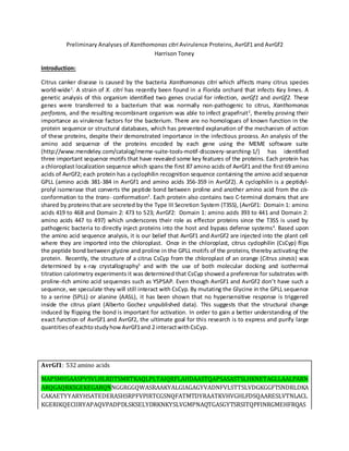
  • 1. Preliminary Analyses of Xanthomonas citri Avirulence Proteins, AvrGf1 and AvrGf2 Harrison Toney Introduction: Citrus canker disease is caused by the bacteria Xanthomonas citri which affects many citrus species world-wide1 . A strain of X. citri has recently been found in a Florida orchard that infects Key limes. A genetic analysis of this organism identified two genes crucial for infection, avrGf1 and avrGf2. These genes were transferred to a bacterium that was normally non-pathogenic to citrus, Xanthomonas perforans, and the resulting recombinant organism was able to infect grapefruit2 , thereby proving their importance as virulence factors for the bacterium. There are no homologues of known function in the protein sequence or structural databases, which has prevented explanation of the mechanism of action of these proteins, despite their demonstrated importance in the infectious process. An analysis of the amino acid sequence of the proteins encoded by each gene using the MEME software suite (http://www.mendeley.com/catalog/meme-suite-tools-motif-discovery-searching-1/) has identified three important sequence motifs that have revealed some key features of the proteins. Each protein has a chloroplast localization sequence which spans the first 87 amino acids of AvrGf1 and the first 69 amino acids of AvrGf2; each protein has a cyclophilin recognition sequence containing the amino acid sequence GPLL (amino acids 381-384 in AvrGf1 and amino acids 356-359 in AvrGf2). A cyclophilin is a peptidyl- prolyl isomerase that converts the peptide bond between proline and another amino acid from the cis- conformation to the trans- conformation3 . Each protein also contains two C-terminal domains that are shared by proteins that are secreted by the Type III Secretion System (T3SS), (AvrGf1: Domain 1: amino acids 419 to 468 and Domain 2: 473 to 523; AvrGf2: Domain 1: amino acids 393 to 441 and Domain 2: amino acids 447 to 497) which underscores their role as effector proteins since the T3SS is used by pathogenic bacteria to directly inject proteins into the host and bypass defense systems4 . Based upon the amino acid sequence analysis, it is our belief that AvrGf1 and AvrGf2 are injected into the plant cell where they are imported into the chloroplast. Once in the chloroplast, citrus cyclophilin (CsCyp) flips the peptide bond between glycine and proline in the GPLL motifs of the proteins, thereby activating the protein. Recently, the structure of a citrus CsCyp from the chloroplast of an orange (Citrus sinesis) was determined by x-ray crystallography5 and with the use of both molecular docking and isothermal titration calorimetry experiments it was determined that CsCyp showed a preference for substrates with proline-rich amino acid sequences such as YSPSAP. Even though AvrGf1 and AvrGf2 don’t have such a sequence, we speculate they will still interact with CsCyp. By mutating the Glycine in the GPLL sequence to a serine (SPLL) or alanine (AASL), it has been shown that no hypersensitive response is triggered inside the citrus plant (Alberto Gochez unpublished data). This suggests that the structural change induced by flipping the bond is important for activation. In order to gain a better understanding of the exact function of AvrGf1 and AvrGf2, the ultimate goal for this research is to express and purify large quantitiesof eachtostudyhowAvrGf1and 2 interactwithCsCyp. AvrGf1: 532 amino acids MAPSMHSAASPVSVLHLRDTSMRTKAQLPLTAIQRFLAHDAASTQAPSASASTSLHKNETAGLLAALPARN ARQGAQRKSGEKEGARQNNGGRGGQWASRAAKYALGIAGAGYVADNFVLSTTSLVDGKGGFTSNDRLDKA CAKAETYYARYHSATEDERASHSRPFVPIRTCGSNQFATMTDYRAATKVHVGHLFDSQAARESLVTNLACL KGERIKQECIIRYAPAQVPADPDLSKSELYDRKNKYSLVGMPNAQTGASGYTSRSITQPFINRGMEHFRQAS
  • 2. QSDKALSLRQCMQSLERALQDTDKLGKQAQHAAGQAILNFRQVYAADEHWGHPEKVIMKTLIANGLLSQE QTDRIDATLMFEDPSISVLKRNTSIAGPLLQKLETKIQSKRLQDQPETLADFMEMAKQKNMEGLPIAHFKL NAEGTGFEDCSGLGDSFTSANAVACINHARLMSGEPRLSKEDVGVVVACLNAVYDDASSIRHSLHEIARGCF VGAGYTTEDADAFYEQICKDAARAFYAGKSMTSSD AvrGf2:508 amino acids MRVAKHNVKPVVSLLVNEASKNSPTSAQTIGGANSLNEILSGLPSFSKNANRTSSSRKTPRRLASLAKTAMK YVAGTAGAAYVYDNTANRFFLSTTSLHDGKQGFTSDARLQEAEKKAEAIYAEYHAHECPDKIEVKRTSLWP KLVGENAFVTMLDFRSATKVHLKELINTKEARDSIALNISCILGERIKPALLTEHGVVQAPVAFDITKQDDFE LKNKYSLLGVPNNDTGSYGYASRSILNPFIEKGEKHHAQAIASDQALAPRDCVAALQPMLKNSQSLIPEAQF RAGQALLILRPLYCGPKTWGDAHKVLMPFLESKGLASTRENQRLGETRPFTPSDMEKGVARRNTSVAGPL LHELNMLIQKSIYKKDEEGMSDLRSKNLREMKYIPISHFRMNDDCTGFEDSSGLADSFTGYNVAAYINHARL LSGEDRLSKQDVVAVVGCLNAVYDNASSERHTLREIAHGCFVGAGYTVEDAEAFYKDVCKDSAREFYGGEA LRAAKSN Box:GPLL domain,Chloroplastlocalizationsequence,PutativeC-Terminal Domain1, Putative C-Terminal Domain2 Figure 1: Amino acidsequencemotifs identifiedin Xanthomonas citriby MEMEanalysis. Materialsand Methods: Expression of recombinant Proteins: Bacterial expression plasmids containing the structural gene for AvrGf1, AvrGf2 or CsCyp ligated into a pET21a vector backbone were obtained from our collaborators in the Department of Plant Pathology at the University of Florida. Aliquots (2µL) of each plasmid were used to transform calcium competent samples of Escherichia coli NiCo BL21(DE3). Aliquots (50 µL) were placed on ice and the plasmid DNA was added to them. The cells were incubated on ice for 10 minutes, removed, and then placed in a 42°C water bath for 2 minutes at which point the cells were placed back on ice for 10 minutes. After the ice incubation, 500µL of Luria-Bertani (LB) broth was added to the cell aliquots which were then incubated for 45 minutes at 37°C with shaking. After the 37°C incubation, the cells were plated onto LB Agar plates containing 100 µg/mL ampicillin and the agar plates were incubated in the 37°C incubator overnight. A colony from each plate was selected and added to 100 mL of Luria-Bertani (LB) medium containing 100μg/mL ampicillin which was then incubated overnight at 37°C with shaking (150 rpm). After 18 hours of growth, each 100 mL culture flask was used to inoculate 1L of LB medium containing 100μg/mL ampicillin and the resulting culture was incubated at 37°C with shaking (150 rpm) until the optical density at 600nm reached 0.6. Once this optical density was reached, protein expression was induced by the addition of IPTG to a final concentration of 0.1 mM in each flask. The flasks were then incubated under the previously given conditions for another 3hrs. The cell cultures were harvested by centrifugation for 30 minutes at 7500 rpm and 4°C. The pellet was collected, and weighed. The cell pellet was suspended in lysis buffer (50 mM NaH2PO4 and 500 mM NaCl pH 7.2) with 5 mL of buffer used to suspend each 1g of cell pellet and 100µL of protease inhibitor cocktail (Amresco
  • 3. Inc). The resulting suspension was placed in a stainless steel beaker on ice and sonicated using a protocol that exposed the sample to 5 seconds of sonication at 30% power and 15 seconds off. This cycle was repeated for 8 minutes and typically resulted in 20 – 30 kJ of energy being transferred. After lysis, the sample was centrifuged for 20 minutes at 15000 rpm and 4°C. The supernatant was collected for purificationandthe pelletwasdiscarded. Purification: The post-lysis supernatants were purifed by Ni2+ -Metal Chelating Affinity Chromatography (MCAC) using a 5 mL HisTrap FF column (GE Healthcare, Inc.). Once the sample was loaded onto the column, the column was washed for 20 column volumes with 50mM Na2HPO4, 500mM NaCl, 10mM imidazole, pH 7.2 (Buffer A) at a flowrate of 2.5 mL/min. Upon completion of the wash step, the imidazole concentration in the buffer was increased to 50 mM by increasing the Buffer B concentration in the mobile phase to 10% (Buffer B: 50mM Na2HPO4, 500mM NaCl, 500mM imidazole, pH 7.2). The column was washed with 10% buffer B for 10 column volumes then a linear gradient ranging from 50mM imidazole to 500mM(10-100% Buffer B) imidazole was applied to the column to elute the bound proteins. The absorbance of the column eluate was monitored at 280 nm and the eluate was collected in fractions (7 mL fraction volumes for the column wash and 2 mL for all gradient steps). Peak fractions determined to contain the recombinant protein of interest by Sodium Dodecyl-Sulfide Polyacrylamide Gel Electrophoresis (SDS-PAGE) and -hexahistidine Western blot were combined and concentrated to 2mL using 10 kDa molecular weight cut off (MWCO) spin concentrators (Centriprep, Amicon, Inc.). Concentrators were loaded with 5 mL of sample and centrifuged for 15 minutes at 4°C and 5000 rpm. This was repeated until the final volume of solution was 2mL. Once concentrated, eluate was further purified by gel filtration chromatography (GFC) using a Superdex 200 column (GE Healthcare, Inc.) equilibrated in 25 mMNa2HPO4, 150 mMNaCl, pH 7.2. The protein was eluted using this same buffer at a flowrate of 0.5 mL/min. The eluate fractions were collected and analyzed by sodium dodecyl sulfate polyacrylamidegel electrophoresis(SDS-PAGE). SDS-PAGE analysis of chromatographic fractions: SDS-PAGE gels were employed to analyze the purity and relative masses of the proteins in the fractions that eluted from the chromatography columns. The 1X SDS-PAGE buffer consisted 16.5mM Tris, 3.5mM SDS and 193mM Glycine. An SDS-PAGE gel was prepared with a final resolving gel percentage of 12%. The resolving gel was prepared by combining 3.75mL of 1M Tris pH 8.8, 50μL of 20% SDS, 3mL of 40% acrylamide, 3.2mL of distilled-deionized H2O, 100μL of 10% ammonium persulfate (APS) and 10μL of Tetramethylethylenediamine (TEMED). The resolving gel was allowed 30-60 minutes for polymerization. Once polymerized, the stacking gel was prepared by combining: 630μL 1M Tris pH 6.8, 50μL 20% SDS, 830μL of 40% Acrylamide, 3.4mL distilled- deionized H2O, 50μL of 10% APS and 5μL of (TEMED). A comb with 10 wells was chosen and added to gel. The stacking gel was allowed 30-60 minutes to polymerize as well. Samples for the SDS-PAGE gel were preparedwith 20μL of protein samples and 5μL of 5x standard SDS dye. Each gel was charged with 160 volts until the dye front reached the buffer line. Upon completion of electrophoresis, the gels were stained using the Gelcode Blue staining reagent. They were stained overnight, and then destained using deionized water. Gels were imaged on a ChemiDoc XRS system (BioRad, Inc.) using the Quantity One 1-D AnalysisSoftware.
  • 4. Expression Test of recombinant AvrGf1 and AvrGf2: Bacterial expression plasmids containing the structural gene for either AvrGf1 or AvrGf2 ligated into a pET21a vector backbone were obtained from our collaborators in the Department of Plant Pathology at the University of Florida. Aliquots (2µL) of each plasmid were used to transform calcium competent samples of Escherichia coli NiCo BL21(DE3) and Escherichia coli Rosetta 2. Aliquots (50 µL) were placed on ice and the plasmid DNA was added to them. The cells were incubated on ice for 10 minutes, removed, and then placed in a 42°C water bath for 2 minutes at which point the cells were placed back on ice for 10 minutes. After the ice incubation, 500µL of Luria-Bertani (LB) broth was added to the cell aliquots which were then incubated for 45 minutes at 37°C with shaking. After the 37°C incubation, the cells were plated onto LB Agar plates containing 100 µg/mL ampicillin and the agar plates were incubated in the 37°C incubator overnight. A colony from each plate was selected and added to 25 mL of Luria-Bertani (LB) medium containing 100μg/mL ampicillin which was then incubated overnight at 37°C with shaking (150 rpm). After 18hrs of incubation 2.5 mL from each culture tube was used to inoculate 25 mL of LB medium containing 100μg/mL ampicillin and the resulting cultures were incubated at 37°C with shaking (150 rpm) until the optical density at 600nm reached 0.6. Once this optical density was reached, protein expression was induced by the addition of IPTG to a final concentration of 0.1 mM in each flask. Aliquots (500 μL) were collected at the time of induction, 1hr, 2hrs and 3hrs post induction. The samples were micro-centrifuged for 1.5 minutes at 14000rpm and 4°C. Following centrifugation, the supernatant was discarded and 200 μL of lysis Buffer (50 mM NaH2PO4 and 500 mM NaCl pH 7.2) and 50 μL of 5x standard SDS dye was added to sample inpreparationforSDS-PAGEanalysis. WesternBlot analysisof SDS-PAGEgel:A westernblotanalysisof the 30°C expressiontestof AvrGf2 containingampicillinandchloramphenicol resistance wasdone todetermineif anyof the expressed proteinsbore the carboxy-terminalhexahistidinetagthatwas engineeredintothe expressed recombinantproteins.The primaryantibodyusedwasraisedagainstahexahistidinetagandan alkaline phosphatase conjugatedsecondaryantibodyraisedagainstthe Fcfragmentof thatsame organism producedinthe primaryantibody.Once the gel wasremovedfromthe electrophoresistank,itwas washedwithddH2Oand soakedinTransferBuffer(48mMTris, 39mM glycine,20% methanol).A polyvinyldifluoride(PVDF) membrane(Millipore)wascutto match the size of the SDS-PAGEgel.The membrane wassoakedin100% methanol towetitand itwas thentransferredtotransferbuffer.A sandwichconsistingof cellulose saturationpads(GelmanInstrumentCompany,Cat#51334), filterpaper (Whatman150 mmcircles,Cat# 1450150), PVDF,SDS-PAGEgel,filterpaperandcellulose saturationpad was assembledonthe bedof the Trans-Blotunit.A 10mL pipette wasusedtoroll outany bubbles.The Trans-blotSDwas connectedtothe powersupplyandthe transferwasperformedatroom temperature at 20 voltsfor 15 minutes.Aftertransfer,the PVDFmembrane wasplacedina5000x dilute solutionof primaryantibodyinTris-bufferedsalineand0.01% Tween-20(TBST) (20mM Tris,150mM Nacl,0.01% (v/v) Tween-20,pH7.5) with20g of powderedmilk andwasthenincubatedinthe coldroomwith shakingovernight.Afterincubation,the primaryantibodysolutionwasdiscardedandthe blotwas washedfourtimesinTBST for15 minuteseachtime inthe coldroom.A 5000x folddilutionof secondary antibody,whichwaschickenanti-mouse,inTBSTwas addedtothe membrane andthe membrane was incubatedfor1 hour withshakinginthe coldroom. Once the hour wascomplete,the secondary antibodysolutionwasdiscardedandthe membrane waswashedwithTBSTfourtimesfor15 minutes
  • 5. each time.Visualizationof the proteinswasperformedbywashingthe blotwithdeionizedwater,adding BCIP/NBTreagent(Sigma-Aldrich) andincubatingatroomtemperature until bandsappeared. The color developmentreactionwasterminatedbywashingthe blotindeionizedwaterafterwhichthe membrane wasdriedona papertowel. Results Purificationofrecombinantproteins CsCyp CsCyp was purified using nickel affinity chromatography. The absorbance by protein at 280 nm was recorded and the representative chromatogram is shown in Fig. 1A. The flow-through peak containing non-bound proteins is seen spanning fractions 2-18. Loosely bound proteins are shown eluting during the second peak which spans fractions 27-37. A final predominant peak is seen eluting at approximately 20% buffer B which spans fractions 37-45. Those fractions were collected and concentrated for GFC. A representative chromatogram of the elution process can be seen in Figure 2. A major peak is seen eluting between fractions 17 and 18. Those fractions from the GFC of CsCyp were collected and analyzed by SDS-PAGE. The chromatogram can be seen in Figure 2 accompanied by the SDS-PAGE gel. Protein bands can be seen for both fractions at approximately 25kDa which is expected due to an amino acid sequence analysiswhichdeterminedthe molecularweighttobe 25,080 Da AvrGf1 Purification of AvrGf1 was attempted using nickel affinity chromatography. The representative chromatogram is shown in Fig. 1B. The flow-through peak containing non-bound proteins is seen spanning fractions 2-12. A predominant second and final peak that spans fractions 28-32 were collected and analyzed by SDS-PAGE which can be seen in Figure 3. It was determined due to a lack of any prominent bands at the calculated mass (57kDa) and the impurity of samples collected that no protein had beenexpressedduringthisprocedure.
  • 6. 1 3 5 A B Figure 1. Purification of Recombinant Proteins. (A) Chromatogram illustrating purification of recombinant CsCyp. (B) Chromatogram illustrating purification of recombinant AvrGf1. The Y-axis is the absorbance of protein at 280 nm and the X-axis is time(min). The lightgreen linedepicts a linear gradientof 10-500mM imidazole. 25kDa 37kDa 50kDa 75kDa Figure 2. GFC Purification of CsCyp. Representative chromatogram illustrating GFC purification of CsCyp with the corresponding SDS-PAGE gel. The Y-axis depicts protein absorbance at 280 nm vs. X-axis which is time. The light green line depicts the gel filtration buffer containing 25 mM NaPO4, 150 mM NaCl, pH 7.2. Lane 1 contains fraction 17 and lane3 contains fraction 18.Molecular weightmarker is shown in lane5.
  • 7. Expressiontest of recombinantproteins AvrGf1 1 2 3 4 6 25kDa 37kDa 50kDa a 75kDa 150kDa Figure 3. SDS-PAGE of AvrGf1 Purification Attempt. Lane 6 contains molecular weight marker. Lane 1=Fraction 28; Lane 2=Fraction 29,Lane 3=Fraction 30, Lane 4=Fraction 31. A B 1 2 3 4 5 6 7 8 9 1 2 3 4 5 6 7 8 9 C Figure 4: Expression Test of AvrGf1. (A) AvrGf1 in E. coli NiCo BL21 (DE3) 25°C, (B) AvrGf1 in E. coli NiCo BL21 (DE3) 37°C, (C) AvrGf1 in E. coli Rosetta 2 37°C. Each gel: Lanes 1&2 0hr-(uninduced, induced); Lanes 3&4 1hr- (uninduced, induced); Lanes 5&6 2hr-(uninduced, induced); Lanes 7&8 3hr-(uninduced,induced); Lane 9 (Ladder) 1 2 3 4 5 6 7 8 9 25kDa 37kDa 50kDa a 75kDa 150kDa 25kDa 37kDa 50kDa a 75kDa 100kDa 150kDa 100kDa
  • 8. AvrGf2 A B 1 2 3 4 5 6 7 8 9 1 2 3 4 5 6 7 8 9 A B 1 2 3 4 5 6 7 8 9 1 2 3 4 5 6 7 8 9 Figure 5: Expression Test of AvrGf2. (A) AvrGf2 in E. coli NiCo BL21 (DE3) @ 30°C (B) AvrGf2 in E. coli NiCo BL21 (DE3) @ 37°C. Each gel: Lanes 1&2 0hr-(uninduced,induced); Lanes 3&4 1hr-(uninduced, induced); Lanes 5&6 2hr-(uninduced,induced); Lanes 7&8 3hr-(uninduced, induced); Lane 9 (Ladder) Figure 6: Expression test of AvrGf2 double antibiotic. (A) AvrGf2 in E. Coli Rosetta 2 @ 30°C (B) AvrGf2 in E. coli Rosetta 2 @ 37°C. Each gel: Lanes 1&2 0hr-(uninduced,induced); Lanes 3&4 1hr- (uninduced, induced); Lanes 5&6 2hr-(uninduced, induced); Lanes 7&8 3hr-(uninduced,induced); Lane 9 (Ladder) 25kDa 37kDa 50kDa a 75kDa 150kDa 25kDa 37kDa 50kDa a 75kDa 150kDa 100kDa 100kDa
  • 9. Discussion Expression of CsCyp was done using the E. coli NiCo BL21(DE3) expression strain. Purification was done using nickel affinity chromatography and gel filtration chromatography. The expression and purification procedures for CsCyp produced high quantities of pure protein which was indicated by an SDS-PAGE analysis after the purification process. The SDS-PAGE gel highlighted prominent protein bands at the expectedmolecularweightof CsCyp(25kDaexpectedmolecularweight). Analysis by SDS-PAGE leads us believe CsCyp has been successfully purified and structural analysis can now be done. Molecular docking experiments between CsCyp and AvrGf1 can also be done to determine how they interact. We can use that information to compare it to previous studies that determined the bindingbetweenCsCypandthe preferential sequence YSPASP5 . AvrGf1 was purified using the same protocol as that used to purify CsCyp, however, the results were not similar. The SDS-PAGE analysis after the MCAC column step revealed many impurities and no protein was seenatthe expectedmolecular weight(57kDaexpectedmolecularweight). I have concluded from the lack of expressed AvrGf1 protein that a problem occurred during either the expression or purification step of the procedure. It is believed that an error occurred during addition of the protease inhibitor during the lysis procedure. The protease inhibitor used contained EDTA which may have interfered with purification by stripping the nickel from the chelating resin. However, it is also possible thatAvrGf1was not expressedin culture in the firstplace. To determine if recombinant AvrGf1 is expressed in culture, tests using small scale cultures of recombinant E. coli NiCo BL21 (DE3) and Rosetta2 expression strains harboring the plasmids encoding AvrGf1 and AvrGf2 in pET21a were performed at 25°C, 30°C, and 37°C. The expression tests compared induced cultures to non-induced cultures at time intervals of 0hr, 1hr, 2hr, and 3hr post-induction. SDS- PAGE analysis of the cultures revealed no detectable expression of AvrGf1 in either strain regardless of growth temperature. Tests on expression of AvrGf2 yielded the same results; a lack of expression regardless of strain or temperature. An anti-hexahistidine western blot of the AvrGf2 was performed to Figure 7: Western Blot of AvrGf2. Molecular weight ladder inlane 1. 100kDa 75kDa 50kDa 37kDa 1
  • 10. determine if the protein was expressed at low levels. The western blot proved that expression was unsuccessful,withnobanddetectedinanyof the samples . We were able to express and purify large quantities of citrus cyclophilin. Unfortunately, we were unable to detect any expression of X. citri AvrGf1 or AvrGf2 in the strains and conditions used. Future work will test the role of IPTG concentration and media type in expression of these novel proteins. Production of recombinant protein is a crucial step in the structure determination of AvrGf1 and AvrGf2. Given the lack of homologous protein sequences in the public databases, the three dimensional structures of AvrGf1 and AvrGf2 and may be the only source of insight into the roles they play during the phytopathogenicprocess. References 1. Almeida, N.; Digiampietri, L.; Potnus, N.; Adi, S.; Bortolossi, J.; Siva de, A.; Moreira, L.; Nalvo, A.F.; Ferraz, A.; Moraes, F.; Oliviera, J.; Souza, R.; Facincani, A.; Ferro, M.; Furlan, L.; Gimenez, D.; Jones, J.; Kitajima, E.; Laia, M.; Leite, R.; Nishiyama, M.; Rodrigues, N.J.; Letícia, N.A.; Norman, D.J.; Ostroski, E.H.; Pereira, H.A.; Staskawicz, B.; Tezza, R.; Ferro, J.; Vinatzer, B.; Setubal, J. Novel Insights into the Genomic Basis of Citrus Canker Based on the Genome Sequences of Two Strains of Xanthomonas fuscans subsp. aurantifolii. BMC Genomics, 2010, 11, 238. 2. Rybak, M. Genetic Determinants of Host Range Specificity Of the Wellington Strain Xanthomonas axonopodis pv. citri. 2005. Doctoral dissertation submitted to the University of Florida. 3. Campos, B.M.; Ambrosio, A.L.; Domingues, M.N.; Brasil de Souza, A.; Barbosa, J.A.; Paes Leme, A.F.; Perez, C.A.; Whittaker, S.B.; Murakami, M.T.; Zeri, A.C.; Benedetti, C.E. A Redox 2-Cys Mechanism Regulates the Catalytic Activity of Divergent Cyclophilins. Plant Physiol., 2013, 162, 1311-1323. 4. Buttner, D.; Sheng Y.; Type III Protein Secretion in Plant Pathogenic Bacteria. Plant Physiol., 2009, 4, 1656-1664. 5. Brunings, A.M; Gabriel, D.W. Xanthomonas citri: Breaking the Surface. Physiol Mol. Plant Path., 2003, 4, 141-157. 6. Bernstein, G.; Davis, T.; Eisenmesser, E.; Finerty Jr, P.; Lee, T.; Mackenzie, F.; Ouyang, H.; Paramanathan, R.; Wolfram, C.S.; Walker, J.; Wen Hwa Lee, S. Structural and Biochemical Characterization of the Human Cyclophilin Family of Peptidyl-Prolyl Isomerases. PLoS Biol., 2010, 8(7): e1000439.
  • 11. 7. Balague, C.; Pontier D.; Roby, D. The Hypersensitive Response: A Programmed Cell Death Associated with Plant Resistance. Comptes Rendus de l'Académie des Sciences, 1998, 321, 721–734. 8. Danal, J.; Morel, J.B. The Hypersensitive response and the Induction of Cell Death in Plants. Cell Death Differ., 1997, 4, 671-683. 9. Gochez, A. 2012. Personal Communication 10. Gottig, N.; Garavaglia, B.S.; Garofalo, C.G.; Zimaro, T.; Sgro, G.G.; Ficarra, F.A.; Dunger, G.; Daurelio, L.D.; Thomas, L.; Gehring, C; Orellano, E.G.; Ottado, J. Mechanism of Infection used by Xanthomonas axonopodis pv. citri in Citrus Canker Disease. Tech. and Educ. Topics in App. Micro. and Micro. Biotech., 2010, 196-204. 11. Howard, B.; Hill, C.; Li, S.; Sundquist, W. Structural Insights into the Catalytic Mechanism of Cyclophilin A. Struct. Biol., 2003, 6, 475-481. 12. Nilsson, J.; Stahl, S.; Lundeberg, J.; Nygren, P.A.; Uhlen, M. Affinity Fusion Stategies for Detection, Purification, and Immobilization of Recombinant Proteins. Protein Expres. Purif., 1997, 11, 1-16. 13. O’Fagain, C.; Cummins, P.; O’Connor, B. Gel-Filtration Chromatography. http://doras.dcu.ie (accessed August 6, 2013).