2. TREYresearch
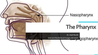
ThePharynx
• Situated behind the nasal cavities, the mouth, and the larynx and may be divided into
nasal, oral, and laryngeal parts.
• The pharynx is funnel shaped, its upper, wider end lying under the skull and its lower,
narrow end becoming continuous with the esophagus opposite the 6th cervical
vertebra.
• The pharynx has a musculomembranous wall, which is deficient anteriorly.
• It is replaced by the posterior openings into the nose (choanae), the opening into the
mouth, and the inlet of the larynx.
• By means of the auditory tube, the mucous membrane is also continuous with that of
the tympanic cavity.
Add a footer 2
3. TREYresearch
MusclesofthePharynx
• The muscles in the wall of the pharynx consist of the superior, middle, and inferior constrictor
muscles whose fibers run in a somewhat circular direction
• The stylopharyngeus and salpingopharyngeus muscles, whose fibers run in a somewhat
longitudinal direction.
• The three constrictor muscles extend around the pharyngeal wall to be inserted into a fibrous
band or raphe that extends from the pharyngeal tubercle on the basilar part of the occipital
bone of the skull down to the esophagus.
• The three constrictor muscles overlap each other so that the middle constrictor lies on the
outside of the lower part of the superior constrictor and the inferior constrictor lies outside the
lower part of the middle constrictor.
• The lower part of the inferior constrictor, which arises from the cricoid cartilage, is called the
cricopharyngeus muscle.
• The fibers of the cricopharyngeus pass horizontally around the lowest and narrowest part of
the pharynx and act as a sphincter.
• Killian’s dehiscence is the area on the posterior pharyngeal wall between the upper propulsive
part of the inferior constrictor and the lower sphincteric part, the cricopharyngeus.
3
4. TREYresearch
InteriorofthePharynx
• The pharynx is divided into three parts: the nasal pharynx, the oral pharynx, and the
laryngeal pharynx.
• Nasal Pharynx; Lies above the soft palate and behind the nasal cavities.
• In the submucosa of the roof is a collection of lymphoid tissue called the pharyngeal
tonsil.
• The pharyngeal isthmus is the opening in the floor between the soft palate and the
posterior pharyngeal wall.
• On the lateral wall is the opening of the auditory tube, the elevated ridge of which is
called the tubal elevation.
• The pharyngeal recess is a depression in the pharyngeal wall behind the tubal
elevation.
• The salpingopharyngeal fold is a vertical fold of mucous membrane covering the
salpingopharyngeus muscle.
Add a footer 4
5. TREYresearch
OralPharynx
• This lies behind the oral cavity.
• The floor is formed by the posterior one third of the tongue and the interval between the
tongue and epiglottis. In the midline is the median glossoepiglottic fold, and on each side the
lateral glossoepiglottic fold.
• The depression on each side of the median glossoepiglottic fold is called the vallecular.
• On the lateral wall on each side are the palatoglossal and the palatopharyngeal arches or folds
and the palatine tonsils between them.
• The palatoglossal arch is a fold of mucous membrane covering the palatoglossus muscle.
• The interval between the two palatoglossal arches is called the oropharyngeal isthmus and
marks the boundary between the mouth and pharynx.
• The palatopharyngeal arch is a fold of mucous membrane covering the palatopharyngeus
muscle.
• The recess between the palatoglossal and palatopharyngeal arches is occupied by the
palatine tonsil.
Add a footer 5
6. TREYresearch
LaryngealPharynx
• This lies behind the opening into the larynx.
• The lateral wall is formed by the thyroid cartilage and the thyrohyoid membrane.
• The piriform fossa is a depression in the mucous membrane on each side of the laryngeal inlet.
• Sensory Nerve Supply of the Pharyngeal Mucous Membrane
• Nasal pharynx: The maxillary nerve (V2)
• Oral pharynx: The glossopharyngeal nerve
• Laryngeal pharynx (around the entrance into the larynx): The internal laryngeal branch of the
vagus nerve
• Blood Supply of the Pharynx
• Ascending pharyngeal, tonsillar branches of facial arteries, and branches of maxillary and lingual
arteries
• Lymph Drainage of the Pharynx
• Directly into the deep cervical lymph nodes or indirectly via the retropharyngeal or paratracheal
nodes into the deep cervical nodes
6
15. TREYresearch
TheProcessofSwallowing(Deglutition)
• Masticated food is formed into a ball or bolus on the dorsum of the tongue and
voluntarily pushed upward and backward against the undersurface of the hard palate.
• This is brought about by the contraction of the styloglossus muscles on both sides,
which pull the root of the tongue upward and backward.
• The palatoglossus muscles then squeeze the bolus backward into the pharynx. From
this point onward, the process of swallowing becomes an involuntary act.
• The nasal part of the pharynx is now shut off from the oral part of the pharynx by the
elevation of the soft palate, the pulling forward of the posterior wall of the pharynx
by the upper fibers of the superior constrictor muscle, and the contraction of the
palatopharyngeus muscles.
• This prevents the passage of food and drink into the nasal cavities.
• The larynx and the laryngeal part of the pharynx are pulled upward by the contraction
of the stylopharyngeus, salpingopharyngeus, thyrohyoid, and palatopharyngeus
muscles.
Add a footer 15
16. TREYresearch
TheProcessofSwallowing
• The main part of the larynx is thus elevated to the posterior surface of the epiglottis,
and the entrance into the larynx is closed.
• The laryngeal entrance is made smaller by the approximation of the aryepiglottic
folds, and the arytenoid cartilages are pulled forward by the contraction of the
aryepiglottic, oblique arytenoid, and thyroarytenoid muscles.
• The bolus moves downward over the epiglottis, the closed entrance into the larynx,
and reaches the lower part of the pharynx as the result of the successive contraction
of the superior, middle, and inferior constrictor muscles.
• Some of the food slides down the groove on either side of the entrance into the
larynx, that is, down through the piriform fossae.
• Finally, the lower part of the pharyngeal wall (the cricopharyngeus muscle) relaxes
and the bolus enters the esophagus.
Add a footer 16
17. TREYresearch
PalatineTonsils
• The palatine tonsils are two masses of lymphoid tissue, each located in the depression on the
lateral wall of the oral part of the pharynx between the palatoglossal and palatopharyngeal
arches.
• Each tonsil is covered by mucous membrane, and its free medial surface projects into the
pharynx.
• The surface is pitted by numerous small openings that lead into the tonsillar crypts.
• The tonsil is covered on its lateral surface by a fibrous capsule.
• The capsule is separated from the superior constrictor muscle by loose areolar tissue, and the
external palatine vein descends from the soft palate in this tissue to join the pharyngeal
venous plexus.
• Lateral to the superior constrictor muscle lie the styloglossus muscle, the loop of the facial
artery, and the internal carotid artery.
• The tonsil reaches its maximum size during early childhood, but after puberty it diminishes
considerably in size.
Add a footer 17
18. TREYresearch
PalatineTonsils
• Blood Supply
• The tonsillar branch of the facial artery. The veins pierce the superior constrictor
muscle and join the external palatine, the pharyngeal, or the facial veins.
• Lymph Drainage of the Tonsil; The upper deep cervical lymph nodes, just below and
behind the angle of the mandible.
• Waldeyer’s Ring of Lymphoid Tissue
• The lymphoid tissue that surrounds the opening into the respiratory and digestive
systems forms a ring.
• The lateral part of the ring is formed by the palatine tonsils and tubal tonsils
(lymphoid tissue around the opening of the auditory tube in the lateral wall of the
nasopharynx).
• The pharyngeal tonsil in the roof of the nasopharynx forms the upper part, and the
lingual tonsil on the posterior third of the tongue forms the lower part.
Add a footer 18
19. TREYresearch
TheEsophagus
• A muscular tube about 10 in. (25 cm) long, extending from the pharynx to the stomach
• It begins at the level of the cricoid cartilage, opposite the body of the sixth cervical vertebra.
• It commences in the midline, but as it descends through the neck, it inclines to the left side.
• Relations in the Neck
• Anteriorly: The trachea; the recurrent laryngeal nerves ascend one on each side, in the groove
between the trachea and the esophagus.
• Posteriorly: The prevertebral layer of deep cervical fascia, the longus colli, and the vertebral
column.
• Laterally: On each side lie the lobe of the thyroid gland and the carotid sheath
• Blood Supply in the Neck
• The arteries of the esophagus in the neck are derived from the inferior thyroid arteries.
• The veins drain into the inferior thyroid veins.
• Lymph Drainage in the Neck; The lymph vessels drain into the deep cervical lymph nodes.
• Nerve Supply in the Neck The nerves are derived from the recurrent laryngeal nerves and from
the sympathetic trunks. 19