
Recomendados
Mais conteúdo relacionado
Mais procurados
Mais procurados (20)
Destaque
Semelhante a Splenic injuries
Semelhante a Splenic injuries (20)
Último
Último (20)
Splenic injuries
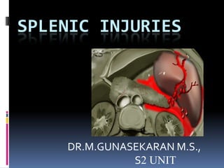
- 1. SPLENIC INJURIES DR.M.GUNASEKARAN M.S., S2 UNIT
- 2. SPLEEN 2nd most commonly injured solid organ in blunt injury abdomen after liver Situated against 9-11 ribs
- 3. SURGICAL ANATOMY Developed from dorsal mesogastrium In children,necessary for both reticuloendothelial and RBC production Pediatric spleen has thicker capsule and tough parenchymal consistency which implies reduced need of operative intervention Adult spleen weight about 100-250g
- 4. Situated posteriorly left upper abdomen Covered by peritoneum except at the hilum Posterior and lateral surface related to left hemidiaphragm and posterolateral lower ribs Lateral surface attached through splenophrenic ligament
- 5. Posteriorly related to left iliopsoas muscle & left adrenal glands Posteriormedial surface related to body & tail of pancreas Antromedially related to great curvature of stomach
- 6. Inferiorly related to distal transverse colon & splenic flexure Lower pole attached to colon through splenicocolic ligament These attachments require devision during mobilisation
- 7. BLOOD SUPPLY Receives blood supply from celiac axis 1.spleenic artery 2.short gastric vessels that connect left gatroepiploic A. & splenic circulation along greater curvature of stomach
- 8. BLOOD SUPPLY
- 9. Drains through splenic vein & confluence with inferior mesentric vein Through short gastric veins into left gastro epiploic vein
- 10. INITIAL ASSESMENT Importance of history- 1.victims located on the left side of car 2.type & nature of weapon is important in penetrating injuries 3.caliber of the gun
- 11. ON EXAMINATION Vitals are most important r/o left lower rib tenderness 14% patients with left lower rib tenderness have splenic injury In children plasticity of chest will have splenic injury without rib # Ecchymoses or abration over LUQ
- 12. SIGNS Kehr sign-is symptom of pain near tip of left shoulder,bcz of reffered pain from the diaphragmatic irritation P/A-generalised tenderness or LUQ tenderness May present with tachycardia ,Tachypnea, anxiety , Hypotension (shock)
- 13. INVESTIGATIONS In unstable patients necesesary investigation is hemoglobin,blood grouping and reservation of blood No specific labaratory studies specific to splenic injuries
- 14. PLAIN RADIOGRAPH The most common finding associated with splenic injury is left lower rib fracture. Rib fractures signify that adequate force has been transmitted to the LUQ to cause splenic pathology. classic triad indicative of acute splenic rupture (ie, left hemidiaphragm elevation, left lower lobe atelectasis, and pleural effusion)
- 15. DIAGNOSTIC PERITONEAL LAVAGE In the past Mainstay of diagnostic technique for abdominal trauma Peritoneal lavage useful when USG not available 10ml of blood or enteric contents (stool, food, etc.) constitutes a positive DPL,
- 16. Other positive findings include more than 100,000 RBCs/mm3, 500 WBCs/mm3, amylase 175 IU, and detection of bile, bacteria or food fibers. Levels of 10,000 RBCs/mm3 are typically used in cases of penetrating trauma Sensitivity-97-98% for blood Complication rate 1%
- 17. FAST (FOCUSED ABDOMINAL SONOGRAPHY IN TRAUMA) 1.non invasive procedure 2.quickly asseses viceral injuries,intra/retro peritoneal fluid collections 3.sensitivity varies from 42-93% due to operator dependency 4.specificity 90-98%
- 19. DISADVANTAGES 1.not reliably detect less than 100ml of blood 2.not identify injured hollow viscus 3.cannot reliably exclude in penetrating trauma
- 20. CT SCAN IOC ,even for clinically unstable patients Sensitivity-100% Specificity-98% “blush” which is due to ongoing blood loss and extravasation of contrast Pseudo aneurysms
- 21. MRI has also been used,in unstable patients which is less important Radio isotope scintigraphy & angiography are also used Diagnostic laparoscopy
- 22. AMERICAN ASSOCIATION FOR THE SURGERY OF TRAUMA SPLENIC INJURY GRADING SCALE
- 24. MANAGEMENT SPLENIC INJURY STABLE UNSTABLE GR 5- GR 1-4- SPLENECTOMY/ STABILISE THE CONSERVATIVE ART PATIENT EMBOLISATION LAPAROTOMY SPLENORRAPH ART Y/SPLENECTOM EMBOLISATION Y
- 25. Indications for initial nonoperative management hemodynamic stability absence of peritonitis CT scan No contrast extravasation absence of other injuries Transfusions - >2 PRBC’s
- 26. CONSERVATIVE Gr 1-4(stable)-hospitalisation -strict bed rest -vitals monitoring -serial USG &CT monitoring -tranfuse blood if necessary Measures taken to find out delayed splenic rupture, (48-72 hrs) in 4% of patients
- 27. SPLENORRHAPHY Parenchyma saving surgery of spleen The technique is dictated by the magnitude of the splenic injury Nonbleeding grade I splenic injury may require no further treatment. 1.superficial hemostatic strategies like fibrin glue,gel foam,argon beem coagulation,diathermy,topical thrombin 2.non absorbable suture repair 3.absorbable mesh wrap(poly galactin) 4.resectional debridement
- 28. SPLENORRHAPHY
- 29. SPLENECTOMY indications -Gr 5 injury -delayed rupture -increasing hematoma -clinically unstable of any grade -actively bleeding Open splenectomy with midline incision prefered
- 30. AUTOTRANSPLANTATION implanting multiple 1-mm slices of the spleen in the omentum after splenectomy. This technique remains experimental role controversial
- 31. EMBOLISATION Tc99/sulphur colloid labeled contrast angiogram to detect vascular damage Presence of extravasation of contrast in arterial phase (blush sign) Pseudo aneurysm pattern needs transarterial embolisation using polyvinyl alcohol/silicone/acrylic embolic spheres Can be given to reduce blood loss preoperatively
- 33. POST OPERATIVE COMPLICATIONS INTRAOPERATIVE EARLY POST OP LATE POST OP • haemorrhage • Hematoma/seroma • OPSI • Pancreatic injury • Wound infection • splenosis • Bowel • Subphrenic abscess injury(stomach & • Lung complication colon) • Atelectasis • Diaphragmatic • Pneumonia injury • Pl effusion • Portal vein thrombosis • DVT • Paralytic ileus
- 34. OPSI(OVERWHELMING POST SPLENECTOMY INFECTION) A rapidly fatal infection following removal of spleen Incidence-0.23-0.42% per year Occurs 1st few years after splenectomy Common organisms 1.s.pneumonia 2.h.influenza 3.n.meningitis Mortality rate -50-80%
- 35. Mechanism-organism with polysaccharide capsules need OPSONIZATION with IGg3 or C3B which attaches to special macrophages found in the spleen Post splenectomy patients lack of macrophages
- 36. SYMPTOMS Starts with flu like symptoms Meningitis or sepsis Rapidly progressive 12-48 hrs
- 37. OPSI
- 38. MANAGEMENT PREVENTION- pneumococcal vaccine(>2 yrs) administered within 24 – 48 hrs after splenectomy Meningococcal & H.influenza vaccine only in endemic areas Antibiotics- PENICILIN V 125mg bd(<3 yrs),250mg bd(3-14 yrs),500 mg bd (adults)