
Recomendados
Recomendados
Mais conteúdo relacionado
Mais procurados
Mais procurados (20)
Semelhante a EMBOLISM AND FILTERS USED IN CARDIOPULMONARY BYPASS
Semelhante a EMBOLISM AND FILTERS USED IN CARDIOPULMONARY BYPASS (20)
Último
Último (20)
EMBOLISM AND FILTERS USED IN CARDIOPULMONARY BYPASS
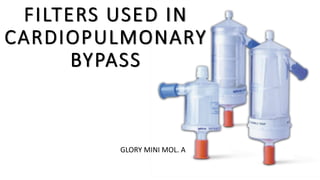
- 1. FILTERS USED IN CARDIOPULMONARY BYPASS GLORY MINI MOL. A
- 2. EMBOLISM DEFINITION: obstruction of an artery, by a clot of blood or an air bubble. • This emboli is categorized to oBiological emboli oForeign emboli oGaseous emboli
- 3. BIOLOGICAL EMBOLI FOREIGN EMBOLI GASEOUS EMBOLI BLOOD • CLOTS • FAT • BONE FRAGMENTS • FIBRIN • FOAM • TINY TUBING FRAGMENTS DUE TO SPALLINATION OF ROLLER • THREADS • CANULATION SITE • OPEN ATRIUM OR VENTRICLE MICRO MACRO EMBOLI EMBOLI < 40 MICRON >40 MICRON
- 4. Non gaseous micro emboli reported during cardiopulmonary bypass • Microthrombi(fibrin) • Platelet aggregates • Neutrophil aggregates • Red cell aggregates • Denatured protein • Fat or lipids • Cold-reacting antibodies • Bone fragments • Muscle fragments • Calcium particles • Cotton fibers • Plastic particles • Filter materials • Tubing materials • Metal • Talc • Thread • Bone wax • Microfibrillar collagen • Silicon antifoam
- 5. There are current technologies to decrease this embolic event delivered to patient • Membrane oxygenators •FILTER • Blood surface coating • Bubble traps • Emboli detection system
- 6. FILTERS USED IN CARDIOPULMONARY BYPASS
- 7. Blood Filters Depth filters • Consist of packed fibers of Dacron wool or polyurethane foam . • No defined pore size • These filters have large wetted surface areas to filter the blood by absorption , they are effective in trapping gross bubbles. Screen filters • composed of a woven mesh of polyester fibers • defined pore sizes • From 20 -40 μm • all of the arterial line filters used are the screen type
- 8. FILTER TYPE FILTER FUNCTION PORE SIZE FIGURE ARTERIAL LINE FILTER Used to remove micro emboli including gas emboli, fat emboli and cellular debris. 20 - 40 microns Pre-CPB filters Used during the priming to remove particulate contamination from circuit primer (Bacterial , microbial ,and endotoxin) 0.2 μm. (μm = micrometer = 1 (1/1000 millimeter Gas line filters Used to remove particulate and microbial contamination from gas lines 0.02 μm
- 9. Leukocyte depletion filters Reduces the levels of leukocytes from the arterial line or cardioplegia system . >40μm Cardioplegic Solution Filters By filtration of crystalloid solution 0.2-5 μm. Blood Transfusion filters used for more effective elimination of harmful blood components, micro- aggregates, and non-blood component particulate matter SCREEN FILTER OR DEPTH FILTER OR COMBINATION OF BOTH 10-40 μm Cell sever (Cell salvage) Used to remove debris such as foreign matter, fibrin, and cell clumps from salvaged blood . 20- 40μm.
- 10. ARTERIAL FILTER • Important component that limits the sources of embolic load to the patient through the perfusion circuit. • Arterial line filters are screen type composed of a woven mesh
- 11. HEMOCONCENTRATOR OR ULTRAFILTER • consist of hollow fiber semipermeable microporous membrane that allows the passage of water and electrolytes from the blood back to the circuit and waste to filtrate bag. • Pore size: 180-200 μm in diameter and pores of 5-10 nm.
- 12. CYTOSORB • CytoSorb cartridge consists of a highly porous high-tech polymer • The tiny beads can bind to a wide range of inflammatory mediators such as cytokines, chemokines, and anaphylline toxins by virtue of their structure, provided there is a corresponding gradient between whole blood and polymer.
- 14. WHEN TO USE SEPSIS: • hyperinflammation Onset of shock within the last 24 hrs Signs of capillary leak – e.g. positive fluid balance • Development of at least one more organ dysfunction Kidney, lung, liver, coagulation, neurologic impairment • Systemic markers of infection PCT > 3 ng/l in case of bacterial or fungal sepsis High IL-6 levels (e.g. >500 pg/ml)
- 15. INTRAOPERATIVE USE At the start of CPB in case of one or more of the following risk factors: Age > 75 yrs Preoperative activation of the immune system • Endocarditis • Cardiac failure with inotropic therapy • Preoperative leukocytosis (> 12,000/μl) • Organ dysfunctions, e.g. kidney or liver Long CPB duration expected (>120 min) High risk for postoperative need for ECMO Procedures with higher risk for complications and/or SIRS • Combination procedures (valve repair/- replacement, CABG) • Redo procedures • Aortic surgery with hypothermic circulatory arrest • LVAD implant Anytime during CPB Patients with low primary risk but unexpected course • Unexpected, significant prolongation of anticipated CPB time • Intraoperative development of a severe SIRS • Intraoperative complications with expected development of severe SIRS
- 16. POSTOPERATIVE USE Immediately upon arrival in ICU • Postoperative continuation of intraoperative CytoSorb treatment • Manifest severe SIRS upon arrival Postoperative (0-48h) development of SIRS with or without proof of infection • Patient cannot be stabilized clinically with standard medical treatment • Impaired hemodynamics (shock) Onset of shock (Norepinephrine > 0,3μg/kg/min or rapidly increasing) Signs of capillary leak – e.g. positive fluid balance Onset of at least one more organ dysfunction • Mechanical ventilation • Acute kidney failure with need for RRT Systemic markers of infection • PCT > 3ng/l in case of bacterial or fungal sepsis – • High IL-6 levels (e.g. > 500 pg/ml)