![Date Patient ISex IAge
Initials
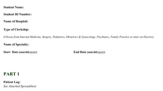
PATIENT LOG
Subjective I Objective I Assessment
C.ut. •" 1,l...
Plan Final Diagnosis
o11-'iLS.·f. TM ~~~JS25, (JQ~~~q, 11~1)1£-, Sol~. ICHF r,::.i~:b'.'-l Io;.t.+;t.S- - l"'fy,"'s,pj~ Htc..-y+(Hr
lt>,./t.S]:f.t>·I/V lll~l~~~~~~-r;L1~h•Le~ . IC'AII..1;-h·r ' lt~.~JNe-.3tfoi~P).! #..
Observations
~ 1::-..fh.rJ "'- c.kn.....·, '>W..
lv' /" I7. s. lf1J:q.5l.Jh.;b, ~J... ' I Wq~c..s c:k1>iQ~~'.r;-rl1~q~ -l ~=~.l>,-z., f,/f.A~,~~ .
1"--,.--.--'-+__._,_~--+--'~~,1,4-1....._-!J.-!-.:.=....;.;..-~-1 ~~ IL l...elito MbJ: r
lol Q ]T:f... TMT9CUK~-~~J.t~r l I.J~OI'j;,.;.~ ~t>MI C....~;..h$' I .,.~t,'E.•. M~ l lic.k b;-k.
1~ ro I 1 ,-- :-~.TMT'3{. I"J"o~~:;--~ - It ~.l-S .r.~l1"';~ r>._~;o. I'Pi~"lcl.r . I'P.(>x~~"'a~s- ----~~.t. o.!'M O..C.~ ~
~:~~~-~~~~~~~~~~~~~~~~~~~~~~~~-+~~~~~~r.
J (.}
~~~~ I~·.~~.:.·_+-L+~~~.J....!Ill-~+--~t':!!:!Ll~~--~~lU:<~~!:L_~ll.l~~l.Ju!4-!~~...12.::1ill<.~-+-A-~-~---:.'ic-
OIO~ I "'1 ...... 1 M i b~ I Soe,"".."'~· I !..&-1-.Ao1."'1.~$ I P~ 1~~[_~. J r~...~ I I (
lto,o:' l firs l f l l,o l f,H~ 1 -f" B~,-f~~.t...v I K:r~t...... F..1~ I Dl~~tii.lS' l A~b
o/O&{ IT. U. I M 1-:trl~c Vt-..cJ.J..I.....~~.~-le. ..,.,J..-Ju.ie ls~~~~~l or~Jit;c. ,~ ;~J..
ID 0 y IL.:J· IM 1 4~ I D;~~ - --UTfo-OJe..2-CV'IAt ,;t reg,&.:~ "?I I LC.k.f ~ ~e.:J.~ I f'tl'~.-i!"A
( t-7- lfv,G. 1Ml:_.._llill:_~~~~"'"'-:::l0rv~~U)~c!IJ~ -lj:"-']t;,;{k'it'. !"~ -~"'1 NSA1.0~ I fo,~o~C.. ~~
l0 1t l- ]<- · M IM Ir , Ip~, "'~'~ " Ib~~~~~~l»>tV I~ ~~ ' I~ft ·Q..T.~ _~~""""' I MB-"'1-W .
1) ((!~ ~~.~' I n 1"5 I s~~t:P 0~/ IS·-ts-hll.Zfc. (IIi I~~c. ~iiT .o..~~ ~ lh..-~c.. S~s·i'~ l(~~..,..t:
to 'of I r.V.. IVI' 1 ~8 I .~.:a I IRl.Q,__ .l..-~C).5 I kki?O'l'll l t>ttr~.. ~-4 ~lubUR~ H.tJ..~ !~; l-.
lklr l :r~~- I M 1 '-S I ~of.>.M'(~Loa.i... I..V e._ ~.'t I P.Jt~OI, l ~rz{ti~· INoSOCC1M-· ~ Vi Jht&t-.t.¥0. p
tQ rtv IP. J~ l F I ~ f J.~v'{ o;o.;I-~ I ~ c&er, ~ Jri. fP~~-:-~1. l uJ..,h•5~~r 1 Ic.Jctp. ~~. I Q
toff; I A :t~-l r Y l !'t l ~hillf'f"V" ~ ~~jO' lr't1 oJ~~t--'l:sT~IoJ -~~Ju -.lt~'~;~t_ ~"'hc.-·1[)"'1
~~ lA.A If l , I f<.A-~ lf:.'~-.,= -~lht:k..,~-'1'-Sh:U~)~'~l fA"~{ -- It'1h~14~ -"' 1~ £~.
'o'.I r I ~- . '1. I~ IS I ~{' ~, ~, '""] k, J tMt.o ~~~ I(J -~o~' I L'ifl'o,..~ I""-'qplt. ~~ •
tolr> IOM. IM It v ~-"1,)~~ I I [;,~,CJS-.t'!.Q., I H~l)f_rj,..f, ~11 A+o~t:h ~-T~ .... t;l7.~{r._...;,'
,p I ,( I <, J I;; lt.tt IL.f':. ~,..... P·~tl s~l l(~uc.e.d RoM lt"'kle e..~...i: "'l~(Q. eo....l'(_,fttlA'~kl.t ·~o roo.;"'
luf(~ I L.C· IM I?h-olb)l'ooe •JA#~ il Pw~c-1111 lt·~·~ ] 1-cc._i'""~.ll"''l ~ ~~~~ .... ~~. I L~~o~"'~·kl.c'~~"' Ls
.of~~-S ~L I~1- ll.l I ~Y [ ft'";~~~ -,v" ~+· ·l "t~t1'~11'. ] S~~".S. I"~~eo~ lv-t· ~"'-
olZ~~.LJ, MlM I"' I C.O~<V ~riJ.~ ~a. ,n,.~ leo""l~~ ~·--J Kr..~~,-~~. r t.~i:. • •
) )p ...~ d u
,
~
t
....J)
G
j](data:image/gif;base64,R0lGODlhAQABAIAAAAAAAP///yH5BAEAAAAALAAAAAABAAEAAAIBRAA7)
Recomendados
Recomendados
Mais conteúdo relacionado
Mais procurados
Mais procurados (20)
Semelhante a Clinical Student Portfolio - Gagandeep Singh Anand - LINKEDIN
Semelhante a Clinical Student Portfolio - Gagandeep Singh Anand - LINKEDIN (13)
Clinical Student Portfolio - Gagandeep Singh Anand - LINKEDIN
- 1. AMERICAN UNIVERSITY OF ANTIGUA College of Medicine Student Portfolio Student Name: Student ID Number: Name of Hospital: Type of Clerkship: (Choose from Internal Medicine, Surgery, Pediatrics, Obstetrics & Gynecology, Psychiatry, Family Practice or enter an Elective) Name of Specialty: Start Date (mm/dd/yyyy): End Date (mm/dd/yyyy): PART 1 Patient Log: See Attached Spreadsheet GAGANDEEP SINGH ANAND NORTHSIDE MEDICAL CENTER 5TH SEMESTER INTERNAL MEDICINE (3) + FAMILY PRACTICE (2) 09/23/2013 11/01/2013
- 2. Date Patient ISex IAge Initials PATIENT LOG Subjective I Objective I Assessment C.ut. •" 1,l... Plan Final Diagnosis o11-'iLS.·f. TM ~~~JS25, (JQ~~~q, 11~1)1£-, Sol~. ICHF r,::.i~:b'.'-l Io;.t.+;t.S- - l"'fy,"'s,pj~ Htc..-y+(Hr lt>,./t.S]:f.t>·I/V lll~l~~~~~~-r;L1~h•Le~ . IC'AII..1;-h·r ' lt~.~JNe-.3tfoi~P).! #.. Observations ~ 1::-..fh.rJ "'- c.kn.....·, '>W.. lv' /" I7. s. lf1J:q.5l.Jh.;b, ~J... ' I Wq~c..s c:k1>iQ~~'.r;-rl1~q~ -l ~=~.l>,-z., f,/f.A~,~~ . 1"--,.--.--'-+__._,_~--+--'~~,1,4-1....._-!J.-!-.:.=....;.;..-~-1 ~~ IL l...elito MbJ: r lol Q ]T:f... TMT9CUK~-~~J.t~r l I.J~OI'j;,.;.~ ~t>MI C....~;..h$' I .,.~t,'E.•. M~ l lic.k b;-k. 1~ ro I 1 ,-- :-~.TMT'3{. I"J"o~~:;--~ - It ~.l-S .r.~l1"';~ r>._~;o. I'Pi~"lcl.r . I'P.(>x~~"'a~s- ----~~.t. o.!'M O..C.~ ~ ~:~~~-~~~~~~~~~~~~~~~~~~~~~~~~-+~~~~~~r. J (.} ~~~~ I~·.~~.:.·_+-L+~~~.J....!Ill-~+--~t':!!:!Ll~~--~~lU:<~~!:L_~ll.l~~l.Ju!4-!~~...12.::1ill<.~-+-A-~-~---:.'ic- OIO~ I "'1 ...... 1 M i b~ I Soe,"".."'~· I !..&-1-.Ao1."'1.~$ I P~ 1~~[_~. J r~...~ I I ( lto,o:' l firs l f l l,o l f,H~ 1 -f" B~,-f~~.t...v I K:r~t...... F..1~ I Dl~~tii.lS' l A~b o/O&{ IT. U. I M 1-:trl~c Vt-..cJ.J..I.....~~.~-le. ..,.,J..-Ju.ie ls~~~~~l or~Jit;c. ,~ ;~J.. ID 0 y IL.:J· IM 1 4~ I D;~~ - --UTfo-OJe..2-CV'IAt ,;t reg,&.:~ "?I I LC.k.f ~ ~e.:J.~ I f'tl'~.-i!"A ( t-7- lfv,G. 1Ml:_.._llill:_~~~~"'"'-:::l0rv~~U)~c!IJ~ -lj:"-']t;,;{k'it'. !"~ -~"'1 NSA1.0~ I fo,~o~C.. ~~ l0 1t l- ]<- · M IM Ir , Ip~, "'~'~ " Ib~~~~~~l»>tV I~ ~~ ' I~ft ·Q..T.~ _~~""""' I MB-"'1-W . 1) ((!~ ~~.~' I n 1"5 I s~~t:P 0~/ IS·-ts-hll.Zfc. (IIi I~~c. ~iiT .o..~~ ~ lh..-~c.. S~s·i'~ l(~~..,..t: to 'of I r.V.. IVI' 1 ~8 I .~.:a I IRl.Q,__ .l..-~C).5 I kki?O'l'll l t>ttr~.. ~-4 ~lubUR~ H.tJ..~ !~; l-. lklr l :r~~- I M 1 '-S I ~of.>.M'(~Loa.i... I..V e._ ~.'t I P.Jt~OI, l ~rz{ti~· INoSOCC1M-· ~ Vi Jht&t-.t.¥0. p tQ rtv IP. J~ l F I ~ f J.~v'{ o;o.;I-~ I ~ c&er, ~ Jri. fP~~-:-~1. l uJ..,h•5~~r 1 Ic.Jctp. ~~. I Q toff; I A :t~-l r Y l !'t l ~hillf'f"V" ~ ~~jO' lr't1 oJ~~t--'l:sT~IoJ -~~Ju -.lt~'~;~t_ ~"'hc.-·1[)"'1 ~~ lA.A If l , I f<.A-~ lf:.'~-.,= -~lht:k..,~-'1'-Sh:U~)~'~l fA"~{ -- It'1h~14~ -"' 1~ £~. 'o'.I r I ~- . '1. I~ IS I ~{' ~, ~, '""] k, J tMt.o ~~~ I(J -~o~' I L'ifl'o,..~ I""-'qplt. ~~ • tolr> IOM. IM It v ~-"1,)~~ I I [;,~,CJS-.t'!.Q., I H~l)f_rj,..f, ~11 A+o~t:h ~-T~ .... t;l7.~{r._...;,' ,p I ,( I <, J I;; lt.tt IL.f':. ~,..... P·~tl s~l l(~uc.e.d RoM lt"'kle e..~...i: "'l~(Q. eo....l'(_,fttlA'~kl.t ·~o roo.;"' luf(~ I L.C· IM I?h-olb)l'ooe •JA#~ il Pw~c-1111 lt·~·~ ] 1-cc._i'""~.ll"''l ~ ~~~~ .... ~~. I L~~o~"'~·kl.c'~~"' Ls .of~~-S ~L I~1- ll.l I ~Y [ ft'";~~~ -,v" ~+· ·l "t~t1'~11'. ] S~~".S. I"~~eo~ lv-t· ~"'- olZ~~.LJ, MlM I"' I C.O~<V ~riJ.~ ~a. ,n,.~ leo""l~~ ~·--J Kr..~~,-~~. r t.~i:. • • ) )p ...~ d u , ~ t ....J) G j
- 3. Case 1: (500 word maximum) Case 2: (500 word maximum) PART 2 List 3 cases and discuss in depth: Pathophysiology Differential Diagnosis Findings to Support Final Diagnosis Treatment Options Mr. S is a 64 year-old male who presents with an improvement in shortness of breath since the day before. His condition improves when he sits up in his bed than lying down flat. He reports generalized fatigue relieved upon resting. He recently got a CABG, before which he had surgery for a toe amputation. He has had some trouble with moving bowels for 2 days and feels bloated. Though this patient experiences shortness of breath, this patient has had a previous coronary artery bypass graft intervention. In this case the pathophysiology is pertaining to atherosclerosis of coronary arteries. Atherosclerosis do not occur in a random fashion, hemodynamic factors interact with activated vascular endothelium. Atherosclerotic plaques, may require 10 – 15 years for its full development. The major branches of the thoracic and abdominal vessels as well as the large conduit vessels of the lower extremities are in high risk. One study suggests that low shear segments in the coronary arteries develop greater necrotic core progression and constrictive remodeling whereas high shear segments develop greater necrotic core and calcium progression, regression of fibrous and fibrofatty tissue, and excessive extensive remodeling. The first phase of atherosclerosis histologically presents as focal thickening of the intima with an increase in smooth muscle cells and extracellular matrix. Lipids accumulate early in fatty streak formation, this yields both intracellular and extracellular deposits. A small dermatan sulfate proteoglycan called by Biglycan, is detected in the artherosclerotic coronary artery segments. This binds and traps lipoproteins, including apolipoprotein E, VLDL remnants, LDL, and HDL. The fatty streak also consists of macrophages with a variable number of T lymphocytes. Macrophages are engorged with lipids are called foam cells, the hallmark of the early atheroma. As these lesions expand, more smooth muscle cells migrate into the intima. The smooth muscle cells within the deep layer of the fatty streak are susceptible to apoptosis, which is associated with further macrophage accumulation and micro vesicles that can calcify, perhaps contributing to the transition of fatty streaks into atherosclerotic plaques. Some of the differential diagnosis includes: angina pectoris, Buerger disease, dilated cardiomyopathy, coronary artery vasospasm, diabetes mellitus [type1, type 2], giant cell arthritis, familial hypercholesterolemia, hypertension, myocardial ischemia, myocarditis, and many more. A proper work up of atherosclerosis requires lipid studies, echo, nuclear imaging studies, CT, serum markers, C– reactive protein,coronary angiography, ultrasonography. Proper treatment requires a multitude of pharmacological strategies. It requires the treatment of low HDL levels and high triglyceride levels in patients with diabetes, the use of ace inhibitors to reduce blood pressure, antiplatelet agents for acute coronary events, pharmacological treatment of angina, revascularization procedures, and hormone therapy concerns. http://emedicine.medscape.com/article/153647-differential Ms. J is a pleasant 77 year-old female who presents with persistent diarrhea for 3 weeks, at 4-5 times/day. It is of watery consistency, brown color, foul smell, with traces of blood – though the patient reports of internal hemorrhoids as a possible reason – no mucus, and of undigested food. The patient reports increased urge to void after consuming a meal and a coinciding BM with micturation, in addition, she reports of a progressively worse frequency. She does not report of any active pain, but of slight lower abdominal discomfort for 15-20mins after a BM. Ms. J reports of having a previous episode with similar symptoms which was diagnosed as pseudo membranous colitis; she reports of no nausea, vomiting, sick contact or recent travel. Ms J also reports of acute stress due to a sickness in the family, this could also present with GI symptoms. She was prescribed Flagyl (10 days), for which she is on her 10th day. She was advised by her PCP to consume bananas and cheese to help with the diarrhea with no improvement. Ms. J reports having a dental procedure one month ago in which amoxicillin (4 tabs) were used. She also stopped taking her monthly Simponi injections for her RA because she believed it was the cause. The pathophysiology of the C. Difficile is particularly specific to toxin A and B. once the normal colonic flora is disturbed, Clostridium difficile is able to release toxins that cause you close of inflammation and damage.The toxins produced by Clostridium difficile are toxin a and toxin B, both are high molecular weight proteins capable of binding to specific receptors on the intestinal mucosal hello there cells. These toxins gain entry within the cell by catalyzing an alteration of the Rho proteins. These proteins help with actin polymerization, cytoskeleton architecture, and cell movement. Once this protein is glycosylated the pathogenesis of Clostridium difficile appears. Some differentials are Chron disease, diverticulitis, viral gastroenteritis, intra-abdominal sepsis, irritable bowel syndrome, malabsorption, salmonellosis, shigellosis, ulcerative colitis, and vibrio. Findings support clustered index of infection are leukocytosis, acute kidney injury, electrolyte imbalances, dehydration, hypoalbuminemia, and ansarca. Guidelines indicate GDH EIA, and stool cytotoxin test. In addition, endoscope he can also be performed,wherein yellowish white 2 to10 mm plaques overlying erythematous, edematous mucosa. In addition CT scanning can also be performed, where marked colonic wall thickening is the most common or finding. Other features include ascites, irregularity of the bowel wall, and pericolonic stranding. Treatment options include pharmacological management, fecalmicrobiota transplantation, and surgical intervention. Pharmacologically speaking in mild to moderate infection patients should receive oral metronidazole or oral vancomycin, 10 – 14 days. In severe or severe – complicated infection, oral vancomycin should be used for 10 – 14 days. In relapsed management the first recurrence requires metronidazole while the second recurrence requires vancomycin. Though probiotics are not recommended treatment for active Clostridium difficile, it can be a supplement during preventative care. http://emedicine.medscape.com/article/186458-overview
- 4. Case 3: (500 word maximum) Ms. J is a pleasant 42 year-old female who presents with an acute left ankle sprain and reported through the ER this morning. She describes spraining her ankle while walking down the stairs and laterally rolling the ankle. She reports a gradually improving, “on-and-off” pain with inflammation, which is aggravated by lateral stretching and relieved by compression and elevation. She reports of using Ultram (synthetically acting opiod analgesic) but has SE of itching. She also taking Celecoxib for her previous diagnosis of tenosynovitis. The patient also reports of bed bug bites on her left arm and right thigh. The lateral ankle complex is composed mainly of the anterior talofibular, calcaneofibular, and posterior talofibular ligaments, which is the most commonly injured site. About 85% of such sprains aren inversion sprains of the lateral ligaments, while 5% are eversion sprains of the deltoid or medial ligament. In addition 10% are syndesmotic sprains. An ATFL is the most likely component of the lateral angle complex to be injured in a lateral ankle sprain. As well as during force dorsiflexion the PTFL can rupture. In the case of extreme external rotation this disrupts the deep deltoid on the medial side and adduction in neutral and dorsiflexed position can disrupt the CFL. In the case of plantarflexion, the ATFL can be injured. Ankle spurs may occur at any of the bony ligament attachments. It is quite common to see an anterior spur at the neck of the Tallus, where the anterior ankle capsule attaches. This is a result of ossification of a hematoma organization associated with anterior ligament sprains. Some differential diagnoses included a fracture where an individual with an ankle sprain can almost always walk on the foot albeit carefully and with pain. Another diagnosis is neurovascular compromise, here we look for patient complaining of a cold foot and paresthesia. If pain persists after rehabilitation, consider intra-articular meniscoid lesions, peroneal tendon subluxation, talar dome fracture, anterior process fracture, complex regional pain syndrome. In addition Achilles tendon ruptures can also mimic ankle sprains. Other factors could be ankle impingement syndrome, ankle fractures, gout ,navicular fracture, as well as stress fractures. To do a work up consider plain radiographic imaging, stress view radiographic imaging, CT scanning, MRI, bone scanning,arthrographic imaging. Pain reduction is essential, but improvement of any loss of motion, strength, and or proprioception is equally important. A meta-analysis found that neuromuscular rehabilitation results in more rapid improvements and function. For re-current lateral ankle sprains, treatment should begin with trial of conservative therapy for approximately 2 to 3 months. http://emedicine.medscape.com/article/1907229
- 6. ' ACADEMIC ACTIVITIES (lECTURES/GRAND ROUNDS/CONFERENCES ETC.) Date -:fntWAtion " ~~~0<>. ~OJ''"'lo.-hevt Topic l-'4/~' D~. <.~r""OI." ~In' 1~ Mov~·,~ R.a..tu~Y+. l7.b.lb' 1 "1'1o'(' ~ ~ "&"/o4 floo n c;. o"'--uU'-tt. ~ "1.A. ~~~ ULt ~ ~v4 ft }.,1-[0' ~QQV_tA.~ - ~t. ~~ -?....;... 'J 7../ (' '"I.t1 ~'"··· o... -...1. zn~ ltaMOlA.J-- U~~~ • gJ h> ·c,~AtJO ~wos , 'iI1) 'IM -1'1r~~~ Rt.wr4- ~I to t:.S f.xllt" E l>r. ·~, ~ ~1b 1c,oOt.lC.. ~)(ltM - Dr . ~ )vv 2.~Jo' H<L' r.s - ~.- ·r--· N.-~~ Q l-S ~~~ 1- ~w·a.~~,.4.~ 'to~t:¥~P$ ?.olo' ~- _W{_()____::_ ~._l~<x , lol'> 0~,e-U n.~~:.-~ t"'- -~M Lb liD M~.a.. (}~ h!.wo -~1~.-. ~-bv-n • J,S/lv £f-NT ~lo Ak. -~ "' F...~ ~"'""' · _j_(lo t,lh;.;L D. ~'~ It ~(lt. ) 0...(r!.. ft.v'r -p~""" ' '2..fu M.~r lA ~o, £'<~"""'- 3o!o· I ~oon ConWeV!d...- C.onf'-1.'- ;on ...
- 7. Week Faculty Initial PART3 Procedure Log: See attached Spreadsheet PART4 Academic Activities: See attached Spreadsheet Formal Case Presentations (If Applicable): ~e attached Spreadsheet L,~c~ur~s/Gr;m{,i Rounds/ Conferel'lce Atte!lded: See attached Spreadsheer Faculty Comments ---__.... Weekly Faculty Sign-off 4 5 6 7 5_5 8 9 10 11 12