
Recomendados
Mais conteúdo relacionado
Semelhante a Blood supply of the brain (1).pptx
Semelhante a Blood supply of the brain (1).pptx (20)
Último
Último (20)
Blood supply of the brain (1).pptx
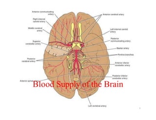
- 1. Blood Supply of the Brain 1
- 2. 2 Arterial supply • The brain is supplied by 2 pairs of arteries • rt & lt internal carotid artery (ICA) • rt & lt vertebral arteries A. Internal carotid artery • arises in the neck as one of the 2 terminal branches of the C.C.A. • has four parts –Cervical part –Interapetrous part –Intracavernous part –Intracranial part
- 3. 3
- 4. 4 • Each I.C.A leaves the cavernous sinus by piercing the dura mater and ascends in the subarachnoid space lat. to the optic chiasma • In the subarachnoid space each gives off the following branches: – Ophthalmic: which enters the optic canal below optic nerve to supply the orbit – Posterior communicating: which Joins the post. cerebral to establish the circle of Willis – Anterior choroidal: which supplies the choroid plexus of the lat. ventricle. • It ends below the ant. perforated substances of the brain by dividing into 2 terminal branches: – The ant. cerebral a (the smaller branch) – The middle cerebral a (the larger branch)
- 5. 5
- 6. Clinical importance • the A.C.A supplies 3 important regions; – The motor & sensory areas of the lower limb in the paracentral lobule – The septal region where a small lesion may result in prolonged unconsciousness – The corpus callosum obliteration of its blood supply may result in apraxia (inability to perform purposeful movements in spite of intact muscle
- 7. 7 Middle Cerebral artery (M.C.A) • arises below the ant. perforated substance as the larger of the 2 terminal branches of I.C.A • ends on the surface of the insula by breaking up into many terminal branches
- 8. 8
- 9. 9
- 10. Clinical importance: • the M.C.A supplies: – the motor & sensory areas for the whole body except the lower limbs – the auditory area in the sup. temporal gyrus – motor speech area in the inf. frontal gyrus – most of the internal capsule Obstruction of its blood supply----Hemiplegia
- 11. 11
- 12. 12 B. The Vertebro-basilar System • Vertebral artery – each vertebral a arises in the root of the neck as a branch from the 1st part of subclavian – enters the cranial cavity through the foramen magnum • Inside the skull, the rt & lt arteries unite at the lower border of the pons to form the basilar artery. • The vertebral arteries enter the cranial cavity & give rise to 2 terminal branches: – a medial terminal br. supplying the inf. vermis of the cerebellum – a lateral terminal br. supplying the post. part of inf. surface of cerebellum
- 13. Clinical importance of P.C.A: • its supplies: – the centre of smell in the uncus. – the whole visual cortex in the occipital lobe – most of thalamus – most of the midbrain – most of the choroids plexus of the 3rd & lat. ventricles. 13
- 14. 14 Circulus arteriosus of Willis • it is an arterial anastomosis b/n arteries supplying the brain • located in the interpeduncular cistern around the interpeduncular fossa.
- 15. Desalegn Tadesse, Jimma University 15
- 16. Desalegn Tadesse, Jimma University 16
- 17. • The following arteries enter in the formation: – Rt. & Lt. anterior cerebral arteries – the ant. communicating a . connecting the 2 cerebral arteries – the Rt. & Lt. ICA – the Rt. & Lt. post communicating aa. branches of ICA – The rt and lf post cerebral arteries
- 18. 18 Venous Drainage of the Brain (the veins are thin walled & valveless) • The superior cerebral veins drain into the superior sagittal sinus • Inferior and superficial middle cerebral veins drain into the straight, transverse, and superior petrosal sinuses. • The great cerebral vein (of Galen) is a single, midline vein formed inside the brain by the union of two internal cerebral veins; – it ends by merging with the inferior sagittal sinus to form the straight sinus • The superior and inferior cerebellar veins drain from the cerebellum into the transverse and sigmoid sinuses.
- 19. 19