SlideShare uma empresa Scribd logo
1 de 1
PPARα Expression in Postprandial Python Liver
Introduction
Eric Cobb, MCDB: 4202-800
Background:
PPARα (Peroxisome Proliferator-Activated Receptor ahlpa) is a
type II nuclear receptor has been identified as the master
regulator of hepatic lipid metabolism. Additionally, PPARα has
been shown to regulate: fatty acid β-oxidation, glucose
metabolism, amino acid metabolism, bile acid synthesis and
hepatocyte proliferation. Similar to other members of the
nuclear receptor superfamily PPARα heterodimerizes with
retinoid X receptor (RXR) and binds to a DNA response element,
in this case the peroxisome proliferator response element
(PPRE). Corepressors are recruited to the PPARα/RXR complex,
preventing transcription of target genes until the a ligand is
present to bind PPARα. Natural ligands of PPARα include a
variety of dietary fatty acids (FA) and other molecules
resembling FA structures including Acetyl-CoAs, Enoyl-CoAs,
and endocannabinoids. Synthetic ligands such as fibrates is
used for the treatment of dyslipidemia associated with type II
diabetes mellitus. Upon ligand binding causes a conformational
change in the PPARα/RXR complex resulting in the dissociation
of corepressor. Thus allowing for essential coactivators such as
PBP/MED1, to promote transcription of genes such as CD36,
Scd1, and Cyp7a1 to name a few. PPARα has also shown to
influence other lipogenic genes such by regulating transcription
factors SREBP-1c and LXRα. Deficiency or absence of PPARα
results in hepatic steatosis and hypolipedemia, indicating that
PPARα is an essential gene for the regulation of lipid levels in
the liver.
Hypothesis:
Considering the high levels of TAGs and FAs in python serum
after 1 day post fed, genes involved in lipid uptake and break
down should increase in order to decrease serum lipid levels
back to normal by 6-10 days post fed. Because PPARα activate
plays such a large role in the regulation of lipid metabolism I
expect to see an increase in PPARα expression between 1 and 6
days post fed. As this is the range in time points showing a
decrease in serum lipid levels.
Department of Molecular, Cellular, and Developmental Biology
University of Colorado-Boulder
Quantitative PCR Results
The Burmese Python:
Burmese Python undergo drastic physiological changes after
feeding. Firstly, the python can digest a meal 1.6 times it’s own
body weight which would be equivalent to a 145lb man eating a
232lb meal. Despite the size of their meal, digestion is
completed only 6-10 days later. After only 1 day post fed, the
python shows an substantial increase in glucose metabolism, a
rapid decrease in pH, and increase an increase in triacylglyceride
(TAGs) levels that would be fatal to humans. Furthermore,
organs such as the liver and the heart nearly double in size
through hyperplasia and hypertrophy respectively. The python
proves to be an extreme example of regulation of physiology
that is worth exploring to uncover ways to improve lipid
regulation and healthy organ hyperplasia and hypertrophy in
humans. Which could be used for target therapies for diseases
like diabetes and heart disease.
Postprandial Python Serum Lipid LevelsIntroduction
PPARα Null Mice Results in Hepatic Steatosis
PPARα -/- fasted mice results in hepatic steatosis: (A) Image (Left) shows WT liver,
image (Right) shows 66hr fasted PPARα -/- liver. Figures (B,D,F) are Fed mice and
(C,E,G) are fasted 48 hr mice, all of which are stained with Oil Red O-stains (stains
neutral triglycerides and lipids. Figures (B,C) are PPAR +/+, Figures (D,E) are PPAR -
/-, and Figures (F,G) is a double knockout PPAR -/- and AOX -/-.
Methods
Primer Design:
• Obtained mRNA sequence of PPARα from NCBI
and mapped exons
• Ran a Nucleotide Blast of mRNA sequence and
assembled python PPARα transcript from
overlapping sequences
• Validated gene product using Expasy Translate and
Protein Blast
• Used Primer3 to design optimal primers and
validated them with Primer Blast
Forward Primer: 5’-CTTGTGGGGAAAGCCAGTAA-3’
Reverse Primer: 5’-CACTGGCAGCAGTGGAAAAT-3’
Experimental Design:
• Isolated RNA from liver tissue homogenates from different
time points (Fasted-15 days post fed) using a Quiagen
RNeasy Mini Kit protocol
• Determined the concentration and purity of our isolated
RNA using spectrophotometry to obtain 260/280
absorbance ratio
• Conducted an analysis of the integrity of our isolated RNA
using gel electrophoresis
• Synthesized cDNA from isolated RNA using Superscript III
reverse transcriptase
• Conducted PCR using our designed Primers and synthesized
cDNA and ran the products on a gel to determine if our
primers are amplifying products of the same size as the
target gene
• Assessed relative gene expression of target genes compared
to reference genes using quantitative PCR (qPCR)
Conclusion
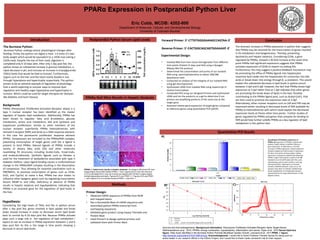
Quantitative PCR Relative Expression of
PPARα: A) Expression of PPARα in python liver
(Fasted-15dpf) relative to GAPDH reference
gene expression. At 3dpf there is a 52 fold
increase in PPARα expression. Fasted expression
showed roughly one fold increase in expression.
All other time points all show higher relative
expression compared to GAPDH indicating that
PPARα is high in abundance in python liver. B)
Standard Curve for qPCR plate with the y-axis
representing cycle number and the x-axis Log
Starting Quantity. Target gene of unknown
samples amplified between cycles 16-21. All
samples were done in triplicate. C) Melt Peak
shows amplification of a single gene product
represented by the single peak between 75-80
degrees Celsius.
A)
B) C)
The dramatic increase in PPARα expression in python liver suggests
that PPARα may be essential for the transcription of genes involved
in FA metabolism and lipogenenesis. Thereby, preventing
lipotoxicity and hepatic steatosis. Considering CD36, a gene
regulated by PPARα, showed a 36 fold increase at the same time
point PPARα had significant expression suggests that PPARα
activates expression of CD36 to import circulating FAs.
Furthermore, this may suggest a positive feedback mechanism may
be promoting the efflux of PPARα ligands into hepatocytes
maximize lipid intake into the hepatocytes for conversion into bile
acids or break down into energy through β, ω-oxidation. This would
explain the substantial decrease in lipid levels in python serum
between 3 dpf and 6 dpf. However, the fact that PPARα shows high
expression at 3 dpf rather than at 1 dpf indicates that other genes
are promoting the break down of lipids in the liver. Possibly
contributing to the PPARα ligand pool, such as Acetyl-CoA’s, that
are then used to promote the expression seen at 3 dpf.
Alternatively, other nuclear receptors such as LXR and FXR may be
expressed earlier resulting in decreased levels of RXR available for
PPARα to heterodimerize with which could explain the decreased
expression levels at theses other time points. Further studies of
genes regulated by PPARα and genes that compete for binding to
RXR would help further solidify PPARα as a key regulator of lipid
metabolism in the python liver.
Sources and Acknowledgements: Background information: Peroxisome Proliferator-Activated Receptor Alpha Target Genes,
Rakhshandehroo et al., 2010; PPARα: energy combustion, hypolipidemia, inflammation and cancer, Pyper et al., 2010 Serum lipid leve
figure: Fatty Acids Identified in the Burmese Python Promote Beneficial Cardiac Growth, Leinwand et al., 2011:PPAR -/- figure:
http://www.jbc.org/content/275/37/28918.long. Acknowledgments: I would like to thank Professor Pamela Harvey for being such an
active leader in our research efforts in the Python Project, and I would like to thank Leslie Leinwand’s lab for their support.

Mais conteúdo relacionado

Mais procurados

G protein coupled receptor
G protein coupled receptorG protein coupled receptor
G protein coupled receptorSumit Kumar
 
Cellular aspects of molecular pharmacology
Cellular aspects of molecular pharmacologyCellular aspects of molecular pharmacology
Cellular aspects of molecular pharmacologyAbhinav Sawhney
 
G protein coupled receptor
G protein coupled receptorG protein coupled receptor
G protein coupled receptormahdi zarei
 
protein phosphorylation
protein phosphorylationprotein phosphorylation
protein phosphorylationSachin kumar
 
7 regulation of metabolism
7   regulation of metabolism7   regulation of metabolism
7 regulation of metabolismMUBOSScz
 
G protein–coupled receptor
G protein–coupled receptorG protein–coupled receptor
G protein–coupled receptorHarsha Chowdary
 
Non receptor tyrosine kinases
Non receptor tyrosine kinasesNon receptor tyrosine kinases
Non receptor tyrosine kinasesshaffain
 
G-protein receptors - BUGEMA UNIVERSITY
G-protein receptors - BUGEMA UNIVERSITY G-protein receptors - BUGEMA UNIVERSITY
G-protein receptors - BUGEMA UNIVERSITY Muunda Mudenda
 
Tyrosine kinase report
Tyrosine kinase reportTyrosine kinase report
Tyrosine kinase reportjinx11
 
Assignment on Secondary messengers and intracellular signaling
Assignment on Secondary messengers and intracellular signalingAssignment on Secondary messengers and intracellular signaling
Assignment on Secondary messengers and intracellular signalingDeepak Kumar
 
Unit 2 & 4 GPCRs WHOLE UNITS (GENERAL PHARMACOLOGY)
Unit  2 & 4 GPCRs WHOLE UNITS (GENERAL PHARMACOLOGY)Unit  2 & 4 GPCRs WHOLE UNITS (GENERAL PHARMACOLOGY)
Unit 2 & 4 GPCRs WHOLE UNITS (GENERAL PHARMACOLOGY)Priyansha Singh
 

Mais procurados (19)

Peroxisomes in dermatology
Peroxisomes in dermatologyPeroxisomes in dermatology
Peroxisomes in dermatology
 
Receptors
ReceptorsReceptors
Receptors
 
PI3 kinase pathway
PI3 kinase pathwayPI3 kinase pathway
PI3 kinase pathway
 
G- Protein Coupled Receptors
G- Protein Coupled ReceptorsG- Protein Coupled Receptors
G- Protein Coupled Receptors
 
G protein coupled receptor
G protein coupled receptorG protein coupled receptor
G protein coupled receptor
 
Cellular aspects of molecular pharmacology
Cellular aspects of molecular pharmacologyCellular aspects of molecular pharmacology
Cellular aspects of molecular pharmacology
 
G protein coupled receptor
G protein coupled receptorG protein coupled receptor
G protein coupled receptor
 
protein phosphorylation
protein phosphorylationprotein phosphorylation
protein phosphorylation
 
7 regulation of metabolism
7   regulation of metabolism7   regulation of metabolism
7 regulation of metabolism
 
G protein–coupled receptor
G protein–coupled receptorG protein–coupled receptor
G protein–coupled receptor
 
Non receptor tyrosine kinases
Non receptor tyrosine kinasesNon receptor tyrosine kinases
Non receptor tyrosine kinases
 
Insulin
InsulinInsulin
Insulin
 
Gpcr signalling
Gpcr signallingGpcr signalling
Gpcr signalling
 
G-protein receptors - BUGEMA UNIVERSITY
G-protein receptors - BUGEMA UNIVERSITY G-protein receptors - BUGEMA UNIVERSITY
G-protein receptors - BUGEMA UNIVERSITY
 
Cell physiology1
Cell physiology1Cell physiology1
Cell physiology1
 
G protein signal
G protein signalG protein signal
G protein signal
 
Tyrosine kinase report
Tyrosine kinase reportTyrosine kinase report
Tyrosine kinase report
 
Assignment on Secondary messengers and intracellular signaling
Assignment on Secondary messengers and intracellular signalingAssignment on Secondary messengers and intracellular signaling
Assignment on Secondary messengers and intracellular signaling
 
Unit 2 & 4 GPCRs WHOLE UNITS (GENERAL PHARMACOLOGY)
Unit  2 & 4 GPCRs WHOLE UNITS (GENERAL PHARMACOLOGY)Unit  2 & 4 GPCRs WHOLE UNITS (GENERAL PHARMACOLOGY)
Unit 2 & 4 GPCRs WHOLE UNITS (GENERAL PHARMACOLOGY)
 

Semelhante a PPAR alpha poster

Python Poster (1)
Python Poster (1)Python Poster (1)
Python Poster (1)Anne Cox
 
Digestive Disease Week 2017 Discovery of dual LXR/GPBAR1
Digestive Disease Week 2017 Discovery of dual LXR/GPBAR1Digestive Disease Week 2017 Discovery of dual LXR/GPBAR1
Digestive Disease Week 2017 Discovery of dual LXR/GPBAR1Attività scientifica
 
structure, substrate specificityand roles in tumorigenesis
structure, substrate specificityand roles  in tumorigenesisstructure, substrate specificityand roles  in tumorigenesis
structure, substrate specificityand roles in tumorigenesisAleenaKhan778846
 
From Nutrigenomics to nutritional systems biology of fatty acid sensing
From Nutrigenomics to nutritional systems biology of fatty acid sensingFrom Nutrigenomics to nutritional systems biology of fatty acid sensing
From Nutrigenomics to nutritional systems biology of fatty acid sensingNorwich Research Park
 
Brown San Diego99
Brown San Diego99Brown San Diego99
Brown San Diego99toluene
 
Peroxisomes in dermatology
Peroxisomes in dermatologyPeroxisomes in dermatology
Peroxisomes in dermatologygueste13bd2
 
keto reductases.pdf
keto reductases.pdfketo reductases.pdf
keto reductases.pdfNazishTariq4
 
Quorum Sensing y Estrés Oxidativo en Bacillus cereus (revisión)
Quorum Sensing y Estrés Oxidativo en Bacillus cereus (revisión)Quorum Sensing y Estrés Oxidativo en Bacillus cereus (revisión)
Quorum Sensing y Estrés Oxidativo en Bacillus cereus (revisión)Manuel García-Ulloa Gámiz
 
Digestive Disease Week 2017 BAR704 reduces liver fibrosis
Digestive  Disease  Week 2017    BAR704   reduces liver fibrosisDigestive  Disease  Week 2017    BAR704   reduces liver fibrosis
Digestive Disease Week 2017 BAR704 reduces liver fibrosisAttività scientifica
 
Seminario de Biologia Molecular
Seminario de Biologia MolecularSeminario de Biologia Molecular
Seminario de Biologia MolecularJulianaTllezGmez
 

Semelhante a PPAR alpha poster (20)

Python Poster (1)
Python Poster (1)Python Poster (1)
Python Poster (1)
 
Digestive Disease Week 2017 Discovery of dual LXR/GPBAR1
Digestive Disease Week 2017 Discovery of dual LXR/GPBAR1Digestive Disease Week 2017 Discovery of dual LXR/GPBAR1
Digestive Disease Week 2017 Discovery of dual LXR/GPBAR1
 
2015-7A1 (1)
2015-7A1 (1)2015-7A1 (1)
2015-7A1 (1)
 
PARP Poly ADP Ribose
PARP Poly ADP RibosePARP Poly ADP Ribose
PARP Poly ADP Ribose
 
Ppar
PparPpar
Ppar
 
keto reductases 7.pdf
keto reductases 7.pdfketo reductases 7.pdf
keto reductases 7.pdf
 
structure, substrate specificityand roles in tumorigenesis
structure, substrate specificityand roles  in tumorigenesisstructure, substrate specificityand roles  in tumorigenesis
structure, substrate specificityand roles in tumorigenesis
 
nutrigenomics
nutrigenomicsnutrigenomics
nutrigenomics
 
Drug transporters
Drug transportersDrug transporters
Drug transporters
 
SURF Poster
SURF PosterSURF Poster
SURF Poster
 
From Nutrigenomics to nutritional systems biology of fatty acid sensing
From Nutrigenomics to nutritional systems biology of fatty acid sensingFrom Nutrigenomics to nutritional systems biology of fatty acid sensing
From Nutrigenomics to nutritional systems biology of fatty acid sensing
 
Brown San Diego99
Brown San Diego99Brown San Diego99
Brown San Diego99
 
Peroxisomes in dermatology
Peroxisomes in dermatologyPeroxisomes in dermatology
Peroxisomes in dermatology
 
keto reductases.pdf
keto reductases.pdfketo reductases.pdf
keto reductases.pdf
 
article_wjpps_1441018221
article_wjpps_1441018221article_wjpps_1441018221
article_wjpps_1441018221
 
Shp and liver fibrosis ddw 2016
Shp and liver fibrosis  ddw 2016Shp and liver fibrosis  ddw 2016
Shp and liver fibrosis ddw 2016
 
Quorum Sensing y Estrés Oxidativo en Bacillus cereus (revisión)
Quorum Sensing y Estrés Oxidativo en Bacillus cereus (revisión)Quorum Sensing y Estrés Oxidativo en Bacillus cereus (revisión)
Quorum Sensing y Estrés Oxidativo en Bacillus cereus (revisión)
 
Digestive Disease Week 2017 BAR704 reduces liver fibrosis
Digestive  Disease  Week 2017    BAR704   reduces liver fibrosisDigestive  Disease  Week 2017    BAR704   reduces liver fibrosis
Digestive Disease Week 2017 BAR704 reduces liver fibrosis
 
Seminario de Biologia Molecular
Seminario de Biologia MolecularSeminario de Biologia Molecular
Seminario de Biologia Molecular
 
Astaxanthin_Liver_Diet_Regulation_Peroxisome_Proliferator_Activated_Receptors...
Astaxanthin_Liver_Diet_Regulation_Peroxisome_Proliferator_Activated_Receptors...Astaxanthin_Liver_Diet_Regulation_Peroxisome_Proliferator_Activated_Receptors...
Astaxanthin_Liver_Diet_Regulation_Peroxisome_Proliferator_Activated_Receptors...
 

PPAR alpha poster

  • 1. PPARα Expression in Postprandial Python Liver Introduction Eric Cobb, MCDB: 4202-800 Background: PPARα (Peroxisome Proliferator-Activated Receptor ahlpa) is a type II nuclear receptor has been identified as the master regulator of hepatic lipid metabolism. Additionally, PPARα has been shown to regulate: fatty acid β-oxidation, glucose metabolism, amino acid metabolism, bile acid synthesis and hepatocyte proliferation. Similar to other members of the nuclear receptor superfamily PPARα heterodimerizes with retinoid X receptor (RXR) and binds to a DNA response element, in this case the peroxisome proliferator response element (PPRE). Corepressors are recruited to the PPARα/RXR complex, preventing transcription of target genes until the a ligand is present to bind PPARα. Natural ligands of PPARα include a variety of dietary fatty acids (FA) and other molecules resembling FA structures including Acetyl-CoAs, Enoyl-CoAs, and endocannabinoids. Synthetic ligands such as fibrates is used for the treatment of dyslipidemia associated with type II diabetes mellitus. Upon ligand binding causes a conformational change in the PPARα/RXR complex resulting in the dissociation of corepressor. Thus allowing for essential coactivators such as PBP/MED1, to promote transcription of genes such as CD36, Scd1, and Cyp7a1 to name a few. PPARα has also shown to influence other lipogenic genes such by regulating transcription factors SREBP-1c and LXRα. Deficiency or absence of PPARα results in hepatic steatosis and hypolipedemia, indicating that PPARα is an essential gene for the regulation of lipid levels in the liver. Hypothesis: Considering the high levels of TAGs and FAs in python serum after 1 day post fed, genes involved in lipid uptake and break down should increase in order to decrease serum lipid levels back to normal by 6-10 days post fed. Because PPARα activate plays such a large role in the regulation of lipid metabolism I expect to see an increase in PPARα expression between 1 and 6 days post fed. As this is the range in time points showing a decrease in serum lipid levels. Department of Molecular, Cellular, and Developmental Biology University of Colorado-Boulder Quantitative PCR Results The Burmese Python: Burmese Python undergo drastic physiological changes after feeding. Firstly, the python can digest a meal 1.6 times it’s own body weight which would be equivalent to a 145lb man eating a 232lb meal. Despite the size of their meal, digestion is completed only 6-10 days later. After only 1 day post fed, the python shows an substantial increase in glucose metabolism, a rapid decrease in pH, and increase an increase in triacylglyceride (TAGs) levels that would be fatal to humans. Furthermore, organs such as the liver and the heart nearly double in size through hyperplasia and hypertrophy respectively. The python proves to be an extreme example of regulation of physiology that is worth exploring to uncover ways to improve lipid regulation and healthy organ hyperplasia and hypertrophy in humans. Which could be used for target therapies for diseases like diabetes and heart disease. Postprandial Python Serum Lipid LevelsIntroduction PPARα Null Mice Results in Hepatic Steatosis PPARα -/- fasted mice results in hepatic steatosis: (A) Image (Left) shows WT liver, image (Right) shows 66hr fasted PPARα -/- liver. Figures (B,D,F) are Fed mice and (C,E,G) are fasted 48 hr mice, all of which are stained with Oil Red O-stains (stains neutral triglycerides and lipids. Figures (B,C) are PPAR +/+, Figures (D,E) are PPAR - /-, and Figures (F,G) is a double knockout PPAR -/- and AOX -/-. Methods Primer Design: • Obtained mRNA sequence of PPARα from NCBI and mapped exons • Ran a Nucleotide Blast of mRNA sequence and assembled python PPARα transcript from overlapping sequences • Validated gene product using Expasy Translate and Protein Blast • Used Primer3 to design optimal primers and validated them with Primer Blast Forward Primer: 5’-CTTGTGGGGAAAGCCAGTAA-3’ Reverse Primer: 5’-CACTGGCAGCAGTGGAAAAT-3’ Experimental Design: • Isolated RNA from liver tissue homogenates from different time points (Fasted-15 days post fed) using a Quiagen RNeasy Mini Kit protocol • Determined the concentration and purity of our isolated RNA using spectrophotometry to obtain 260/280 absorbance ratio • Conducted an analysis of the integrity of our isolated RNA using gel electrophoresis • Synthesized cDNA from isolated RNA using Superscript III reverse transcriptase • Conducted PCR using our designed Primers and synthesized cDNA and ran the products on a gel to determine if our primers are amplifying products of the same size as the target gene • Assessed relative gene expression of target genes compared to reference genes using quantitative PCR (qPCR) Conclusion Quantitative PCR Relative Expression of PPARα: A) Expression of PPARα in python liver (Fasted-15dpf) relative to GAPDH reference gene expression. At 3dpf there is a 52 fold increase in PPARα expression. Fasted expression showed roughly one fold increase in expression. All other time points all show higher relative expression compared to GAPDH indicating that PPARα is high in abundance in python liver. B) Standard Curve for qPCR plate with the y-axis representing cycle number and the x-axis Log Starting Quantity. Target gene of unknown samples amplified between cycles 16-21. All samples were done in triplicate. C) Melt Peak shows amplification of a single gene product represented by the single peak between 75-80 degrees Celsius. A) B) C) The dramatic increase in PPARα expression in python liver suggests that PPARα may be essential for the transcription of genes involved in FA metabolism and lipogenenesis. Thereby, preventing lipotoxicity and hepatic steatosis. Considering CD36, a gene regulated by PPARα, showed a 36 fold increase at the same time point PPARα had significant expression suggests that PPARα activates expression of CD36 to import circulating FAs. Furthermore, this may suggest a positive feedback mechanism may be promoting the efflux of PPARα ligands into hepatocytes maximize lipid intake into the hepatocytes for conversion into bile acids or break down into energy through β, ω-oxidation. This would explain the substantial decrease in lipid levels in python serum between 3 dpf and 6 dpf. However, the fact that PPARα shows high expression at 3 dpf rather than at 1 dpf indicates that other genes are promoting the break down of lipids in the liver. Possibly contributing to the PPARα ligand pool, such as Acetyl-CoA’s, that are then used to promote the expression seen at 3 dpf. Alternatively, other nuclear receptors such as LXR and FXR may be expressed earlier resulting in decreased levels of RXR available for PPARα to heterodimerize with which could explain the decreased expression levels at theses other time points. Further studies of genes regulated by PPARα and genes that compete for binding to RXR would help further solidify PPARα as a key regulator of lipid metabolism in the python liver. Sources and Acknowledgements: Background information: Peroxisome Proliferator-Activated Receptor Alpha Target Genes, Rakhshandehroo et al., 2010; PPARα: energy combustion, hypolipidemia, inflammation and cancer, Pyper et al., 2010 Serum lipid leve figure: Fatty Acids Identified in the Burmese Python Promote Beneficial Cardiac Growth, Leinwand et al., 2011:PPAR -/- figure: http://www.jbc.org/content/275/37/28918.long. Acknowledgments: I would like to thank Professor Pamela Harvey for being such an active leader in our research efforts in the Python Project, and I would like to thank Leslie Leinwand’s lab for their support.