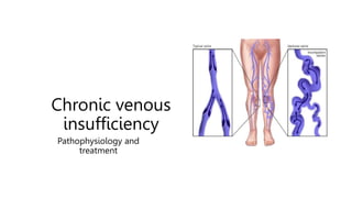
Chronic Venous insufficiency Overview.pptx
- 2. Introduction to the Human Cardiovascular System
- 3. INTRODUCTION The cardiovascular system is transport system of body It comprises blood, heart and blood vessels. The system supplies nutrients to and remove waste products from various tissue of body.
- 4. The cardiovascular system includes 2 circuits • The cardiovascular system includes 2 circuits: 1. Pulmonary circuit which circulates blood through the lungs • The pulmonary artery carries Deoxygenated blood from the right ventricle to the lungs where blood becomes oxygenated • The pulmonary veins returned oxygenated blood to the left side of the heart
- 5. Introduction to Cardio Vascular System • Components of Cardiovascular system 1.Blood 2.Blood Vessels and Types of Blood Vessels 3.Heart
- 6. BLOOD VESSELS • Blood Vessels ,a closed network of tubes •These includes: Arteries Capillaries Veins
- 7. Heart • Hollow, cone shaped muscle • Heart composed of 4 heart chambers 1.2 Atria (atrium) Upper chambers that receive blood Separated by interatrial septum 2.2 Ventricles Lower chambers that
- 8. Structure of Heart Wall 1.PERICARDIUM Outer layer which is a protective serous membrane made of connective tissue 2.MYOCARDIUM Middle layer oMade up of cardiac muscle oPump blood 3. ENDOCARDIUM Inner layer made up of epithelium and connective
- 9. Wall of Arteries and Veins composed of 3 Layers • Tunica Externa or Adventitia Connective tissue-collagen • Tunica Media Muscular tissue and elastic fibers • Tunica Intima Simple squamous epithelium (endothelium) and elastin
- 10. Arteries • Thick-walled vessels that transport blood via the aorta from the heart to the tissues • Types of Arteries 1. Large size arteries (Aorta) 2. Middle size Arteries 3. Small size arteries (Arterioles)
- 11. Veins • Transport Blood from the capillaries back to the right side of the heart • There is little Vascular smooth muscle & connective tissue makes the veins more distensible as they accumulate large volumes of blood • Veins have one-way Valves 1. The venous valves are abundant in the distal lower extremity and decreases proximally with no valves in superior and inferior vena cava
- 12. Veins Vein Carries unoxygenated blood towards the heart • Types of Veins 1.Large size Veins (Vena cava) 2.Middle size Veins 3.Small size Veins (Venules)
- 13. Capillaries • Capillaries “Microcirculation” 1.blood flow between arterioles, capillaries and venules 2.Exchange of Gases, Nutrients, and Waste Between Blood and Tissue Occurs in the Capillaries 3.Exchange is controlled by 2 forces Hydrostatic and Colloidal Osmotic pressures
- 14. Capillary Exchange • At the arterial end of a capillary, the blood pressure is higher than the osmotic pressure; therefore, water tends to leave the bloodstream. • In the midsection, oxygen and carbon dioxide follow their concentration gradients. • At the venous end of a capillary, the osmotic
- 15. Lymphatic system • The lymphatic system works with the circulatory system , The two systems are closely linked to each other • Functions of Lymphatic system 1. Return of plasma I. Extra plasma that has been filtered out of the blood and is in the interstitial fluid may need to be returned to the blood and this can be done through the lymphatic system (lymph vessels)
- 16. Lymphatic system • Lymph capillaries • Lymphatic vessels • Lymph nodes 1. Filtration sites along the lymphatic system 2. Produce white blood cells to fight viruses and other infections (immune defense) • Lymphatic trunks • Collecting ducts
- 17. Chronic Venous Diseases (CVD) Lower limb
- 18. Lower Extremity Venous System 1. Superficial venous system run between the dermis and the muscle fascia • Greater saphenous vein medially • lesser saphenous vein laterally 2. Deep venous system intramuscular or Intermuscular ,named after the artery they accompany • Posterior Tibial vein • Anterior Tibial vein • Join to form the popliteal vein • Become the superficial femoral vein • Joined by the deep femoral vein to form the common femoral vein • Renamed the common Iliac vein
- 19. Perforator veins 1. Connect superficial to deep veins at various levels. 2. The direction of blood flow :from superficial to deep veins. 3. Guarded by valves so that the flow is unidirectional, i.e. Towards deep veins. 4. Reversal of flow occurs due to incompetence of perforators which will lead to varicose veins
- 20. Factors Affecting Venous Return • Blood is drained from superficial to deep veins of legs through perforating veins and through deep veins it is carried to heart. Back flow of blood is impossible. • Intrinsic Factors 1. Venous wall contraction (Venous tone) 2. Valve Integrity 3. Musculovenous Pump 4. Abdomino -thoracic Pump and Cardiac Pump • Extrinsic Factors 1. Gravity 2. External Compression 3. Temperature
- 21. MUSCULO-VENOUS PUMP • Muscular contraction expel blood from the deep venous system • As the muscles relax Blood is drawn from the superficial to the deep system, thereby lowering the superficial venous pressure. • Competent valves are required to prevent
- 22. Venous valves • Delicate structures • The venous valves are abundant in the distal lower extremity and number of valves decreases proximally, with no valves in superior and inferior vena cava • Prevent reverse flow in the veins Ensure that the blood is pumped from the superficial to the deep
- 23. Abdomino - thoracic Pump and Cardiac Pump • Abdomino - thoracic Pump 1.Inspiration decreases intra thoracic pressure promoting venous return 2.Expiration reverses the process • Cardiac Pump Normal cardiac pulsations cause negative pressure in RA and RV that have aspiration effect on the venous
- 24. Varicose Vein pathophysiology • Venous Distensibility The venous wall differs from the arterial wall in that it is highly distensible • The accumulation of blood in the veins of the lower limbs upon standing is limited by I. Venous tone (Contraction of venous wall) II.presence of valves III.Effective contraction of the calf muscles • Causes of Varicose veins Venous stasis (Venous hypertension) is the main cause ,either 1. Primary varicose veins , without known cause (develop for no obvious reason)
- 25. Varicose Vein pathophysiology • In varicose veins, the primary defect is an exaggerated distensibility of the veins: 1. Genetics Abnormal connective tissue composition of the venous wall. 2. Age 3. Obesity 4. Hormones, Female sex Hormones (estrogens and progesterone) The media of human saphenous veins expresses progesterone receptors, suggesting that the hormone can act directly on the venous wall and cause relaxation of the vein wall 5. Sedentary life/ occupation (sitting or standing position at work) leads to
- 26. Varicose Vein pathophysiology - Venous hypertension • Laminar Blood flow keeps leukocytes in Central layer of blood stream away from endothelium and prevents endothelium from expression of cellular adhesion molecules • Venous hypertension induces Turbulence in blood flow which will result in 1. leukocytes become in contact with endothelium. 2. Endothelium expression
- 27. Varicose Vein pathophysiology - Remodeling of Venous wall • Inflammation and damage of venous wall due to Leukocyte migration , activation and production proteolytic enzymes and Oxygen free radicals • Vein wall show Hypertrophic areas alternated by Atrophic areas • Hypertrophic areas with Accumulated 1. Extra cellular matrix
- 28. Varicose Vein pathophysiology - Venous Valve destruction (a partially mobile leaflet or a completely “frozen valve.”) • Valve dysfunction can occur due to inflammation as result of Leukocyte infiltration , activation and migration into the endothelium of proximal surfaces of the vein valves and promote destruction and remodeling of the valves which results in valvular insufficiency and Venous Reflux (Varicose
- 29. Varicose Vein pathophysiology - Microcirculation (Capillaries) • Sustained hypertension at the capillary level is associated with elongation and dilation of the capillary bed • Expression of ICAM 1 and VCAM 1 results in leukocyte adhesion and migration through the vessel wall into the extravascular tissues , neutrophils become activated and
- 30. Varicose Vein pathophysiology - Skin changes (Ulceration/ Venous ulcer) • White cell trapping theory: 1. Venous hypertension expression of leukocyte adhesion molecules adherence of leukocytes to the capillary endothelial cells. 2. The trapped cells become activated releasing proteolytic enzymes and oxygen free radicals which produce tissue destruction. • Fibrin cuff theory : 1. Capillary hyper permeability will allow Fibrin leakage and deposition around capillary
- 31. Varicose Vein pathophysiology - Skin changes (Ulceration/ Venous ulcer) • Cutaneous iron overload: 1. extravasations of red blood cells. 2. The iron is released from the hemoglobin and deposited within the tissue which promote the production of oxygen free radicals and lipid peroxides which in turn can lead to tissue destruction
- 32. Varicose Vein pathophysiology - Lymphatic system (Lymphatic overload ) 1. The increased permeability of capillaries causes leakage of excess fluid and interstitial edema. 2. Lymphatic vessels transport capacity is limited, After they are overloaded, they become insufficient and gradually they become damaged. 3. Lymph vessels are hypertrophic and
- 33. Varicose Vein pathophysiology 1.HIGH TEMPERATURE 2.PROLONGED Standing or sitting 3.HORMONAL FACTORS 4.GENETIC FACTORS Increased Venous wall distensibil ity Valvular incompetence Increased venous pressure Increased Capillary pressure Inflamm ation Venous wall remolding Valvular destruction Fluid leakage and edema Inflamm ation Ulcer ation Venous Reflux (perforating and Superficial veins vein)
- 34. Symptoms of CVD • Venous disease is symptomatic as soon as wall damage occurs Accompanied early by feelings of 1.Pain 2.Itching 3.Heaviness 4.Swelling 5.Cramps
- 35. Progression of chronic venous disease (Venous hypertension is key) MACRO circulatio n Valve damage Vein wall remodeling Reflux Capillary damage Skin Changes (C4) Edema (C3) Capillary leakage Varicose Veins (C2) MICRO circulatio n Venous Ulcer (C5,6) Venous Hypertension
- 36. Classification and severity scoring of chronic venous disease • The CEAP classification was proposed and subsequently adopted worldwide as a basis for improved patient description 1. Clinical 2. Etiology • Ec: congenital. • Ep: primary. • Es: secondary (post thrombotic). 3. Anatomy • S: superficial veins. • P: perforator veins. • D: deep veins 4. Pathology • Pr: reflux. • Po: obstruction. • Pr,o: reflux and obstruction. • Pn: no venous pathophysiology
- 39. Symptoms of Varicose veins • Telangiectasia(spider veins) : A confluence of dilated intradermal venules of less than 1 mm in caliber.. • Reticular veins (blue veins) : Dilated bluish sub dermal veins usually from I mm to less than 3 mm in diameter. They usually are tortuous. • Varicose veins : Subcutaneous dilated , tortuous veins equal to or more than 3 mm in diameter
- 40. Symptoms of Varicose veins • Edema : An increase in volume of fluid in the skin and subcutaneous tissue characteristically indenting with pressure. Venous edema usually occurs in the ankle region, but it may extend to the leg and foot. • Pigmentation : A brownish darkening of the skin resulting from extravasated blood, which usually occurs in the ankle
- 41. Symptoms of Varicose veins • Lipodermatosclerosis (LDS): Localized proliferation of the dermal capillaries and fibrosis on subcutaneous tissue It is a combination of induration , pigmentation and inflammation • Atrophic blanche or white atrophy : Localized, often circular whitish and atrophic skin area surrounded by
- 42. Symptoms of Varicose veins • venous ulcer : Full thickness defect of the skin most frequently in the ankle region that fails to heal spontaneously and is sustained by CVD. • Most common site medial aspect of ankle & lower 1/3rd of leg • characteristic features: • Vertically oval • Painless • Irregular, Ragged ,Sloping edges
- 43. Treatment • OBJECTIVE OF TREATMENT OF CVD 1. 1st to rapidly and powerfully relieve patients from symptoms and pain in order to help them recover a better quality of life. 2. 2nd to protect VV patients from further complications. • Leg elevation and regular exercise • Use of elastic compression stockings. • Injection sclerotherapy. • Endovenous laser treatment and radiofrequency ablation • Saphenous vein ligation and stripping
- 44. Use of elastic compression stockings. Injection sclerotherapy. Endovenous laser treatment and radiofre Saphenous vein ligation and stripping surgical excision using the "stab avuls
- 46. Anatomy of the Anal Canal • Anatomical anal canal, extending from dentate line to anal verge to is only 2 cm in length • The anal canal is lined with squamous epithelium below the dentate line, and columnar epithelium above • Internal Anal Sphincter 1. Involuntary 2. Circular muscle layer • External Anal Sphincter 1. Voluntary 2. Striated muscle layer
- 47. Hemorrhoids • The term “hemorrhoids” refers 2 different vascular structures: 1. Internal haemorrhoidal plexus, which is sub mucosal (Typically, there are 3 major cushions located in right anterior, right posterior, and left lateral aspect of the anal canal) 2. External haemorrhoidal plexus, which is subcutaneous. • Internal Hemorrhoidal
- 48. Hemorrhoids (Arteriovenous Shunt) • Within each Internal Hemorrhoid Plexus , there is an Arteriovenous plexus formed by direct communication between the terminal branches of rectal arteries and their corresponding veins • Arteriovenous shunt contains smooth muscle
- 49. Internal Hemorrhoids function • The function of the internal sphincter itself is not sufficient to ensure complete closure of the anal canal. • Internal hemorrhoids play an important role to keep intestinal contents in the rectum. 1. At rest, vascular are filled with blood and are in contact with each other causing the anal canal to close and the pressure in the sphincter to increase. 2. During defecation Internal hemorrhoids are squeezed and emptied out of blood which allow passage of rectal content
- 50. Causes of Haemorrhoidal diseases • Acute intrarectal or abdominal pressure are predisposing factors (Straining , Chronic constipation /diarrhea , Prolonged sting on Toilet , Pregnancy ) • The origin of haemorrhoidal disease can be either 1. Mechanical theory, the supportive structure of the haemorrhoidal plexus undergoes excessive
- 51. Degeneration of Connective tissue which fix the internal hemorrhoid to rectal wall Favors stagnation and stasis of blood in vascular plexus of internal hemorrhoid Leukocytes marginated and become in contact with endothelial cells Leukocytes adhesion , migration and activation releasing proteolytic enzymes and Capillary hyper permeability , Fragility and necrosis of capillary wall internal hemorrhoid easily traumatized by passage of stool and bleed Internal Hemorrhoids pathogenesis Chronic Constipation / Diarrhea Pregnancy Prolonged sitting on toilets Straining Vascular abnormalities Increased flow in AV plexus
- 52. Types and complications • Types 1. Internal 2. External 3. Mixed • Complications 1. Strangulation of internal Hemorrhoids 2. Anemia 3. Perianal dermatitis 4. Thrombosis of external Hemorrhoids
- 53. Classified according to origin of hemorrhoid. External hemorrhoid Internal hemorrhoid Below dentate line Above dentate line Lined by squamous epithelium Lined by columnar epithelium Painful Not painful Prone to thrombosis if vein ruptures (Thrombosed May prolapse outside anal canal (prolapsed
- 54. Symptoms of Internal Hemorrhoids 1. Bleeding most common and earliest symptom Bright red painless bleeding especially at the end of defecation 2. Prolapse 3. Tenesmus 4. Pruritus 5. Pain and discomfort Hemorrhoids are usually painless. 6. Discharge (Soiling) A constant mucous
- 55. Internal Hemorrhoids : • Internal Hemorrhoids Disease Manifested by two main symptoms 1. Painless Bleeding 2. Protrusion 3. Pain is rare as they originate above dentate line Gr I Gr
- 56. Treatment of hemorrhoids has to achieve 3 objectives: • Eliminate mechanical and local triggering factors (lifestyle modification) I. Dietary measures, must include fiber-rich food and abundant liquids II.Mechanical laxatives, such as Vaseline or liquid paraffin, III.Avoiding consumption of stimulating drinks (tea, coffee), alcohol and spices. IV.avoiding smoking, a sedentary lifestyle, and a sitting position for prolonged periods of time.
- 57. Treatment • Conservative: Medical Invasive therapy 1. Injection sclerotherapy 2. Rubber band ligation 3. Cryotherapy 4. Photocoagulation • Surgical: 1. Open haemorrhoidectomy 2. Closed haemorrhoidectomy 3. White head haemorrhoidectomy 4. Laser haemorrhoidectomy 5. Diathermy haemorrhoidectomy
- 58. Chronic venous disease during pregnancy Haemorrhoidal disease during pregnancy Dysfunctional Uterine Bleeding
- 60. Female reproductive system • Ovaries • Fallopian tubes • Uterus 1. Perimetrium - external serosa layer 2. Myometrium - middle muscular layer ,smooth muscle 3. Endometrium inner layer simple columnar epithelium • Cervix • Vagina
- 61. Menstrual cycle • Averages 28 days 1. Follicular phase (2 weeks) • Menstruation (Blood, serous fluid and endometrial tissue are Discharged) occurs during first 3 to 5 days of cycle Uterus replaces lost endometrium and follicles grow 2. Luteal phase (2
- 62. Dysfunctional uterine bleeding • DUB is defined as abnormal uterine bleeding in the absence of organic disease. • Dysfunctional uterine bleeding is the most common cause of abnormal vaginal bleeding during a woman's reproductive years. • A normal menstrual cycle is characterized by an approximate flow
- 63. Abnormal uterine bleeding • DUB refers to abnormal bleeding from the uterus and can be characterized clinically by amount, duration, and periodicity: 1. Menorrhagia - Prolonged (>7 d) or excessive (>80 mL daily) uterine bleeding occurring at regular intervals 2. Metrorrhagia - Uterine bleeding occurring at irregular and more frequent than normal intervals 3. Menometrorrhagia - Prolonged or excessive uterine bleeding occurring at irregular and more frequent than normal intervals 4. Intermenstrual bleeding - Uterine bleeding of
- 64. CAUSES Disruption of normal hormonal regulation of peri ods e.g. excess Oestrogen Intrauterine Devices (IUD’s) Dysfunctional Uterine Bleeding Damage of the microcirculation Leukocytes migration Venous stasis Leukocytes adhesion Leukocytes activation Bleeding Hormonal disturbances
- 65. Mechanical trauma Inflammation Capillary fragility Menorrhagia Dysmenorrhea IUCD-INDUCED BLEEDING • Abnormal uterine bleeding is the most common complication of IUD use. • Minor Metrorrhagia during the insertion and the initial 2 or 3 cycles is common and has no pathological significance. • The true complications
- 66. Treatment of Menorrhagia Medical therapy for menorrhagia may include: • Nonsteroidal anti-inflammatory drugs (NSAIDs). NSAIDs, such as ibuprofen or naproxen sodium , help reduce menstrual blood loss. NSAIDs have the added benefit of relieving painful menstrual cramps (dysmenorrhea). • Oral contraceptives. Aside from providing birth control, oral contraceptives can help regulate menstrual cycles and reduce episodes of excessive or prolonged menstrual bleeding. • Oral progesterone. The hormone progesterone can help correct hormone imbalance and reduce menorrhagia.
- 67. Thanks