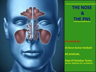
ANATOMY, PHYSIOLOGY,APPLIED ANATOMY AND AYURVEDIC NASA SHARIRA WITH REFERENCE. EASY TO REMEMBER AND UNDERSTAND WITH LOTS OF PICTORIAL PRESENTATIONS. BRIEFLY DESCRIBED.
5. The nose is the door of head. The drugs that is
administered to the Nasal cavity reaches to the
sringataka Marma then spread to murdha, netra,
karna ,kantha and siramukha then scartches the
morbidly attached doshas and expels them out.
Dr.Tarun Kumar Dwibedi / PG SCHOLAR /
dr.tarun52@gmail.com / SJG AMC & H,
KOPPAL
6. • Drugs administered into the nose goes
directly in to the CNS through CRIBRIFORM
PLATE by ...
1.through OLFACTORY NEURONS
2. Through supporting cells and the
surrounding capillary bed.
Dr.Tarun Kumar Dwibedi / PG SCHOLAR /
dr.tarun52@gmail.com / SJG AMC & H,
KOPPAL
7. नासा शारीरं-3
द्वे अंर्ुलातन बृषणचिबुकदशन नासापुटभार् कणगमूल भृनयन अन्तराणी
,
ितुरंर्ुलातन मेिनबदनअंतरनाsaकणगललाटग्रीब I
Su.su.35/12
• Nasaputa (NOSTRIL) is 2 ANGULO.
• NOSE IS 4 ANGULO.
Dr.Tarun Kumar Dwibedi / PG SCHOLAR /
dr.tarun52@gmail.com / SJG AMC & H,
KOPPAL
8. नासा शारीरं-4
फणावु भयतो घ्राणमार्ं श्रत्रपथानर्ौ ,अन्तर्गल
स्थतौ वेर्ात्र्न्ध ववज्ञान िाररणौ।
A.h.Sa.4/30
• On the both side of the nasal canal ,towards
the ear canal and in the throat ,we have 2
FANA marma . Injury to this marma causes
loss of smell sense.
Dr.Tarun Kumar Dwibedi / PG SCHOLAR /
dr.tarun52@gmail.com / SJG AMC & H,
KOPPAL
10. PYRAMDAL IN SHAPE – ROOT
UPWARD AND BASE DOWN WARD.
THE EXTERNAL PART HAS
OSTEOCARTELAGENOUS FRAMEWORK
COVERED BY SKIN AND MUSCLE .
THE NOSE
Dr.Tarun Kumar Dwibedi / PG SCHOLAR /
dr.tarun52@gmail.com / SJG AMC & H,
KOPPAL
24. Skin over the nasal bone & over the lateral
cartilage part is thin, loose and freely movable .
The skin on the alar cartilage part is thick &
adherent which contains many sebaceous
glands.
The hypertrophy of those glands causes
Rhinophyma .
Dr.Tarun Kumar Dwibedi / PG SCHOLAR /
dr.tarun52@gmail.com / SJG AMC & H,
KOPPAL
32. VESTIBULITIS
• Vestibulitis is an inflammation of this skin and
the mucous secreting glands found in the skin.
.
• When it is in ant. nasal cavity it will be nasal
vestibulitis.
Dr.Tarun Kumar Dwibedi / PG SCHOLAR /
dr.tarun52@gmail.com / SJG AMC & H,
KOPPAL
36. Rhinosporidiosis
• Rhinosporidiosis is a chronic granulomatous
infection of the mucous membranes that
usually manifests as vascular friable polyps
that arise from the nasal mucosa .
• Organism..RHIISPORODIUM SEEBERI
Dr.Tarun Kumar Dwibedi / PG SCHOLAR /
dr.tarun52@gmail.com / SJG AMC & H,
KOPPAL
44. Largest among all the nasal meatus.
Space below the inferior concha.
It is thin curved & independent.
Naso-lacrimal duct is opens in to it.
The end of naso lacrimal duct is guarded by
Hasner’s valve ,a mucosal valve.
INFERIOR MEATUS
Dr.Tarun Kumar Dwibedi / PG SCHOLAR /
dr.tarun52@gmail.com / SJG AMC & H,
KOPPAL
46. LACRIMAL GLAND & FLOW OF TEAR
• paired and almond shaped
• Present upper lateral part of each eye ( upper
outer part of orbit)
• secrets the aqueous layer of the tear film.
• This liquid moist the corneal surface.
• The tears lacrimal sac through sup. & inf.
Lacrimal canal.
• From this sac ,tears drains through a passage ie.
Naso-lacrimal duct into the inferior meatus of
lateral wall of the nasal cavity.
Dr.Tarun Kumar Dwibedi / PG SCHOLAR /
dr.tarun52@gmail.com / SJG AMC & H,
KOPPAL
50. UNCINATE PROCESS-
Its a hook like structure present
anteroposteriorly to the
posterior direction in the middle
meatus.
BULLA ETHMOIDALIS-
Its the elevation above the
uncinate process in the middle
meatus of lateral wall.
HIATUS SEMILUNARIS-
Its a groove between the
uncinate process & the bulla
ethmoidalis .
STRUCTURES IN MID. MEATUS , WHILE CONCHAE ARE REMOVED
Dr.Tarun Kumar Dwibedi / PG SCHOLAR /
dr.tarun52@gmail.com / SJG AMC & H,
KOPPAL
51. SUPERIOR MEATUS
Space below the sup. Concha.
Its a process of ethmoidal bone.
Smallest among nasal meatus.
Post. Eth. Sinus opens into it.
Sphenoidal sinus also opens
into it.
Dr.Tarun Kumar Dwibedi / PG SCHOLAR /
dr.tarun52@gmail.com / SJG AMC & H,
KOPPAL
56. ARTERIES OF THE NASAL CAVITY
Dr.Tarun Kumar Dwibedi / PG SCHOLAR /
dr.tarun52@gmail.com / SJG AMC & H,
KOPPAL
57. NASAL BLEED / EPISTAXIS
Bleeding from the nose.
May be venous or Arterial.
It could be from anterior part of nose or
posterior part of nose.
Dr.Tarun Kumar Dwibedi / PG SCHOLAR /
dr.tarun52@gmail.com / SJG AMC & H,
KOPPAL
66. NERVE SUPPLY OF NOSE
1.OLFACTORY N.
2.Nerves of general sensation
-ophthalmic N.
-ant. Ethmoidal N.
-Maxillary N.
-Nasopalatine N.
- Palatine branch of Ganglion.
Dr.Tarun Kumar Dwibedi / PG SCHOLAR /
dr.tarun52@gmail.com / SJG AMC & H,
KOPPAL
68. Nasal myiasis
Myiasis is the parasitic infestation of
the body of a live mammal by fly-
larvae (maggot )that grow inside the
host while feeding on its tissue .
Dr.Tarun Kumar Dwibedi / PG SCHOLAR /
dr.tarun52@gmail.com / SJG AMC & H,
KOPPAL
71. MELANOMA
Melanoma is a cancer that develops in
melanocytes.
When the melanoma present on skin of nose
it would b the Nasal melanoma.
Usually those are malignant .
Dr.Tarun Kumar Dwibedi / PG SCHOLAR /
dr.tarun52@gmail.com / SJG AMC & H,
KOPPAL
73. THE PNS
THE PARA NASAL SINUS
Dr.Tarun Kumar Dwibedi / PG SCHOLAR /
dr.tarun52@gmail.com / SJG AMC & H,
KOPPAL
74. The air filled cavities in the bone , surrounding the nose.
PNS are on Frontal ,Ethmoidal , Sphenoidal , Maxilla.
Helps in resonance of voice , Lightening of skull , Air
conditioning.
Appears after birth & grow with age.
Maxillary and sphenoid sinus appear at birth.
Maxillary sinus appear first.
THE PNS
Dr.Tarun Kumar Dwibedi / PG SCHOLAR /
dr.tarun52@gmail.com / SJG AMC & H,
KOPPAL
75. MUCOUS MEMBRANE OF PNS
THE SINUSES ARE LINED WITH MUCOUS MEMBRANE WHICH IS
THE CONTINUATIONS OF NASAL CAVITY.
COMPARATIVELY LESS VASCULAR AS COMPARED TO
NASAL CAVITY.
IT CONTAINS CILLIATED COLUMNER EPITHELLIUM .
IT CONTAINS GOBLET CELLS THAT SECRETS MUCOUS.
CILLIA IS MORE NEAR THE OSTEA OF SINUS WHICH HELPS IN
DRAINING THE MUCOUS TO THE RESPECTIVE MEATUS.
Dr.Tarun Kumar Dwibedi / PG SCHOLAR /
dr.tarun52@gmail.com / SJG AMC & H,
KOPPAL
82. FRONTAL SINUS
Second Largest among all PNS.
2 In no. 2- 2.5 cm .
Roughly triangular in shape.
Separated from each by a bony septum.
Opens into middle meatus by a canal called
fronto-nasal duct.
Dr.Tarun Kumar Dwibedi / PG SCHOLAR /
dr.tarun52@gmail.com / SJG AMC & H,
KOPPAL
86. SIZE OF FRONTAL SINUS C AGE
Dr.Tarun Kumar Dwibedi / PG SCHOLAR /
dr.tarun52@gmail.com / SJG AMC & H,
KOPPAL
87. SPHENOIDAL SINUS
IN THE BODY OF SPHENOID BONE.
2 IN NO SEPARATED BY A THIN BONY
SEPTUM.
BELOW THE SELLA TURSICA.
VOL.-7ML.
Dr.Tarun Kumar Dwibedi / PG SCHOLAR /
dr.tarun52@gmail.com / SJG AMC & H,
KOPPAL
92. ETHMOIDAL SINUS
PRESENT WITH IN THE UPPER THIRD OF LATERAL NASAL
WALL AND MEDIAL WALL OF ORBIT .
3-18 IN No. .
CLINICALLY IT HAS 2 GROUPS,
ANTERIOR &
POSTERIOR GROUP .
ANTE. GROUP DRAINS IN TO MIDDLE MEATUS &
POSTERIOR GROUP DRAINS IN TO SUPERIOR MEATUS AND
SPHENO-ETHMOIDAL RECESS.
Dr.Tarun Kumar Dwibedi / PG SCHOLAR /
dr.tarun52@gmail.com / SJG AMC & H,
KOPPAL