Pelvic mass panel discussion
•Transferir como PPTX, PDF•
23 gostaram•7,498 visualizações

4 cases of pelvic mass are discussed .Adnexal mass invilves masses arisinf from ovary,fallopian tube,uterus,bowel and some miscellenious masses.USG is used to detect its size and the origin.Histopathological findings are diagnostic.

Recomendados
Mais conteúdo relacionado
Mais procurados
Mais procurados (20)
Semelhante a Pelvic mass panel discussion
Semelhante a Pelvic mass panel discussion (20)
Mais de Niranjan Chavan
Mais de Niranjan Chavan (20)
Último
Último (20)
Pelvic mass panel discussion
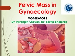
- 1. Pelvic Mass in Gynaecology MODERATORS Dr. Niranjan Chavan, Dr. Sarita Bhalerao
- 2. PANELISTS Dr.Amita Maheshwari Dr. Susan Sodder Dr. Shilpa Sankhe Dr. Sarabjee Kaur Dr. Kinjal Shah Dr. Sachin Ajmera Dr. Sachin Dalal Dr. Bhumika Kotecha Dr. Nidhi Gandhi
- 5. What differential diagnosis comes to your mind when you hear ADNEXAL MASS?
- 7. CASE 1 Mrs XYZ 45yr old P2L2 (FTND), with complaints of Heaviness in abdomen Irregular heavy menses Dysmenorrhea , since 2 – 3 months On Examination: P/A – soft, non tender P/S - cervix , vagina healthy P/V – uterus bulky firm mobile, AV, soft to cystic mass 4x5 cm in right and posterior fornix separate from uterus. Left fornix free and non tender. P/R – bogginess felt anteriorly, rectal mucosa and parametrium free. Tumour Marker - WNL
- 8. Investigations: USG Pelvis: solid, hypoechoic, well-circumscribed right adnexal mass of size 3.6x4.6 cm
- 9. What is your diagnosis? -Open to all the Panelist.
- 10. BROAD LIGAMENT FIBROID
- 11. How will You differentiate between a True and False Broad Ligament Fibroid ? What will be relation of the Ureter to this fibroid?
- 12. True Broad Ligament False Broad Ligament Originates from the muscle fibres normally found in the mesometrium (in the round ligament, ovario-uterine ligament, and the connective tissue around the uterine and ovarian vessels) Arises from the lateral wall of the uterine corpus or of the cervix, and bulges outward between the layers of the broad ligament. Ureter is medial to mass Ureter is lateral to mass No groove felt between mass and uterus Groove felt between mass and uterus
- 13. What will be your approach in this case? Is there any role of ureteric stenting?
- 14. In this case , the right ureter was safeguarded by dissection and enucleation could be carried out rather easily.
- 17. CASE 2 A 14 years old girl , unmarried c/o large abdominal mass pain in abdomen backache for 2-3 days. Menstrual history : LMP : 27/12/11 . Menarche attained 5 days back. Patient had bleeding for 5 days , changing 2 pads per day . On Examination: Per Abdomen: 34 – 36 weeks size mass arising from pelvis, Cystic in consistency . Mobile from side to side. Smooth surface. Lower margin of the mass could not be made out. Upper border of mass was 1-2 cm below the intercostal margin. Mass was extending towards both the flanks. Per Rectal: Lower margin of the mass felt. No involvement of the rectal mucosa. Mass was free, mobile, non tender. No nodularity or induration felt.
- 18. Investigation: Tumour markers- Ca125- 19.1 u/mL AFP- 1.06 B-HCG-< 1.2 CEA- 1. 34 USG Abdomen & Pelvis: Uterus 6 x 3.1 x 2.7 cm. Normal endometrial echoes. ET 5 mm. B/L ovaries not visualized. A large complex cystic lesion seen extending from pelvis to epigastric region with multiple thick septae & multiple echoes within noted. No vascularity noted within. The mass displacing the bowels loops peripherally and uterus inferiorly. No calcification or mass lesion within. Impression: Mucinous Cystadenoma of the ovary of benign etiology.
- 19. CT Scan Abdomen (Plain & Contrast): Large 18 x 15 x 11 cm sized, well defined , multiloculated , predominantly cystic , with multiple enhancing septae within noted in the pelvis, extending into the lower abdomen, abutting the anterior abdominal wall upto L1 – L2 vertebral level. No calcification or solid component noted within the mass. The lesion causing mass effect on the uterus displacing it postero – inferiorly , on the urinary bladder displacing it inferiorly and on the surrounding bowel loops displacing them peripherally. Mass effect on lower ureter with resultant mild proximal b/l hydroureter & hydronephrosis. The fat plane of this lesion with the surrounding structures appeared well maintained. Rt ovary seen separate from the lesion. Left ovary not appreciated on the CT . Impression: LT ovarian cyst adenoma of benign etiology.
- 21. Intraoperative finding: Patient underwent a successful ovariectomy with partial salpingectomy on the Left side with removal of a multi lobulated mucinous cyst adenoma. About 300 cc of mucinous fluid was also aspirated from the cyst. Post operative course of the patient in the ward was uneventful.
- 23. Role of Tumour Markers? What is ROMA test and its importance?
- 25. ROMA: RISK OF MALIGNANCY ALGORITHM Clinical Use: Assess the likelihood that an ovarian mass is malignant in women whose pre-surgical assessment did not indicate malignancy. Assess the need to refer the patient to a gynecologic oncologist for treatment. used as a supplement to the standard presurgical evaluation to further assess the likelihood of malignancy before surgery when the presurgical evaluation does not indicate malignancy. The pre-surgical evaluation should include menopausal status, physical examination, transvaginal ultrasonography, CA 125 concentration, and family history of breast or ovarian cancer in a first-degree relative.
- 26. It combines the results of human epididymis protein 4 (HE4) enzyme immunometric assay (EIA), ARCHITECT CA 125 , and menopausal status to generate a single numerical score that correlates with the likelihood of malignancy being seen at surgery. The ROMA test is intended for use in women who meet the following criteria: Are over 18 years of age Have an ovarian mass Surgery is planned Not yet referred to an oncologist The ROMA test should not be used in women who have a rheumatoid factor concentration >250 IU/mL.
- 27. Should we do Open Laparotomy knowing fully well before hand that’s Malignant or Laparoscopic Treatment ? Management of Germ Cell Tumour? Role of Neoadjuvant Chemotherapy? Debulking or Cytoreductive Surgery ? Followup & Survelliance in Stage 1A? Fertility preservation surgery? -.
- 28. Management of Germ Cell Tumour Role of Neoadjuvant Chemotherapy Debulking or Cytoreductive Surgery Follow-up & Surveillance in Stage 1A
- 30. Ovarian Germ Cell Tumours Dysgerminoma Endodermal sinus tumour Embryonal Carcinoma Polyembryoma Choriocarcinoma Teratoma-Mature/Immature Immature – low grade/high grade Surgical Principles Suspect diagnosis Pre-operative markers Conservative- fertility preserving surgery Staging-controversial- careful inspection of peritoneum, omentum, contralateral ovary and nodes with washings and biopsies of suspicious areas adequate Unilateral oophorectomy with debulking if advanced stage
- 31. Surgery Importance of staging in earl of staging in early disease Fertility-sparing surgery often required Can preserve uterus for future IVF, even if BSO Debulking improves outcome Chemotherapy BEP Bleomycin 20 U/m2 weekly x 9 Etoposide 100 mg/m2 days 1-5 q 3 weeks x 3 Cisplatin 20 mg/m2 days 1-5 q 3 weeks x 3 VAC Vincristine 105 mg/m2 weekly x 12 Act D 0.5 mg days 1-5 q 4 weeks Cytoxan 5-7 mg/kg days 1-5 q 4 weeks VBP Vinblastine 12 mg/m2 q 3 weeks x 4 Bleomycin 20 U/m2 weeks x 7, 8 on week 10 Cisplatin 20 mg/m2 days 1-5 q 3 weeks x 3
- 32. Germ Cell Tumour Surgery Chemotherapy Dysgerminoma USO staging if possible BEP x 3 cycles if stage II-IV Endodermal sinus tumor Debulk preserve fertility BEP x 3-4 cycles Embryonal carcinoma Debulk preserve fertility BEP x 3-4 cycles Malignant teratoma Debulk preserve fertility BEP or VAC x 3-4 cycles Granulosa cell tumor USO if young o/w TAH/BSO BEP x 3-4 cycles GnRH agonists for advanced ds Sertoli-leydig cell USO if young o/w TAH/BSO BEP or VAC x 3-4 cycles
- 33. Surveillance: Markers HCG AFP LDH CA125 1st year every 2 weeks x 6 m and then monthly x 6 2nd year monthly 3rd year every 3/12 4th year every 4/12 Subsequent years 6/12 Clinical exam 1st year monthly 2nd year every 2 months 3rd year every 3 months 4th year every 4 months 5-10 every 6 months Imaging Chest X ray alternate visits Abdo-pelvic US every 3rd visit for first 2 years followed by annual abdo-pelvic ultrasound subsequent years
- 34. Modalities of fertility preservation strategies Strategy Modality Preserving uterus and at least one ovary Conservative cancer surgery Reducing radiation exposure to ovary Ovarian transposition Reducing chemotherapy related damage Ovarian Suppression Cryopreservation Freezing of embryos, oocytes and ovarian tissue
- 36. CASE 3 Mrs. ABC, 30/F, m/s 5 yrs came with the c/o infertility progressive cyclical dysmenorrhea since last 1 year. Patient gives no h/o any menstrual irregularities. On examination: Per Abdomen: Soft, NO guarding/rigidity/distention/tenderness P/S: Cervix / vagina healthy P/V: Uterus bulky, Anteverted, Firm , restricted mobility Right fornix free. E/o bogginess in left and posterior fornix measuring 5x5cm USG – there is e/o left adnexal mass of 6x5 cm with thin septations with no vascularity. CA-125 – 105 IU/ml Rest tumour makers - WNL
- 37. USG- Pelvis: Low level internal echoes. „„Thick walled „„Homogeneous “ground glass” appearance „„Multilocular „„,Cystic „Show varying degrees of echogenicity in locules „„Round Shape „„Regular Margins
- 39. On diagnostic laparoscopy - There is e/o dense adhesions in the pelvic cavity. uterus – restricted mobility. POD obliterated. Left tubo ovarian chocolate cysts seen with dense adhesions to the bowel.
- 40. What is your diagnosis? What are various management options and what would be your approach in this case? Future fertility and their management options ?
- 41. CASE 4 28 yr P2 L2 (FTND) with CuT 380A in situ with c/o Dull lower abdominal pain with White foul smelling pv discharge Low grade fever. On and off since 6 months Dyspareunia since 10 days Menstrual history – menarche attained at 12 yrs. Past cycles were regular/mod flow/painless/4/30 days On Examination: G/E: GC – average Pallor mild Febrile 38⁰Celscius Pulse 100 b/min BP-90/60mm Hg Per Abdomen-Soft, Tenderness in the left iliac fossa and hypogastrium, No guarding, rigidity, distention, No Organomegaly.
- 42. Pelvic examination Per speculum – cervix shows sign of cervical erosion circumferentially around the external os White discharge seen in the vaginal wall On Bimanual Examination Uterus anteverted, normal size Left Fornix full, 4x4 cm cystic mass palpable Bilateral fornices tender Cervical motion tenderness + Mobility restricted
- 43. Investigations – Hb – 10.5 gm/dl/ TLC -20,000/cm, DLC-neutrophils 70, lymphocytes-26,eosinophil-4 urine routine – pus cells 10 – 15/hpf High vaginal swab sent CA-125 – 42.5IU/ml. CXR – NAD USG abdomen and pelvis – Uterus normal size , ET – 3mm Right fallopian tubes and ovary normal. Left fallopian tube enlarged , distended filled with low echogenic material. Tubal wall can be delineated. There is e/o 4 by 3 cm multiloculated cystic left adnexal mass Rest USG - NAD
- 44. What is your diagnosis? What are the causes Of PID? Approach towards such patients? Indication for In patient care? Is there any role of Laparoscopy in this case? Treatment of sexual partner and their follow up?
- 45. Pelvic Inflammatory Disease (PID) comprises a spectrum of inflammatory disorders of the upper female genital tract, including any combination of endometritis, salpingitis, tubo- ovarian abscess, and pelvic peritonitis • Sexually transmitted organisms, especially N. gonorrhoea and C. trachomatis, are implicated in many cases • However, microorganisms that comprise the vaginal flora (e.g., anaerobes, G. virginals, Haemophilus influenzae, enteric Gram-negative rods, and Streptococcus agalactiae) also have been associated with PID • In addition, M. [Mycoplasma] hominis and U. [Ureaplasma] urealyticum might be etiological agents of PID
- 46. ACCEPTED PROPOSED 1. Menstruating teens 1. Low socio-economic status 2. Multiple sex partners 2. Early age of sexual activity 3. Prior H/O PID 3. Urban living 4. Sexually Transmitted Infection 4. High frequency of coitus 5. Non-use of barrier contraceptive 5. Use of IUCD 6. Cigarette smoking 7. Substance abuse 8. Douching Risk Factors
- 47. Acute PID : CDC DiagnosisCriteria
- 48. Acute PID : Hospital admission 2. Patient meeting following criteria a. Surgical emergencies (e.g., appendicitis) cannot be excluded b. Pt. is pregnant c. Pt. does not respond clinically to oral antimicrobial therapy d. Pt. is unable to follow or tolerate an outpatient oral regimen e. Pt. has severe illness, nausea and vomiting, or high fever f. Pt. has a tubo-ovarian abscess 1. Judgment of the provider
- 49. Management: relief of acute symptoms eradication of current infection minimalization of the risk of long term consequences antibiotics surgery (remove or drain a tubo-ovarian abscess)
- 50. Acute PID : Management (Antibiotics for specific pathogen) Organism Antibiotics N. gonorrhea Cephalosporins, Quinolones Chlamydia Doxycycline, Erythromycin & Quinolones (Not to cephalosporins) Anaerobic organisms Flagyl, Clindamycin & in some cases to Doxycycline ß-Haemolytic streptococci. & E. coli Penicillin derivatives, Tetracyclines, and Cephalosporins., E. Coli is most often treated with the penicillins or gentamicin
- 51. Inpatient treatment Regimen A: Administer cefoxitin 2 g IV q6h or cefotetan 2 g IV q12h plus doxycycline 100 mg PO/IV q12h.. Continue this regimen for 24 hours after the patient remains clinically improved, and then start doxycycline 100 mg PO bid for a total of 14 days. Administer doxycycline PO when possible because of pain associated with infusion. Bioavailability is similar with PO and IV administrations. If tubo- ovarian abscess is present, use clindamycin or metronidazole with doxycycline for more effective anaerobic coverage. Regimen B: Administer clindamycin 900 mg IV q8h plus gentamicin 2 mg/kg loading dose IV followed by a maintenance dose of 1.5 mg/kg q8h. IV therapy may be discontinued 24 hours after the patient improves clinically, and PO therapy of 100 mg bid of doxycycline should be continued for a total of 14 days. If tubo- ovarian abscess is present, use clindamycin or metronidazole with doxycycline for more effective anaerobic coverage.
- 52. Outpatient treatment Regimen A: Administer ceftriaxone 250 mg IM once as a single dose plus doxycycline 100 mg PO bid for 14 days, with or without metronidazole 500 mg PO bid for 14 days. Metronidazole can be added if there is evidence or suspicion for vaginitis or gynecologic instrumentation in the past 2-3 weeks. Regimen B: Administer cefoxitin 2 g IM once as a single dose and probenecid 1 g PO concurrently in a single dose or other single dose parenteral third- generation cephalosporin (ceftizoxime or cefotaxime) plus doxycycline 100 mg PO bid for 14 days with or without metronidazole 500 mg PO bid for 14 days. Metronidazole can be added if there is evidence or suspicion of vaginitis or gynecological instrumentation in the past 2-3 weeks.
- 53. Management :Surgery in Acute PID Indications 1. Ruptured abscess 2. Failed response to medical treatment 3. Uncertain diagnosis Type of surgeries 1. Colpotomy 2. Percutaneous drainage/aspiration 3. Exploratory laparotomy Extend of surgeries 1. Conservation - if fertility desired 2. U/L or B/L Sal.-oophorectomy with/without hysterectomy 3. Drainage of abscess at laparotomy
- 54. PID : Specialsituation IUD users Considerations The risk for PID associated with IUD use is primarily confined to the first 3 weeks after insertion and is uncommon thereafter Practitioners might encounter PID in IUD users because it’s a popular method of contraception Management Evidence is insufficient to recommend the removal of IUDs However Caution should be exercised if the IUD remains in place, and close clinical follow-up is mandatory. If improvement is not seen within 72 hrs of starting treatment then removal of IUCD is considered No data have been collected regarding treatment outcomes by type of IUD (e.g., copper or levonorgestrel)