The radial forearm flap is based on the radial artery and its venae comitantes. It can be harvested as a fasciocutaneous or osteocutaneous flap with a long vascular pedicle. The radial forearm flap is commonly used in reconstructive surgery due to its reliable vascular anatomy, long pedicle length allowing for versatile positioning, and ability to provide a hairless skin match. Potential donor site complications include functional impairment and need for skin grafting or local flaps.
Call Girls Ahmedabad Just Call 9630942363 Top Class Call Girl Service Available
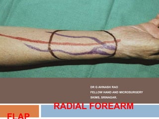
Radial Forearm Flap - Hand Surgery
1. DR G AVINASH RAO
FELLOW HAND AND MICROSURGERY
SKIMS, SRINAGAR.
RADIAL FOREARM
2. HISTORY
Chinese flap
1978-1981 – SHENYANG MILITARY HOSPITAL (60 cases) – DR YANG GUOFAN & DR
YUZHI.
Initial use – free flap in (head and neck) burn contracture release..
French microsurgery mission – visited PRC – used in france after 1981.
GERMAN - introduced to western world.
Modified to use as island pedicle / free flap for hand defects.
Further modifications – adipofascial / osteofasciocutaneous flaps /
vascularized tendon in fascia graft / interpositional graft / prefabricated flaps.
3. Anatomy
Immediately below (2cms) the elbow crease just distal to the
bicipital aponeurosis the brachial artery divides into its two terminal
branches-the radial and ulnar arteries.
The radial àrtery courses between the bellies of the brachioradialis
and pronator teres muscles in the upper half of the forearm and
between the tendons of the brachioradialis and flexor carpii radialis
in the lower half of the forearm.
The brachioradialis muscle lies radial and superficial to the artery,
pronator teres, and flexor carpii radialis on its ulnar side.
4. Initially the artery lies deep to the brachioradialis muscle, but as it courses
distally along the forearm, it gradually becomes more superficial until it lies in
the subcutaneous plane at the wrist.
At this level the artery winds dorsally across the radial border of the distal
wrist onto the dorsal surface of the hand passing deep to the tendons of the
abductor pollicis longus and extensor pollicis brevis.
It then proceeds through the anatomic snuff-box and into the interspace
between the first and second metacarpal bones, where it forms the deep
palmar arch through an anastomosis with the deep branch of the ulnar artery
6. BRANCHES OF RADIAL
ARTERY
RADIAL RECURRENT ARTERY – Just below branching.
Radial artery courses down the LATERAL INTERMUSCULAR SEPTUM and branches in
DEEP FASCIA to supply skin, subcutaneous tissue, flexor muscles, nerves and periosteum of
distal radius.
PERFORATORS (9-17) – PROXIMAL (0-10) & DISTAL GROUP (4-14)
PROXIMAL(large and well spread) - INFERIOR CUBITAL ARTERY (0.5mm)– Large
Septocutaneous Perforator 4 cms below Inter-epicondylar line – form basis for
ANTECUBITAL FLAP.
DISTAL (small and grouped) - 6 to 10 Septocutaneous perforators near anatomic snuff box.
Large-caliber perforator - THE DORSAL SUPERFICIAL BRANCH - found within 2–4 cm
proximal to the radial styloid.
PALMAR CARPAL BRANCH,
SUPERFICIAL PALMAR BRANCH,
DEEP PALMAR BRANCH.
8. CORMACK AND LAMBERTY
TYPE - C
Type C The flap is based on the fascial plexus supplied by multiple small
perforators along the length of a fascial septum. The supplying artery is
taken in continuity with the fascial septum and integument. It may be
pedicled, based distally or proximally or used as a free flap; for example,
the radial forearm flap.
Type D Similar to type C, the type D flap is based on multiple small
perforators, but it is raised as an osteomyofasciocutaneous flap. The
fascial septum and the source artery are taken in continuity with the bone
and adjacent muscle; for example, the radial forearm flap with half of the
radius longitudinally
13. Fasciocutaneous vascular
territory
The fascial plexus on the anterior (volar) aspect is orientated
predominantly along the longitudinal axis in the proximal two-thirds
of the forearm and is orientated more transversely in the distal third
of forearm
15. VENOUS DRAINAGE OF FLAP
DEEP – 2 venae comitantes (valved) but has inter-connecting channels
(ladder pattern) – flow is retrograde in DISTALLY PEDICLED FLAPS.
Drain into Median Cubital Vein (via – Constant Branch near elbow)
SUPERFICIAL – Cephalic V , Basilic V , Median Cubital Vein.
16.
17. Nerve supply to flap
Lateral cutaneous nerve of forearm (c5,c6) /(MCN) – its anterior
branch runs along with cephalic vein.
Medial cutaneous nerve of forearm (c8,T1) (Medial Cord) – via its
anterior branch.
19. ADVANTAGES
Relatively constant and reproducible anatomy based on Large vessels with
Long pedicle & large Diameter vessels.
Two team approch of reconstruction (often)
Ability to use as a Conduit / Interpositional Grafts / Flow through Flap
Communicating superficial and deep veins of flap allow for flexibility of
drainage.
Used as free / pedicle flap proximally (or) distally. (prolonged immobilization
not required)
Allow for variation in size, shape, design & composition with thin pliable and
hairless skin,(no need for secondary thinning).
Harvested under regional block.
20. DISAVANTAGES
Donar site – functional and Cosmetic disadvantage.
Requirement for Local Rotational / Advancement flaps / Skin
Grafting.
Radius Fracture due to weakening of radius after harvesting
osteofasciocutaneous flaps.
Sacrifice major artery of forearm (exception-perforator flaps)
21. Preoperative evaluation
ALLENS TEST – Integrity of Palmar arch.
Looks for any previous injuries on forearm.
In elderly pts - look for atheroma in distal radial artery.
Nondominant > Dominant Limb.
No need for MRA / Angiography.
Xray of forearm – in osteocutaneous flaps to look for size, shape of
radius and ruleout old fractures.
Contraindicated in - post CABG, post A-V fistula, Peripheral
vascular diseases.
22. FLAP DESIGN
Large flaps can be harvested – (risk of impaired lymphatic drainage)
Consider leaving at least 3 cms of skin on extensor aspect and ulnar
subcutaneous border of fore arm.
Proximally designed flaps (thicker) - Subfascial dissection (more
subcutaneous fat) – perforators are sparely distributed – pedicle length is
small (antegrade) – direct wound closure not posssible – graft can be done
on exposed muscles. Retrograde pedicle length – large (when radial artery +
Lateral IMS included)
23. Small flaps – designed distally – numerous perforators
Distally designed flaps (thin) – supra/subfascial dissection – need to
elevate deep fascia at perforators – protects tendon from exposure – long
vascular pedicle – primary closure possible with ulnar transposition flap
(7*4cms)
Central flaps from forearm – few perforators – so include wide cuff of
fascia proximally and distally.
Bone harvesting – Max 12 cms in adults – from pronator insertion to radial
styloid.
Distal flaps – used as pedicled or free flaps
Proximal flaps – retrograde flow based on ulnar artery – hand defects.
24. Surgical steps
Pt supine on OT table with arm abducted, elbow extended and
supinated forearm rested on a hand table.
Supraclavicular or Axillary Block.
Markings of Flap, Radial artery, Cephalic and Basilic vein are made.
Method of closure - designed as part of skin incision.
Exsanguination by elevation and torniquet control (not to
exsanguinate completely)
28. Step 1
The skin is incised at the ulnar border through the subcutaneous fatty tissue
until the forearm fascia is reached. The fascia, which has a dense and tight
structure, is bluntly undermined above the flexor carpi ulnaris tendon
29. Step 2
The fascia is incised and elevated, until the tendon of the flexor carpi
ulnaris muscle is exposed. The paratenon, which envelopes the tendon, is
left untouched. The cut margin of the fascia is clearly visible.
30. Step 3
The further dissection is performed strictly underneath the fascia, and the
tendons of the FDS, PL muscles become visible. The fibrous attachments
between the undersurface of the forearm fascia and the paratenon are
carefully transected. The paratenon itself is to be preseved.
31. Step 4
Now the strong tendon of the flexor carpi radialis muscle is reached and
subsequently isolated from the forearm fascia in its distal portion.
32. Step 5
Directly radial to this tendon, the radial artery is palpated, which
runs into the septum between the FCR & BR. The superficial branch
of the radial nerve is identified over the tendon of the brachioradialis
and is carefully preserved.
33. Step 6
The radial artery is divided (after clamping and checking for vascularity) at the
distal border of the flap. In the perfused arm, the pulsation of the distal stump of
the radial artery, caused by the intact circulation through the palmar vessel
arches, is visible.
34. Step 7
Now the skin incision is made 1 cm radial to the artery down to the forearm
fascia. The cephalic vein and the superficial branches of the radial nerve are
left intact.
35. Step 8
The tendon of the BR muscle is exposed and retracted laterally. Intermuscular
septum contains the radial artery. The artery is carefully elevated together with
the flap and remains firmly connected to the forearm fascia. The deep
dissection plane is above the flexor pollicis longus muscle.
36. Step 9
It can be clearly seen that the undersurface of the flap is built by the forearm
fascia and that the vascular bundle is securely attached to the fascia by the
intermuscular septum.
37. Step 10
The skin incision is made at the proximal border of the flap, where one or more
cutaneous veins, which run superficial to the fascia, can be observed. If a vein
is identified coming from the central part of the flap, it can be left intact for
additional venous drainage. A wave-like skin incision is made for exposure of
the proximal segment of the vascular pedicle.
38. Step 11
Prior to incision of the forearm fascia, the superficial cutaneous vein is traced
proximally by careful subcutaneous dissection. The cutaneous antebrachial
nerve becomes visible, giving the opportunity to create a sensate flap.
39. Step 12
The forearm fascia is now incised between the bellies of the brachioradialis
and flexor digitorum muscles, and the vascular pedicle is exposed by
retracting the brachioradialis muscle.
40. Step 13
The vascular pedicle is traced proximally so that sufficient length for a safe
anastomosis is obtained. Excess pedicle length can lead to kinking of the
pedicle at the recipient site and cause vascular occlusion.
41. Step 14
At the end of flap raising, residual connections between the flap and the FCR
tendon are transected at the flap hilum, and the vascular pedicle is completely
freed from the donor site.
42. Step 15
Ligation of the pedicle is not performed until the recipient vessels are ready for
anastomosis..
43. For reliable perfusion of the flap, anastomosing the radial artery and one of the
deep radial veins is always safe and sufficient. Venous anastomosis requires
microsurgical experience. If a superficial vein is included, it can be used as
additional venous drainage
44. Flap Modifications
Reverse forearm flap – flap designed proximally, preserve wide fascial
attachments distally for good flap perfusion – preserve soft tissue distally for
adequate venous drainage.
Suprafascial flap – need experience, preserve fascia distally to protect
paratenon – dissection proceed above deep fascia – allow easy graft uptake
– include deep fascia near perforators. Cuff of fascia radially near vascular
pedicle is maintained intact.
Musculo & Tendinocutaneous flaps – plane of dissection is deep and lies on
fascia over PQ and FPL, which include PL tendon and its sheet (palmar
aponeurosis). (facial sling reconstruction)
45. Osteocutaneous flaps – preserve attachment of lateral intermuscular septum to
periosteum of radius.
Radial border of radius from insertion of PT to radial styloid (no muscle
attachments).
Length 10-12 cms in adults.
Dissection – proceed over radial border of PL, deepen plane to expose PQ and
FPL – incise close to their attachment – deep to Lateral intermuscular septum.
Osteotomy performed.
Wedge of bone – BOAT shaped removed.
46.
47. Tissue expanded flap - Two staged procedure.
Provide large flaps and closes donar defect primarily.
Expanders are placed in subfascial plane without disturbing lateral
intermuscular septum.
Sensate flap – preserve lateral and medial cutaneous nerve of forearm
Split flap – skin and subcutaneous fat can be split preserving fascia intact, as
perforator (or) group of perforators can supply a skin island,
Flap can be split into several segments based on septocutaneous
perforators.
48. Fascial and adipofascial flaps - elevate skin flaps in subcutaneous plane –
mark flap including radial artery – used as free soft tissue fillers or pedicled
flaps.
Perforator based flaps –
o Antecubital flap - proximally – based on inferior cubital artery.
o Dorsal superficial branch of radial artery – distally – turnover flap for hand
reconstruction.
Conduit flap – through flow flaps – in limb revascularization / to piggy back a
second free flap.
50. ARC OF ROTATION
Standard Flap: This flap has a long arc of rotation that includes the anterior
and posterior forearm, elbow, and upper arm. When the distal forearm skin is
elevated and the radial artery and associated veins of the superficial system
are elevated up to the level of the bifurcation, the flap has a very long pedicle
and wide arc of rotation.
Reverse Flap: Based on the retrograde flow through the deep palmar arch
and associated venae comitantes with the rotation point of the reverse flap at
the level of the wrist, the arc of rotation will allow coverage of defects of the
palmar and dorsal surfaces of the hand and thumb reconstruction.
51.
52. Defect closure
FTG > STSG
Local advancement flaps based on ulnar artery (v-y)
Bilobed flap based on ulnar artery
use drain in these case and splint wrist in flexion for 5-7 days.
Osseous flap – a/e plaster for 3 wks F/B b/e for further 3 weeks.
53. Surgical considerations
Females – small venae comitantes – difficult to repair - consider superficial
veins.
Preserve paratenon – to prevent tendon tethering to graft / Graft failure.-
consider suprafascial dissection of flap.
Not more than 25% (<40%) of circumference of radius – If more than 25%
consider grafting / plating to prevent fractures.
Redo allens test before dividing radial artery – consider venous graft.
Donot fully exanguinate the limb – difficult vein identification.
Anamolous Superficial Ulnar artery – TRAP ( high origin and courses over
flexor muscles)
54. Flap uses
Pedicled (Arc of rotation 180 degrees)
Defects of hand, Antecubital fossa, Upper arm, elbow.
Reconstruction of thumb and phalanges
Free flaps
HEAD AND NECK - oropharangeal mucosal reconstruction, esophagus
reconstruction, resurfacing of face and neck,
TRUNK – penile reconstruction, lower limb reconstruction
complex facial and nasal reconstruction.
55. Typical indications for RFF
MAJOR
INDICATIONS
COMPOSITE
TISSUE
TRANSFER
VASCULAR
PEDICLE
PREFABRICATION
SOFT
PLIABLE
TISSUE
57. COMPLICATIONS
Tendon tethering (5%)
Infection (2%)
Graft loss (partial 10% - complete 1%)
Restricted forearm function (12-16 %)
Subjective decreased strength (10-16%)
Hematoma (6%)
Seroma (6%)
Decrease hand Vascularity – sacrifising radial artery
Injury Sup.sensory branch of Radial nerve (27%) – painful neuroma
Chronic pain (9%)
Cosmetic deformity
SUMA YALAMANCHILI, ROBERT M ROTATORI et al (2020), Radial Forearm Flap Donar Site Morbidity, A
Systemic Review . J Aesthet Reconstr Surg Vol.6 No.3:9.
58. Optimising Cosmetic Result of
Donor Site
Attempt to advance skin to cover the exposed tendons.
Skin grafts – preseve tendon sheath over tendons.
Bury tendons by oversewing with deeper muscles.
Always fix and immobilise skin graft with sutures and appropriate dressings.
Use volar splint to restrict movement of flexor tendons beneath skin graft.
Negative pressure wound therapy (NPWT) of the donor site for 1 week may
improve the graft bed by improving the bed vascularity, achieving granulation
tissue cover over exposed tendons and a smoother graft bed. A drawback is
that a 2 nd surgical procedure is required for skin grafting
59. Suprafascial flap elevation
Flap reconstruction of smaller round or more horizontally orientated defects may
be achieved with an ulnar based V-Y rotation-advancement flap which is based on
perforators from the ulnar artery.
Avoid using meshed skin grafts on the forearm. They leave an unsightly,
permanent "patterned" scar