SlideShare uma empresa Scribd logo
1 de 57
Acute and Chronic Inflammation
Inflammation is a protective response intended to eliminate the cause of cell
injury as well as the necrotic debris resulting from the original insult. It then heal and
reconstitute the sites of injury. Although inflammation helps clear infections and,
along with repair, both inflammation and repair have considerable potential to
cause harm. For example inflammation in the peritoneum leading to fibrous bands
that cause intestinal obstruction, or pericardial inflammation resulting in dense
encasing scar that impairs cardiac function.
The inflammatory players including circulating cells (neutrophils, eosinophils,
basophils, lymphocytes, monocytes and platelets), plasma proteins (clotting
factors, kininogens, and complement components), vascular wall cells
(endothelial cells and smooth muscle cells) and extracellular matrix (structural
proteins (e.g., collagen and elastin), gel-forming proteoglycans, and adhesive
glycoproteins (e.g., fibronectin) that are the cell-ECM and ECM-ECM
connectors). The connective tissue cells include sentinel cells such as mast cells,
macrophages, and lymphocytes, in addition to the fibroblasts that synthesize the
extracellular matrix and can proliferate to fill in a wound.
All of these players interact to resolve a local injury and restore normal tissue
function.
The components of acute and chronic inflammatory responses and their
principal functions.
1-Acute inflammation is of relatively short duration, lasting from a few minutes
up to a few days, and is characterized by fluid and plasma protein exudation
and a predominantly neutrophilic leukocyte accumulation.
2-Chronic inflammation is of longer duration (days to years) and is typified by
influx of lymphocytes and macrophages with associated vascular proliferation
and scarring.
Acute inflammation
This process has two major components:
1-Vascular changes: alterations in vessel caliber resulting in increased blood
flow (vasodilation) and increased vascular permeability that permit plasma
proteins to leave the circulation.
2-Cellular events: emigration of the leukocytes from the microcirculation into
the focus of injury (cellular recruitment and activation).
The five classic cardinal signs of acute inflammation are: heat (calor), redness
(rubor), and swelling (tumor), pain (dolor) and loss of function (functio laesa).
Vascular Changes
After transient (seconds) vasoconstriction, arteriolar vasodilation occurs,
resulting in locally increased blood flow and engorgement of the capillary beds
causing redness (erythema) and warmth.
Subsequently, the microvasculature becomes more permeable, resulting in
leakage of protein-rich fluid into the extravascular tissues causing
concentrated red blood cells increasing blood viscosity and slowing the
circulation (Stasis). Then, leukocytes (principally neutrophils) settling out of
the flowing blood and accumulate along the vascular endothelial surface,
(Margination). Then, the leukocytes adhere to endothelial cells, squeeze
between them and migrate through the vascular wall into the interstitial tissue.
Transudate: is an extravasation of blood plasma contains little protein.
Exudate: is an extravasation of protein-rich fluid and even cells into the
interstitium. This fluid accumulation is called edema.
The major local manifestations of acute inflammation, compared to normal. (1)
Vascular dilation and increased blood flow (causing erythema and warmth), (2)
extravasation and deposition of plasma fluid and proteins (edema), and (3) leukocyte
(mainly neutrophil) emigration and accumulation in the site of injury.
-Endothelial cell contraction leads to intercellular gaps in
venules and is a reversible process elicited rapidly by binding of
histamine, bradykinin, leukotrienes and other chemical
mediators to specific receptors. The main sites for this rapid
increase in vascular permeability are postcapillary venules.
-Endothelial cell retraction is another reversible mechanism
resulting in increased vascular permeability by effect of tumor
necrosis factor (TNF) and interleukin 1(IL-1).
-Direct endothelial injury results in vascular leakage by causing
endothelial cell necrosis and detachment. This effect is usually
seen after severe injuries (e.g., burns or infections).
Formation of transudates and exudates. A, Normal hydrostatic pressure (blue arrows) is
about 32 mm Hg at the arterial end of a capillary bed and 12 mm Hg at the venous end; the
mean colloid osmotic pressure of tissues is approximately 25 mm Hg (green arrows), which is
equal to the mean capillary pressure. Therefore, the net flow of fluid across the vascular bed
is almost nil. B, A transudate is formed when fluid leaks out because of increased hydrostatic
pressure or decreased osmotic pressure. C, An exudate is formed in inflammation because
vascular permeability increases as a result of increased interendothelial spaces.
Cellular Events
The sequence of events in the extravasation of leukocytes from the vascular
lumen to the extravascular space is divided into (1) margination and rolling,
(2) adhesion and transmigration between endothelial cells, and (3) migration
in interstitial tissues toward a chemotactic stimulus.
Rolling, adhesion, and transmigration are mediated by the binding of
complementary adhesion molecules on leukocytes and endothelial surfaces.
Endothelial Molecule Leukocyte Molecule Major Role
P-selectin Sialyl-Lewis X-modified
proteins
Rolling (neutrophils,
monocytes, lymphocytes)
GlyCam-1, CD34 L-selectin Rolling* (neutrophils,
monocytes)
E-selectin Sialyl-Lewis X-modified
proteins
Rolling and adhesion
(neutrophils, monocytes, T
lymphocytes)
VCAM-1 (immunoglobulin
family)
VLA-4 integrin Adhesion (eosinophils,
monocytes, lymphocytes)
ICAM-1 (immunoglobulin
family)
CD11/CD18 integrins (LFA-1,
Mac-1)
Adhesion, arrest,
transmigration (neutrophils,
monocytes, lymphocytes)
CD31 (PECAM-1) CD31 (PECAM-1) Arrest, transmigration
(neutrophils, monocytes,
lymphocytes)
ICAM-1, intercellular adhesion molecule 1; PECAM-1, platelet endothelial cell
adhesion molecule 1; VCAM-1, vascular cell adhesion molecule 1.w
Endothelial and leukocyte adhesion molecule pairs
Margination and Rolling: the smaller, discoid red cells tend to
move faster than the larger, spherical white cells, therefor,
leukocytes are pushed out of the central axial column (where they
normally flow) and accumulate and interact with endothelial cells
(Margination). Subsequently, leukocytes tumble on the endothelial
surface, transiently sticking along the way, a process called rolling.
The relatively loose and transient adhesions involved in rolling are
accounted for by the selectin family of molecules. Selectins are
receptors expressed on leukocytes and endothelium. These include
E-selectin (CD62E), confined to endothelium; P-selectin (CD62P),
present on endothelium and platelets; and L-selectin (CD62L), on
the surface of most leukocytes. The endothelial selectins are
typically expressed at low levels or are not present at all on normal
cells.
Adhesion and Transmigration: Eventually,
leukocytes firmly stick to endothelial surfaces
(adhesion). This firm adhesion is mediated by adhesion
molecules of the immunoglobulin superfamily (ICAM-1
& VCAM-1) on endothelial cells that interact with
integrins, glycoproteins expressed on leukocyte cell
surfaces. Integrins are normally expressed on leukocyte
plasma membranes but do not adhere to their
appropriate ligands until the leukocytes are activated by
chemotactic agents or other stimuli (produced by
endothelium or other cells at the site of injury).
After margination, The leukocytes first roll, then become activated and adhere to
endothelium, then transmigrate across the endothelium, pierce the basement
membrane, and migrate toward chemoattractants at the source of injury. Different
molecules play predominant roles in different steps of this process - selectins in
rolling; chemokines in activating the neutrophils to increase avidity of integrins;
integrins in firm adhesion; and CD31 (PECAM-1) in transmigration. ICAM-1,
intercellular adhesion molecule 1; IL-1, interleukin 1; PECAM-1, platelet
endothelial cell adhesion molecule 1; TNF, tumor necrosis factor
Phagocytosis and Degranulation: Phagocytosis and the elaboration of
degradative enzymes are two major benefits of having recruited leukocytes at the
site of inflammation. Phagocytosis consists of three distinct but interrelated steps:
(1) recognition and attachment of the foreign particle to the phagocytic leukocyte,
(2) engulfment the foreign particle, with subsequent formation of a phagocytic
vacuole, and (3) killing and degradation of the foreign particle.
Recognition and attachment of leukocytes to most microorganisms is
facilitated by serum proteins generically called opsonins (e.g. IgG, C3b &
collectins); opsonins bind specific molecules on microbial surfaces and in turn
facilitate binding with specific opsonin receptors on leukocytes. The
corresponding receptors on leukocytes are the Fc receptor (FcR) for IgG, the
complement receptors (CR1, 2, and 3) for complement fragments, and C1q for the
collectins.
Binding of opsonized particles triggers engulfment; in addition, IgG binding to
FcR induces cellular activation that enhances degradation of ingested microbes. In
engulfment, pseudopods are extended around the object, eventually forming a
phagocytic vacuole. The membrane of the vacuole then fuses with the membrane
of a lysosomal granule, resulting in phagolysosome and degranulation of the
leukocyte.
Phagocytosis of a particle (e.g., a bacterium) involves (1) attachment and binding of
the particle to receptors on the leukocyte surface, (2) engulfment and fusion of the
phagocytic vacuole with granules (lysosomes), and (3) destruction of the ingested particle.
iNOS, Inducible nitric oxide synthase; NO, nitric oxide; ROS, reactive oxygen species.
The final step in the phagocytosis of microbes is killing and
degradation. Microbial killing is accomplished largely by reactive
oxygen species. Phagocytosis stimulates an oxidative burst
characterized by a sudden increase in oxygen consumption,
glycogen catabolism (glycogenolysis), increased glucose
oxidation, and production of reactive oxygen metabolites.
The generation of the oxygen metabolites is due to rapid
activation of a leukocyte NADPH oxidase, which oxidizes
NADPH (reduced nicotinamide adenine dinucleotide phosphate)
and, in the process, converts oxygen to superoxide ion.
Defects in Leukocyte Function
Since leukocytes play a central role in host defense, it is not surprising that
defects in leukocyte function, both genetic and acquired, lead to increased
vulnerability to infections, often recurrent and life-threatening.
-Defects in adhesion. In leukocyte adhesion deficiency type 1 (LAD-1),
defective synthesis of the CD18 β subunit of the leukocyte integrins LFA-1 and
Mac-1 leads to impaired adhesion, spreading, phagocytosis, and generation
of an oxidative burst. Leukocyte adhesion deficiency type 2 (LAD-2)
resulting in the absence of sialyl-Lewis X on leukocytes that binds to selectins
on activated endothelium.
-Defects in microbicidal activity: An example is chronic granulomatous
disease (CGD), a genetic deficiency in one of the components of the NADPH
oxidase responsible for generating superoxide. In these patients, engulfment of
bacteria does not result in activation of oxygen-dependent killing mechanisms,
despite the fact that the myeloperoxidase activity of the cells is normal.
-Defects in phagolysosome formation: One such disorder, Chédiak-Higashi
syndrome, is an autosomal recessive disease that results from disordered
intracellular trafficking of organelles, ultimately impairing lysosomal
degranulation into phagosomes. The secretion of lytic secretory granules by
cytotoxic T cells is also affected, explaining the severe immunodeficiency seen in
the disorder.
Chemical Mediators of Inflammation
-Mediators may be circulating in the plasma (synthesized by the liver), or may be
produced locally by cells at the site of inflammation. Plasma-derived mediators
(complement, kinins and coagulation factors) circulate as inactive precursors that
must undergo proteolytic cleavage getting the biologic properties. Cell-derived
mediators are normally sequestered in intracellular granules that are secreted upon
activation (e.g., histamine in mast cells) or are synthesized de novo in response to a
stimulus (e.g., prostaglandins).
-Most mediators induce their effects by binding to specific receptors on target
cells. However, some have direct enzymatic and/or toxic activities (e.g., lysosomal
proteases or reactive oxygen species). On the other hand, they may have opposing
functions and act to counter-regulate the initial stimulus.
-Mediators may act on one or few targets, or may have widespread activity.
Disease Defect
Genetic
Leukocyte adhesion deficiency 1 β chain of CD11/CD18 integrins
Leukocyte adhesion deficiency 2 Sialyl -Lewis X (selectin receptor)
Neutrophil-specific granule deficiency Absence of neutrophil-specific granules
Chronic granulomatous disease Decreased oxidative burst
X-linked NADPH oxidase (membrane component)
Autosomal recessive NADPH oxidase (cytoplasmic component)
Myeloperoxidase deficiency Absent MPO-H2O2 system
Chédiak-Higashi syndrome Membrane protein involved in organelle
trafficking
Acquired
Thermal injury, diabetes, sepsis, etc. Chemotaxis
Hemodialysis, diabetes Adhesion
Leukemia, sepsis, diabetes, malnutrition, etc. Phagocytosis and microbicidal activity
MPO, myeloperoxidase; NADPH, reduced nicotinamide adenine dinucleotide
phosphate
DEFECTS IN LEUKOCYTE FUNCTION
The principal chemical mediators of inflammation. EC, Endothelial cells.
-Mediator function is generally tightly regulated. Once activated and released
from the cell, most mediators quickly decay (e.g., AA metabolites), are inactivated
by enzymes (e.g., kininase inactivates bradykinin), are eliminated (e.g.,
antioxidants scavenge toxic oxygen metabolites), or are inhibited (complement
inhibitory proteins).
Vasoactive Amines: Histamine is widely produced by mast cells adjacent to
vessels, although it is also present in circulating basophils and platelets. Preformed
histamine is stored in mast cell granules and is released in response to a variety of
stimuli. Histamine causes arteriolar dilation and is the principal mediator of the
immediate phase of increased vascular permeability, inducing venular
endothelial contraction and interendothelial gaps. Soon after its release,
histamine is inactivated by histaminase.
-Serotonin is also a preformed vasoactive mediator, with effects similar to those
of histamine. It is found primarily within platelet granules (along with histamine,
adenosine diphosphate, and calcium) and is released during platelet aggregation.
-Neuropeptides: Like the vasoactive amines, neuropeptides can initiate
inflammatory responses; these are small proteins, such as substance P, that
transmit pain signals, regulate vessel tone, and modulate vascular permeability.
Nerve fibers that secrete neuropeptides are especially prominent in the lung and
gastrointestinal tract.
Plasma Proteases: Many of the effects of inflammation are mediated by three
interrelated plasma-derived factors: the kinin system, the clotting system, and
complement system. They all linked by the initial activation of Hageman factor
(factor XII of the intrinsic coagulation cascade). Hageman factor is a protein
synthesized by the liver that circulates as inactive form until it encounters
collagen, basement membrane, or activated platelets (at a site of endothelial
injury).
Kinin system activation leads to the formation of bradykinin from its
circulating precursor (high-molecular-weight kininogen). Like histamine,
bradykinin increases vascular permeability, arteriolar dilation, and bronchial
smooth muscle contraction. It also causes pain when injected into the skin.
In the clotting system, the resultant factor XIIa-driven proteolytic cascade
causes thrombin activation, which in turn cleaves circulating soluble fibrinogen
to generate an insoluble fibrin. Factor Xa, an intermediate in the clotting
cascade, causes increased vascular permeability and leukocyte emigration. The
resultant thrombin participates in inflammation by enhancing leukocyte
adhesion to endothelium and by generating fibrinopeptides (during fibrinogen
cleavage) that increase vascular permeability and are chemotactic for
leukocytes.
-The complement system consists of a cascade of plasma proteins
that play an important role in both immunity and inflammation.
They ultimately generate a pore like membrane attack complex
(MAC) that effectively punches holes in the membranes of invading
microbes.
-Complement components (numbered C1 to C9) are present in
plasma as inactive forms. Briefly, the most critical step in the
elaboration of the biologic functions of complement is the activation
of the third component, C3. Cleavage of C3 occurs by the classic
pathway, triggered by antigen-antibody complexes; or through the
alternative pathway, by bacterial polysaccharides (e.g., endotoxin).
-Vascular and cellular effects: C3a and C5a (also called
anaphylatoxins) increase vascular permeability and cause
vasodilation by inducing mast cells to release their histamine. Also,
C5a can activates leukocytes and increases affinity of their integrins,
thereby increasing adhesion to endothelium.
Interrelationships among the four plasma mediator systems triggered by
activation of factor XII (Hageman factor).
The activation and functions of the complement system. Activation of
complement by different pathways leads to cleavage of C3. The functions of the
complement system are mediated by breakdown products of C3 and other
complement proteins, and by the membrane attack complex (MAC).
Arachidonic Acid Metabolites (Prostaglandins, Leukotrienes, and Lipoxins):
Arachidonic Acid is a polyunsaturated fatty acid and present in the body
mainly as a component of cell membrane phospholipids. It is released via cellular
phospholipases that have been activated by injurious stimuli, or by inflammatory
mediators such as C5a. A A metabolism proceeds along one of two major
pathways: cyclooxygenase, synthesizing prostaglandins and thromboxanes, and
lipoxygenase, synthesizing leukotrienes and lipoxins. AA metabolites
(eicosanoids) can mediate virtually every step of inflammation; their synthesis is
increased at sites of inflammatory response, and agents that inhibit their synthesis
also diminish inflammation.
Cyclooxygenase pathway: Products of this pathway include prostaglandin (PG) E2
(PGE2), PGD2, PGF2α, PGI2 (prostacyclin), and thromboxane A2 (TXA2).
platelets contain the enzyme thromboxane synthase, and hence TXA2, a potent
platelet-aggregating agent and vasoconstrictor, is the major product in these cells.
Endothelium possesses prostacyclin synthase, which leads to the formation of
PGI2, a vasodilator and a potent inhibitor of platelet aggregation. PGD2 is the
major metabolite of the cyclooxygenase pathway in mast cells, it causes
vasodilation and potentiates edema formation. The prostaglandins involved in the
pathogenesis of pain and fever in inflammation;
Lipoxygenase pathway: 5-Lipoxygenase (5-LO) is the predominant AA-
metabolizing enzyme in neutrophils.
The first leukotriene generated is leukotriene A4 (LTA4), which in turn gives
rise to LTB4 and LTC4. LTB4 is a potent chemotactic agent and causes
aggregation of neutrophils. LTC4 and its subsequent metabolites, LTD4 and
LTE4, cause vasoconstriction, bronchospasm, and increased vascular
permeability.
Lipoxins
Lipoxins have both pro- and anti-inflammatory actions. For example, LXA4
causes vasodilation and antagonizes LTC4-stimulated vasoconstriction; other
activities, inhibit neutrophil chemotaxis and adhesion while stimulating
monocyte adhesion.
Generation of arachidonic acid metabolites and their roles in inflammation. Note the
enzymatic activities whose inhibition through pharmacologic intervention blocks major
pathways (denoted with a red X). COX-1, COX-2, Cyclooxygenase 1 and 2; HETE,
hydroxyeicosatetraenoic acid; HPETE, hydroperoxyeicosatetraenoic acid.
There are two forms of cyclooxygenase (Cox), called Cox-1 and Cox-2.
Moreover, Cox-1 (but not Cox-2) is expressed in the gastric mucosa, and the
mucosal prostaglandins generated by Cox-1 are protective against acid-
induced damage. Thus, inhibition of cyclooxygenases by aspirin and
NSAIDs (which inhibit both Cox-1 and Cox-2) reduces inflammation by
blocking prostaglandin synthesis but also predisposes to gastric ulceration.
Platelet-Activating Factor: Originally named for its ability to aggregate
platelets and cause degranulation. It is another phospholipid-derived
mediator with a broad spectrum of inflammatory effects. PAF acts directly on
target cells via a specific G-protein-coupled receptor. PAF causes
vasoconstriction and bronchoconstriction and is 100 to 10,000 times more
potent than histamine in inducing vasodilation and increased vascular
permeability.
Cytokines
Cytokines are polypeptide products of many cell types (mainly activated
lymphocytes and macrophages) that modulate the function of other cell types.
These include colony-stimulating factors (CSF), which direct the growth of
immature marrow precursor cells.
Many cell types produce multiple cytokines, and the effects tend to be pleiotropic
(different cells are affected differently by the same cytokine). Cytokines are also
frequently redundant in that the same activity may be induced by many different
Cytokines. They can act on the same cell that produces them (autocrine effect), on
adjacent cell (paracrine effect), or systemically (endocrine effect). These
activities are mediated by binding to specific receptors.
-Cytokines that regulate lymphocyte function such as activation, growth, and
differentiation (e.g., IL-2, which stimulates proliferation, and transforming
growth factor β, which inhibits lymphocyte growth).
-Cytokines involved in innate immunity (TNF and IL-1).
-Cytokines that activate inflammatory cells (in particular, macrophages) such as
interferon-γ (IFN-γ) and IL-12.
-Chemokines that have chemotactic activity for various leukocytes.
-Cytokines that stimulate hematopoiesis, including granulocyte-monocyte colony-
stimulating factor (GM-CSF) and IL-3.
Interleukin 1 and Tumor Necrosis Factor
Both IL-1 and TNF are mainly produced by activated macrophages, and their
secretion is stimulated by injury, or inflammatory mediators. They induce
endothelial activation with increased expression of adhesion molecules,
secretion of additional cytokines and growth factors, production of eicosanoids
and nitric oxide (NO), and increased endothelial thrombogenicity. TNF also
causes aggregation and activation of neutrophils and the release of proteolytic
enzymes from mesenchymal cells, thus contributing to tissue damage. Both
cytokines activate tissue fibroblasts, resulting in increased proliferation and
production of extracellular matrix.
Chemokines
Structurally related proteins that act primarily as activators and
chemoattractants for subsets of leukocytes. Chemokines can stimulate
hematopoietic precursor cells. Many chemokines bind to extracellular matrix;
maintaining the chemotactic gradients necessary for the directed migration of
recruited cells. Chemokines mediate their activities by binding to specific G-
protein-coupled receptors on target cells.
The two major groups of these are CXC and CC chemokines:
-CXC chemokines: act primarily on neutrophils. IL-8 is typical of this group; it
is produced by activated macrophages, endothelium and fibroblasts, mainly in
response to IL-1 and TNF.
-CC chemokines: A monocyte chemoattractant protein 1 (MCP-1) and
macrophage inflammatory protein 1α (MIP-1α) (both chemotactic
predominantly for monocytes), and eotaxin (chemotactic for eosinophils).
Nitric Oxide and Oxygen-Derived Free Radicals
NO is a short-lived, soluble, free radical gas produced by a variety of cells. In
the central nervous system, it regulates neurotransmitter release as well as
blood flow. Macrophages use it (NO) as a cytotoxic metabolite for killing
microbes and tumor cells. When produced by endothelium, it stimulate smooth
muscle relaxation (vasodilation). Since the half-life of NO is measured in
seconds, it can affect only those cells near the source where it is generated. NO
is synthesized de novo from l-arginine, molecular oxygen, and NADPH by the
enzyme nitric oxide synthase (NOS).
(NO) plays multiple roles in inflammation, including (1) relaxation of
vascular smooth muscle (vasodilation), (2) antagonism of all stages of platelet
activation (adhesion, aggregation, and degranulation), (3) reduction of leukocyte
recruitment at inflammatory sites, and (4) action as a microbicidal agent (with or
without superoxide radicals) in activated macrophages.
Oxygen-derived free radicals: are synthesized via the NADPH oxidase pathway
and are released from neutrophils and macrophages after stimulation by
chemotactic agents, immune complexes, or phagocytic activity. Superoxide is
subsequently converted to H2O2, OH·, and toxic NO derivatives. At low levels,
these reactive oxygen species can increase chemokine, cytokine, and adhesion
molecule expression. While, at higher levels, these short-lived molecules are
implicated in a variety of tissue injury mechanisms, including (1) endothelial
damage, with thrombosis and increased permeability; (2) protease activation and
antiprotease inactivation, with increase breakdown of the extracellular matrix; and
(3) direct injury to other cell types (e.g., tumor cells, erythrocytes, parenchymal
cells).
Fortunately, a variety of antioxidant protective mechanisms (e.g., catalase,
superoxide dismutase, and glutathione) are present in tissues and serum to
minimize the toxicity of the oxygen free radicals.
Sources and effects of nitric oxide (NO) in inflammation. NO synthesized by endothelial
cells (mostly via endothelial cell [type III] NO synthase [eNOS]) and by macrophages
(mostly via inducible [type II] NO synthase [iNOS]) causes vasodilation and reduces
platelet and leukocyte adhesion; NO produced in phagocytes is also cytotoxic to microbes.
Lysosomal Constituents.
The lysosomal granules of neutrophils and monocytes contain
multiple molecules that can mediate acute inflammation. These may
be released after cell death,
Neutral proteases, including enzymes such as elastase,
collagenase, and cathepsin, that are activate in the extracellular
matrix and cause destructive tissue injury by degrading elastin,
collagen, basement membrane, and other matrix proteins. These
effects are checked, however, by a series of antiproteases present
in the serum and extracellular matrix. These include α2-
macroglobulin (in the serum) as well as antiprotease α1-
antitrypsin, which is the major inhibitor of neutrophil elastases.
Deficiencies of these inhibitors may result in sustained activation
of leukocyte proteases, potentially resulting in tissue destruction at
sites of leukocyte accumulation.
Vasodilation
Prostaglandins
Nitric oxide
Increased Vascular Permeability
Vasoactive amines (histamine, serotonin)
C3a and C5a (by inducing release of vasoactive amines)
Bradykinin
Leukotrienes C4, D4, E4
Platelet-activating factor
Chemotaxis, Leukocyte Activation
C5a
Leukotriene B4
Bacterial products
Chemokines (e.g., interleukin 8 [IL-8])
Fever
IL-1, IL-6, tumor necrosis factor
Prostaglandins
Pain
Prostaglandins
Bradykinin
Tissue Damage
Neutrophil and macrophage lysosomal enzymes
Oxygen metabolites (Oxygen free radicles)
Nitric oxide
Inflammation-
induced tissue injury effects of
inflammation and their major
mediators.
Outcomes of Acute Inflammation
1-Resolution: When the injury is limited or short-lived resulting
in minimal tissue damage, and the tissue is capable of replacing
any died cells, the usual outcome is restoration to histologic and
functional normalcy. This involves neutralization or removal of
the chemical mediators, normalization of vascular permeability,
and cessation of leukocyte emigration with apoptosis of
extravasated neutrophils. The lymphatic drainage and
macrophage ingestion of necrotic debris lead to the clearance of
the edema fluid and inflammatory cells.
2-Scarring or fibrosis: results after substantial tissue destruction
or when the tissues do not regenerate. Abscess formation may
occur in the setting of extensive neutrophilic infiltrates, or in
certain bacterial or fungal infections (pyogenic, or "pus
forming"), and due to the extensive tissue destruction (including
the extracellular matrix), the only outcome of abscess formation
is scarring.
3-Progression to chronic inflammation may follow acute inflammation,
although signs of chronic inflammation may be present at the onset of injury (e.g.
in viral infections or immune responses to self-antigens). Chronic inflammation
may be followed by regeneration of normal structure and function
(regeneration) or may lead to scarring.
CHRONIC INFLAMMATION
Inflammation of prolonged duration (weeks to months to years) in which
active inflammation, tissue injury, and healing proceed simultaneously. Chronic
inflammation is characterized by:
1-Infiltration with mononuclear ("chronic inflammatory") cells, including
macrophages, lymphocytes, and plasma cells.
2-Tissue destruction, largely directed by the inflammatory cells.
3-Repair, involving new vessel proliferation (angiogenesis) and fibrosis.
Peptic ulcer of the duodenum initially shows acute inflammation followed by
stages of resolution. However, recurrent bouts of duodenal epithelial injury result
in a lesion of both acute and chronic inflammation.
Fibrosis, in particular the proliferation of fibroblasts and accumulation of
excess extracellular matrix is a common feature of many chronic inflammatory
diseases and is an important cause of organ dysfunction.
Outcomes of acute inflammation: resolution, healing by scarring (fibrosis),
or chronic inflammation.
Events in the resolution of inflammation. Phagocytes clear the fluid, leukocytes
and dead tissue, and fluid and proteins are removed by lymphatic drainage.
Chronic inflammation arises in the following settings:
1-Viral infections. Intracellular infections of any kind typically require lymphocytes
(and macrophages) to identify and eradicate infected cells.
2-Persistent microbial infections: including mycobacteria (tubercle bacilli), Treponema
pallidum (causative organism of syphilis), and certain fungi. These organisms are of low
direct pathogenicity, but typically they evoke an immune response called delayed
hypersensitivity ,which may culminate in a granulomatous reaction.
3-Prolonged exposure to potentially toxic agents. Examples include nondegradable
exogenous material such as inhaled particulate silica, which can induce a chronic
inflammatory response in the lungs (silicosis) and endogenous agents such as chronically
elevated plasma lipid components, which may contribute to atherosclerosis.
4-Autoimmune diseases, in which an individual develops an immune response to self-
antigens and tissues. Because the responsible antigens are in most instances constantly
renewed, a self-perpetuating immune reaction results (e.g., rheumatoid arthritis or
multiple sclerosis).
Chronic Inflammatory Cells and Mediators
Macrophages: Constituting the critical mainstay and heart of chronic inflammation.
Macrophages also found in organs such as the liver (Kupffer cells), spleen and lymph
nodes (sinus histiocytes), central nervous system (microglial cells), and lungs (alveolar
macrophages). In these sites, macrophages act as filters for particulate matter, microbes,
and senescent cells (the so-called mononuclear phagocyte system), as well as acting as
sentinels to alert the specific components of the immune system (T and B lymphocytes) to
injurious stimuli.
A, Chronic inflammation in the lung, showing a collection of chronic inflammatory
cells (asterisk), destruction of parenchyma (alveoli are replaced by spaces lined by
cuboidal epithelium, (arrowheads), and replacement by connective tissue (fibrosis,
arrows). B, acute inflammation of the lung, neutrophils fill the alveolar spaces and
blood vessels are congested.
Macrophages may also become activated, increased in its size, increased content
of lysosomal enzymes, more active metabolism, and greater ability to kill
organisms, called (epithelioid macrophages). It is activated by signals include
cytokines secreted by sensitized T lymphocytes (IFN-γ), bacterial endotoxin,
mediators produced during acute inflammation. Activated macrophages secrete a
wide variety of biologically active products that can result in the tissue injury and
fibrosis.
These products include:
-Acid and neutral proteases. Recall that the latter were also implicated as
mediators of tissue damage in acute inflammation. Other enzymes, such as
plasminogen activator, greatly amplify the generation of proinflammatory
substances.
-Complement components Although hepatocytes are the major source of these
proteins, activated macrophages may release significant amounts of these proteins
-Reactive oxygen species and NO.
-AA metabolites (eicosanoids).
-Cytokines, such as IL-1 and TNF, as well as growth factors that influence the
proliferation of smooth muscle cells and fibroblasts and the production of
extracellular matrix.
In chronic inflammatory sites, macrophage accumulation persists, and macrophages can
proliferate. IL-4 or IFN-γ can also induce macrophages to fuse into large, multinucleated
cells called giant cells.
Lymphocytes, Plasma Cells, Eosinophils, and Mast Cells:
Both T and B lymphocytes migrate into inflammatory sites using some of the same
adhesion molecule pairs and chemokines that recruit monocytes. Lymphocytes are
mobilized in the setting of any immune-mediated stimulus (i.e., infections) as well as non-
immune-mediated stimulus (infarction or trauma).
T lymphocytes have a reciprocal relationship to macrophages in chronic inflammation;
they are initially activated by interaction with macrophages presenting antigen
fragments on their cell surface. The activated lymphocytes produce IFN-γ, a major
stimulating cytokine for activating monocytes and macrophages. Activated macrophages
release cytokines, including IL-1 and TNF, that further activate lymphocytes.
Plasma cells are the terminally differentiated end product of B-cell activation; they can
produce antibodies directed against antigens in the inflammatory site or against altered
tissue components.
Eosinophils are characteristically found in parasitic infections or immune reactions
mediated by IgE, associated with allergies. Their emigration is driven by adhesion
molecules similar to those used by neutrophils, and by specific chemokines (e.g., eotaxin)
derived from leukocytes or epithelial cells. Eosinophil-specific granules contain major
basic protein (MBP), that is toxic to parasites but also causes epithelial cell lysis.
The roles of activated macrophages in chronic inflammation. Macrophages are activated by
nonimmunologic stimuli such as bacterial endotoxin or by cytokines from immune-
activated T cells, particularly interferon-gamma; (IFN-gamma;). The products made by
activated macrophages that cause tissue injury and fibrosis are indicated. AA, Arachidonic
acid; PDGF, platelet-derived growth factor; FGF, fibroblast growth factor; TGF-beta;
Macrophage-lymphocyte interactions in chronic inflammation. Activated lymphocytes and
macrophages stimulate each other, and both cell types release inflammatory mediators that
affect other cells. IFN-gamma;, interferon-gamma; IL-1, interleukin 1; TNF, tumor necrosis
factor.
Mast cells are sentinel cells widely distributed in connective tissues throughout the body and can participate in
both acute and chronic inflammatory responses. Mast cells are "armed" with IgE to certain antigens. When these
antigens are subsequently encountered, the prearmed mast cells are triggered to release histamines and AA
metabolites that elicit the early vascular changes of acute inflammation. IgE-armed mast cells are central players in
anaphylactic shock, but they also play a beneficial role in a variety of infections, particularly those involving
parasites. Mast cells can also elaborate cytokines such as TNF, thereby participating in more chronic responses.
An important final point: although neutrophils are the classic hallmarks of acute inflammation, many forms of
chronic inflammation may nevertheless continue to show extensive neutrophilic infiltrates, due either to persistent
microbes or to mediators elaborated by macrophages or necrotic cells. This is sometimes called acute chronic
inflammation.
Granulomatous Inflammation
Granulomatous inflammation is a distinctive pattern of chronic inflammation characterized by aggregates of
activated macrophages that assume a squamous cell-like (epithelioid) appearance. Granulomas are encountered in
relatively few pathologic states; consequently, recognition of the granulomatous pattern is important because of the
limited number of conditions (some life-threatening) that cause it. Granulomas can form in the setting of persistent
T-cell responses to certain microbes (such as Mycobacterium tuberculosis, Treponema pallidum causing the
syphilitic gumma, or fungi), where T-cell-derived cytokines are responsible for persistent macrophage activation.
Tuberculosis is the archetypal granulomatous disease due to infection and should always be excluded as a cause
when granulomas are identified. Granulomas may also develop in response to relatively inert foreign bodies (e.g.,
suture, splinter, breast implant), forming so-called foreign body granulomas. Notably, granuloma formation does
not always lead to eradication of the causal agent, which is frequently resistant to killing or degradation.
Nevertheless, the formation of a granuloma effectively "walls off" the offending agent and is therefore a useful defense
mechanism.
MORPHOLOGY
In the usual H & E preparations, activated macrophages in granulomas have pink, granular cytoplasm with
indistinct cell boundaries. The aggregates of epithelioid macrophages are surrounded by a collar of lymphocytes
secreting the cytokines responsible for ongoing macrophage activation. Older granulomas also develop a
surrounding rim of fibroblasts and connective tissue, due to cytokines elaborated by the activated macrophages;
this rim of scarring is useful in containing the injurious agent that caused formation of the granuloma in the first
place, although it may also be a cause of tissue injury and dysfunction.
Frequently, but not invariably, multinucleated giant cells 40 to 50 μm in diameter are also found in granulomas.
They consist of a large mass of cytoplasm and multiple nuclei and derive from the fusion of 20 or more
macrophages. In granulomas associated with certain infectious organisms (most classically due to the tubercle
bacillus), a combination of hypoxia and free radical injury leads to a central zone of necrosis. Grossly, this has a
granular, cheesy appearance and is therefore called caseous necrosis.
Lymphatics and Lymph Nodes in Inflammation
The system of lymphatics and lymph nodes filters and polices the extravascular fluids. Together with the
mononuclear phagocyte system, it represents a secondary line of defense that is called into play whenever a local
inflammatory reaction fails to contain and neutralize injury.
Lymphatics are extremely delicate channels that are difficult to visualize in ordinary tissue sections because they
readily collapse unless they are engorged with edema fluid and/or recirculating leukocytes. They are lined by
continuous endothelium with loose overlapping cell junctions, scant basement membrane, and no muscular
support except in the larger ducts. Valves are present in the larger collecting lymphatics, allowing lymph content
to flow only in the distal-to-proximal direction. Thin fibrils, attached at right angles to the walls of the lymphatic
vessel, extend into the adjacent tissues and serve to maintain patency of the channel.
A typical granuloma resulting from infection with Mycobacterium tuberculosis
showing central caseous necrosis, activated epithelioid macrophages, many giant cells,
and a peripheral accumulation of lymphocytes.
Caseous center
Bacterial
Tuberculosis (Mycobacterium tuberculosis)
Leprosy (Mycobacterium leprae)
Syphilitic gumma (Treponema pallidum)
Cat-scratch disease (Bartonella henselae)
Parasitic
Schistosomiasis (Schistosoma mansoni, S. haemotobium, S.
japonicum)
Fungal
Histoplasma capsulatum
Blastomycosis
Cryptococcus neoformans
Coccidioides immitis
Inorganic Metals or Dusts
Silicosis
Berylliosis
Foreign Body
Suture, breast prosthesis, vascular graft
Unknown
Sarcoidosis
EXAMPLES OF GRANULOMATOUS INFLAMMATION
Because the junctions of lymphatics are loose, lymphatic fluid eventually equilibrates with extravascular fluid.
Consequently, during inflammation, lymphatic flow is increased and helps drain the edema fluid from the
extravascular space. In addition to fluid, leukocytes and cell debris may also find their way into lymph. Moreover, in
the setting of extensive inflammation, the drainage may also transport the offending (microbial or chemical) agent.
As a result, the lymphatics themselves may become secondarily inflamed (lymphangitis), as may the draining lymph
nodes (lymphadenitis). For example, it is not uncommon in infections of the hand to observe red streaks following
the course of the lymphatic channels and extending along the arm to the axilla, accompanied by painful enlargement
of the axillary lymph nodes. The nodal enlargement is usually caused by the proliferation of lymphocytes and
macrophages in the lymphoid follicles and sinuses, as well as by hypertrophy of the phagocytic cells. This
constellation of nodal histologic changes is termed reactive, or inflammatory, lymphadenitis.
The secondary lymph node barriers usually contain the spread of the infection. However, in some instances they
are overwhelmed, and infectious organisms drain through progressively larger channels, eventually gaining access to
the vascular circulation and resulting in bacteremia. The phagocytic cells of the liver, spleen, and bone marrow
constitute the next line of defense, but in massive infections, bacteria seed distant tissues of the body. The heart
valves, meninges, kidneys, and joints are favored sites of implantation for blood-borne organisms, and in such a
fashion endocarditis, meningitis, renal abscesses, and septic arthritis may develop.
MORPHOLOGIC PATTERNS OF ACUTE AND CHRONIC INFLAMMATION
The severity of the inflammatory response, its specific cause, and the particular tissue involved can all modify the
basic morphologic patterns of acute and chronic inflammation. Such patterns frequently have clinical significance and
are described in greater detail as follows:
-SEROUS INFLAMMATION. This is characterized by the outpouring of a watery, relatively protein-poor fluid
(effusion) that, depending on the site of injury, derives either from the serum or from the secretions of mesothelial
cells lining the peritoneal, pleural, and pericardial cavities. The skin blister resulting from a burn or viral infection
is a good example of a serous effusion accumulated either within or immediately beneath the epidermis of the skin.
-FIBRINOUS INFLAMMATION. This occurs as a consequence of more severe injuries, with a resultant
greater vascular permeability allowing larger molecules (specifically, fibrinogen) to pass the endothelial
barrier. Histologically, the accumulated extravascular fibrin appears as an eosinophilic meshwork of
threads or, sometimes, as an amorphous coagulum. Fibrinous exudates may be degraded by fibrinolysis,
and the accumulated debris may be removed by macrophages, resulting in restoration of the normal tissue
structure (resolution).
However, failure to completely remove the fibrin results in the ingrowth of fibroblasts and blood
vessels, leading ultimately to scarring (organization). For example, organization of a fibrinous pericardial
exudate forms dense fibrous scar tissue that bridges or obliterates the pericardial space and restricts
myocardial function.
-SUPPURATIVE (PURULENT) INFLAMMATION. This is manifested by the presence of large amounts of
purulent exudate (pus) consisting of neutrophils, necrotic cells, and edema fluid. Certain organisms (e.g.,
staphylococci) are more likely to induce this localized suppuration and are therefore referred to as
pyogenic. Abscesses are focal collections of pus that may be caused by deep seeding of pyogenic
organisms into a tissue or by secondary infections of necrotic foci. Abscesses typically have a central,
largely necrotic region rimmed by a layer of preserved neutrophils, with a surrounding zone of dilated
vessels and fibroblastic proliferation indicative of early repair. In time, the abscess may become completely
walled off and eventually replaced by connective tissue.
-ULCERATION. This refers to a site of inflammation where an epithelial surface (skin, gastric epithelium,
colonic mucosa, bladder epithelium) has become necrotic and eroded, often with associated
subepithelial acute and chronic inflammation. This can occur as a consequence of toxic or traumatic
injury to the epithelial surface (e.g., peptic ulcers) or may be due to vascular compromise (as in foot
ulcers associated with the vasculopathy of diabetes). The peptic ulcer of the stomach or duodenum
illustrates the typical findings. There is usually an early intense neutrophilic infiltrate with associated
vascular dilation. In chronic lesions where there has been repeated insult, the area surrounding the ulcer
develops fibroblastic proliferation, scarring, and the accumulation of chronic inflammatory cells.
Serous inflammation. Low-power view of a cross-section of a skin
blister showing the epidermis separated from the dermis by a focal
collection of serous effusion.
Fibrinous pericarditis. A, Deposits of fibrin on the pericardium. B, A pink
meshwork of fibrin exudate (F) overlies the pericardial surface (P).
Purulent inflammation. A, Multiple bacterial abscesses in the lung
(arrows) in a case of bronchopneumonia. B, The abscess contains
neutrophils and cellular debris, and is surrounded by congested blood
The morphology of an ulcer. A, A chronic duodenal ulcer. B, Low-power
cross-section of a duodenal ulcer crater with an acute inflammatory
SYSTEMIC EFFECTS OF INFLAMMATION
Anyone who has suffered through a severe bout of a viral illness (such as influenza) has
experienced the systemic effects of inflammation, collectively called acute-phase reaction. Fever is
only one of the more obvious of these systemic effects of inflammation; others include increased
somnolence, malaise, anorexia, accelerated degradation of skeletal muscle proteins, hypotension,
hepatic synthesis of a variety of proteins (e.g., complement and coagulation proteins), and
alterations in the circulating white blood cell pool.
The cytokines IL-1, IL-6, and TNF are the most important mediators of the acute-phase reaction.
These cytokines are produced by leukocytes (and other cell types) in response to infection, or to
immune and toxic injury, and are released systemically, frequently in a sort of cytokine cascade.
Thus, TNF induces the production of IL-1, which in turn stimulates the production of IL-6. Although
there are some differences, TNF and IL-1 cause similar effects; for example, both act on the
thermoregulatory center of the hypothalamus-via local PGE production-to induce fever (hence the
efficacy of aspirin and NSAIDs in reducing fever). IL-6 stimulates the hepatic synthesis of several
plasma proteins, most notably fibrinogen; elevated fibrinogen levels cause erythrocytes to
agglutinate more readily, explaining why inflammation is associated with a higher erythrocyte
sedimentation rate by objective testing.
Leukocytosis (increased white blood cell count) is a common feature of inflammatory reactions,
especially those induced by bacterial infection. The leukocyte count typically increases to 15,000 or
20,000 cells per μL (normal = 4000 to 10,000 cells per μL) but may climb as high as 40,000 to
100,000 cells per μL, a so-called leukemoid reaction. Leukocytosis initially results from the release
of cells from the bone marrow (caused by IL-1 and TNF) and is associated with an increased
number of relatively immature neutrophils in the blood ("left-shift"). Prolonged infection,
however, also induces proliferation of precursors in the bone marrow, caused by IL-1- and TNF-
driven increases in the production of colony-stimulating factors.
Most bacterial infections induce a relatively selective increase in polymorphonuclear
cells (neutrophilia), while parasitic infections (as well as allergic responses)
characteristically induce eosinophilia. Certain viruses, such as infectious mononucleosis,
mumps, and rubella, engender selective increases in lymphocytes (lymphocytosis).
However, most viral infections, as well as rickettsial, protozoal, and certain types of
bacterial infections (typhoid fever), are associated with a decreased number of circulating
white cells (leukopenia). Leukopenia is also encountered in infections that overwhelm
patients debilitated by, for example, disseminated cancer.
Although this concludes our discussion of the cellular and molecular events in acute
and chronic inflammation, we still need to consider the changes induced by the body's
attempts to heal the damage, the process of repair. The repair begins almost as soon as the
inflammatory changes have started and involves several processes, including cell
proliferation, differentiation, and extracellular matrix deposition.

Mais conteúdo relacionado

Mais procurados

Mais procurados (20)

Cell injury, apotosis and necrosis(1)
Cell injury, apotosis and necrosis(1)Cell injury, apotosis and necrosis(1)
Cell injury, apotosis and necrosis(1)
 
Apoptosis
ApoptosisApoptosis
Apoptosis
 
Cell membrane
Cell membraneCell membrane
Cell membrane
 
Cell Injury Patho
Cell Injury PathoCell Injury Patho
Cell Injury Patho
 
APOPTOSIS AND NECROSIS.pptx
APOPTOSIS AND NECROSIS.pptxAPOPTOSIS AND NECROSIS.pptx
APOPTOSIS AND NECROSIS.pptx
 
Transport across cell membrane
Transport across cell membraneTransport across cell membrane
Transport across cell membrane
 
CELL JUNCTIONS.pptx
CELL JUNCTIONS.pptxCELL JUNCTIONS.pptx
CELL JUNCTIONS.pptx
 
Cell injury. intracellular extracellular accumulation
Cell injury. intracellular extracellular accumulationCell injury. intracellular extracellular accumulation
Cell injury. intracellular extracellular accumulation
 
Active transport
Active transport Active transport
Active transport
 
Cellular ageing
Cellular ageingCellular ageing
Cellular ageing
 
Bio 134 Pathology: Cell injury
Bio 134 Pathology: Cell injuryBio 134 Pathology: Cell injury
Bio 134 Pathology: Cell injury
 
glycogen storage disorders
glycogen storage disordersglycogen storage disorders
glycogen storage disorders
 
SPLEEN AND RETICULOENDOTHELIAL SYSTEM
SPLEEN AND RETICULOENDOTHELIAL SYSTEMSPLEEN AND RETICULOENDOTHELIAL SYSTEM
SPLEEN AND RETICULOENDOTHELIAL SYSTEM
 
Cell junctions ppt
Cell junctions pptCell junctions ppt
Cell junctions ppt
 
Cell communication
Cell communicationCell communication
Cell communication
 
Cell injury
Cell injuryCell injury
Cell injury
 
RBC.pptx
RBC.pptxRBC.pptx
RBC.pptx
 
Cell injury inflammation and repair
Cell injury inflammation and repairCell injury inflammation and repair
Cell injury inflammation and repair
 
Rbc membrane
Rbc membraneRbc membrane
Rbc membrane
 
Extracellllular matrix
Extracellllular matrixExtracellllular matrix
Extracellllular matrix
 

Semelhante a inflammation-3.pptx

Inflammation Part 2
Inflammation Part 2Inflammation Part 2
Inflammation Part 2hussamdr
 
Inflammation Part (2)
Inflammation Part  (2)Inflammation Part  (2)
Inflammation Part (2)hussamdr
 
Gingival inflammation.pptx
Gingival inflammation.pptxGingival inflammation.pptx
Gingival inflammation.pptxSandeep81431
 
Acute Inflammation process in biological system.pptx
Acute Inflammation process in biological system.pptxAcute Inflammation process in biological system.pptx
Acute Inflammation process in biological system.pptxabdulqudus23
 
Mechanism involved in the process of inflammation lecture 5
Mechanism involved in the process of inflammation  lecture 5Mechanism involved in the process of inflammation  lecture 5
Mechanism involved in the process of inflammation lecture 5Archana Mandava
 
pathoinflammation-190307150750.pptx
pathoinflammation-190307150750.pptxpathoinflammation-190307150750.pptx
pathoinflammation-190307150750.pptxDishaThakur53
 
atherosclerosis ppt for education purpose
atherosclerosis ppt for education purposeatherosclerosis ppt for education purpose
atherosclerosis ppt for education purposemedicalcare002
 
Lecture 6 inflammation.pptx
Lecture 6 inflammation.pptxLecture 6 inflammation.pptx
Lecture 6 inflammation.pptxKizito Robert
 
Lecture-inflamation_Engl_.ppt
Lecture-inflamation_Engl_.pptLecture-inflamation_Engl_.ppt
Lecture-inflamation_Engl_.pptBharath Doltade
 
INFLAMMATION PATHOPHYSIOLOGY
INFLAMMATION PATHOPHYSIOLOGYINFLAMMATION PATHOPHYSIOLOGY
INFLAMMATION PATHOPHYSIOLOGYDAWN V TOMY
 
Inflammation (2).pptx
Inflammation (2).pptxInflammation (2).pptx
Inflammation (2).pptxYomif3
 

Semelhante a inflammation-3.pptx (20)

Inflammation
InflammationInflammation
Inflammation
 
INFLAMMATION.pptx
INFLAMMATION.pptxINFLAMMATION.pptx
INFLAMMATION.pptx
 
Acute inflammation
Acute inflammationAcute inflammation
Acute inflammation
 
Inflammation Part 2
Inflammation Part 2Inflammation Part 2
Inflammation Part 2
 
Inflammation Part (2)
Inflammation Part  (2)Inflammation Part  (2)
Inflammation Part (2)
 
Acute inflammation
Acute inflammationAcute inflammation
Acute inflammation
 
Inflammation
InflammationInflammation
Inflammation
 
Inflammation
Inflammation Inflammation
Inflammation
 
Gingival inflammation.pptx
Gingival inflammation.pptxGingival inflammation.pptx
Gingival inflammation.pptx
 
Acute Inflammation process in biological system.pptx
Acute Inflammation process in biological system.pptxAcute Inflammation process in biological system.pptx
Acute Inflammation process in biological system.pptx
 
Mechanism involved in the process of inflammation lecture 5
Mechanism involved in the process of inflammation  lecture 5Mechanism involved in the process of inflammation  lecture 5
Mechanism involved in the process of inflammation lecture 5
 
pathoinflammation-190307150750.pptx
pathoinflammation-190307150750.pptxpathoinflammation-190307150750.pptx
pathoinflammation-190307150750.pptx
 
atherosclerosis ppt for education purpose
atherosclerosis ppt for education purposeatherosclerosis ppt for education purpose
atherosclerosis ppt for education purpose
 
Lecture 6 inflammation.pptx
Lecture 6 inflammation.pptxLecture 6 inflammation.pptx
Lecture 6 inflammation.pptx
 
Inflammation
InflammationInflammation
Inflammation
 
Lecture-inflamation_Engl_.ppt
Lecture-inflamation_Engl_.pptLecture-inflamation_Engl_.ppt
Lecture-inflamation_Engl_.ppt
 
INFLAMMATION PATHOPHYSIOLOGY
INFLAMMATION PATHOPHYSIOLOGYINFLAMMATION PATHOPHYSIOLOGY
INFLAMMATION PATHOPHYSIOLOGY
 
Inflammation
InflammationInflammation
Inflammation
 
Inflammation
InflammationInflammation
Inflammation
 
Inflammation (2).pptx
Inflammation (2).pptxInflammation (2).pptx
Inflammation (2).pptx
 

Último

Micro-Scholarship, What it is, How can it help me.pdf
Micro-Scholarship, What it is, How can it help me.pdfMicro-Scholarship, What it is, How can it help me.pdf
Micro-Scholarship, What it is, How can it help me.pdfPoh-Sun Goh
 
Kodo Millet PPT made by Ghanshyam bairwa college of Agriculture kumher bhara...
Kodo Millet  PPT made by Ghanshyam bairwa college of Agriculture kumher bhara...Kodo Millet  PPT made by Ghanshyam bairwa college of Agriculture kumher bhara...
Kodo Millet PPT made by Ghanshyam bairwa college of Agriculture kumher bhara...pradhanghanshyam7136
 
ICT Role in 21st Century Education & its Challenges.pptx
ICT Role in 21st Century Education & its Challenges.pptxICT Role in 21st Century Education & its Challenges.pptx
ICT Role in 21st Century Education & its Challenges.pptxAreebaZafar22
 
Jual Obat Aborsi Hongkong ( Asli No.1 ) 085657271886 Obat Penggugur Kandungan...
Jual Obat Aborsi Hongkong ( Asli No.1 ) 085657271886 Obat Penggugur Kandungan...Jual Obat Aborsi Hongkong ( Asli No.1 ) 085657271886 Obat Penggugur Kandungan...
Jual Obat Aborsi Hongkong ( Asli No.1 ) 085657271886 Obat Penggugur Kandungan...ZurliaSoop
 
Spellings Wk 3 English CAPS CARES Please Practise
Spellings Wk 3 English CAPS CARES Please PractiseSpellings Wk 3 English CAPS CARES Please Practise
Spellings Wk 3 English CAPS CARES Please PractiseAnaAcapella
 
SKILL OF INTRODUCING THE LESSON MICRO SKILLS.pptx
SKILL OF INTRODUCING THE LESSON MICRO SKILLS.pptxSKILL OF INTRODUCING THE LESSON MICRO SKILLS.pptx
SKILL OF INTRODUCING THE LESSON MICRO SKILLS.pptxAmanpreet Kaur
 
How to Give a Domain for a Field in Odoo 17
How to Give a Domain for a Field in Odoo 17How to Give a Domain for a Field in Odoo 17
How to Give a Domain for a Field in Odoo 17Celine George
 
How to Create and Manage Wizard in Odoo 17
How to Create and Manage Wizard in Odoo 17How to Create and Manage Wizard in Odoo 17
How to Create and Manage Wizard in Odoo 17Celine George
 
Graduate Outcomes Presentation Slides - English
Graduate Outcomes Presentation Slides - EnglishGraduate Outcomes Presentation Slides - English
Graduate Outcomes Presentation Slides - Englishneillewis46
 
ICT role in 21st century education and it's challenges.
ICT role in 21st century education and it's challenges.ICT role in 21st century education and it's challenges.
ICT role in 21st century education and it's challenges.MaryamAhmad92
 
Application orientated numerical on hev.ppt
Application orientated numerical on hev.pptApplication orientated numerical on hev.ppt
Application orientated numerical on hev.pptRamjanShidvankar
 
Accessible Digital Futures project (20/03/2024)
Accessible Digital Futures project (20/03/2024)Accessible Digital Futures project (20/03/2024)
Accessible Digital Futures project (20/03/2024)Jisc
 
Single or Multiple melodic lines structure
Single or Multiple melodic lines structureSingle or Multiple melodic lines structure
Single or Multiple melodic lines structuredhanjurrannsibayan2
 
Interdisciplinary_Insights_Data_Collection_Methods.pptx
Interdisciplinary_Insights_Data_Collection_Methods.pptxInterdisciplinary_Insights_Data_Collection_Methods.pptx
Interdisciplinary_Insights_Data_Collection_Methods.pptxPooja Bhuva
 
Towards a code of practice for AI in AT.pptx
Towards a code of practice for AI in AT.pptxTowards a code of practice for AI in AT.pptx
Towards a code of practice for AI in AT.pptxJisc
 
REMIFENTANIL: An Ultra short acting opioid.pptx
REMIFENTANIL: An Ultra short acting opioid.pptxREMIFENTANIL: An Ultra short acting opioid.pptx
REMIFENTANIL: An Ultra short acting opioid.pptxDr. Ravikiran H M Gowda
 
This PowerPoint helps students to consider the concept of infinity.
This PowerPoint helps students to consider the concept of infinity.This PowerPoint helps students to consider the concept of infinity.
This PowerPoint helps students to consider the concept of infinity.christianmathematics
 
The basics of sentences session 3pptx.pptx
The basics of sentences session 3pptx.pptxThe basics of sentences session 3pptx.pptx
The basics of sentences session 3pptx.pptxheathfieldcps1
 

Último (20)

Micro-Scholarship, What it is, How can it help me.pdf
Micro-Scholarship, What it is, How can it help me.pdfMicro-Scholarship, What it is, How can it help me.pdf
Micro-Scholarship, What it is, How can it help me.pdf
 
Mehran University Newsletter Vol-X, Issue-I, 2024
Mehran University Newsletter Vol-X, Issue-I, 2024Mehran University Newsletter Vol-X, Issue-I, 2024
Mehran University Newsletter Vol-X, Issue-I, 2024
 
Kodo Millet PPT made by Ghanshyam bairwa college of Agriculture kumher bhara...
Kodo Millet  PPT made by Ghanshyam bairwa college of Agriculture kumher bhara...Kodo Millet  PPT made by Ghanshyam bairwa college of Agriculture kumher bhara...
Kodo Millet PPT made by Ghanshyam bairwa college of Agriculture kumher bhara...
 
ICT Role in 21st Century Education & its Challenges.pptx
ICT Role in 21st Century Education & its Challenges.pptxICT Role in 21st Century Education & its Challenges.pptx
ICT Role in 21st Century Education & its Challenges.pptx
 
Jual Obat Aborsi Hongkong ( Asli No.1 ) 085657271886 Obat Penggugur Kandungan...
Jual Obat Aborsi Hongkong ( Asli No.1 ) 085657271886 Obat Penggugur Kandungan...Jual Obat Aborsi Hongkong ( Asli No.1 ) 085657271886 Obat Penggugur Kandungan...
Jual Obat Aborsi Hongkong ( Asli No.1 ) 085657271886 Obat Penggugur Kandungan...
 
Spellings Wk 3 English CAPS CARES Please Practise
Spellings Wk 3 English CAPS CARES Please PractiseSpellings Wk 3 English CAPS CARES Please Practise
Spellings Wk 3 English CAPS CARES Please Practise
 
SKILL OF INTRODUCING THE LESSON MICRO SKILLS.pptx
SKILL OF INTRODUCING THE LESSON MICRO SKILLS.pptxSKILL OF INTRODUCING THE LESSON MICRO SKILLS.pptx
SKILL OF INTRODUCING THE LESSON MICRO SKILLS.pptx
 
How to Give a Domain for a Field in Odoo 17
How to Give a Domain for a Field in Odoo 17How to Give a Domain for a Field in Odoo 17
How to Give a Domain for a Field in Odoo 17
 
How to Create and Manage Wizard in Odoo 17
How to Create and Manage Wizard in Odoo 17How to Create and Manage Wizard in Odoo 17
How to Create and Manage Wizard in Odoo 17
 
Graduate Outcomes Presentation Slides - English
Graduate Outcomes Presentation Slides - EnglishGraduate Outcomes Presentation Slides - English
Graduate Outcomes Presentation Slides - English
 
Spatium Project Simulation student brief
Spatium Project Simulation student briefSpatium Project Simulation student brief
Spatium Project Simulation student brief
 
ICT role in 21st century education and it's challenges.
ICT role in 21st century education and it's challenges.ICT role in 21st century education and it's challenges.
ICT role in 21st century education and it's challenges.
 
Application orientated numerical on hev.ppt
Application orientated numerical on hev.pptApplication orientated numerical on hev.ppt
Application orientated numerical on hev.ppt
 
Accessible Digital Futures project (20/03/2024)
Accessible Digital Futures project (20/03/2024)Accessible Digital Futures project (20/03/2024)
Accessible Digital Futures project (20/03/2024)
 
Single or Multiple melodic lines structure
Single or Multiple melodic lines structureSingle or Multiple melodic lines structure
Single or Multiple melodic lines structure
 
Interdisciplinary_Insights_Data_Collection_Methods.pptx
Interdisciplinary_Insights_Data_Collection_Methods.pptxInterdisciplinary_Insights_Data_Collection_Methods.pptx
Interdisciplinary_Insights_Data_Collection_Methods.pptx
 
Towards a code of practice for AI in AT.pptx
Towards a code of practice for AI in AT.pptxTowards a code of practice for AI in AT.pptx
Towards a code of practice for AI in AT.pptx
 
REMIFENTANIL: An Ultra short acting opioid.pptx
REMIFENTANIL: An Ultra short acting opioid.pptxREMIFENTANIL: An Ultra short acting opioid.pptx
REMIFENTANIL: An Ultra short acting opioid.pptx
 
This PowerPoint helps students to consider the concept of infinity.
This PowerPoint helps students to consider the concept of infinity.This PowerPoint helps students to consider the concept of infinity.
This PowerPoint helps students to consider the concept of infinity.
 
The basics of sentences session 3pptx.pptx
The basics of sentences session 3pptx.pptxThe basics of sentences session 3pptx.pptx
The basics of sentences session 3pptx.pptx
 

inflammation-3.pptx

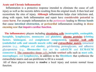
  • 1. Acute and Chronic Inflammation Inflammation is a protective response intended to eliminate the cause of cell injury as well as the necrotic debris resulting from the original insult. It then heal and reconstitute the sites of injury. Although inflammation helps clear infections and, along with repair, both inflammation and repair have considerable potential to cause harm. For example inflammation in the peritoneum leading to fibrous bands that cause intestinal obstruction, or pericardial inflammation resulting in dense encasing scar that impairs cardiac function. The inflammatory players including circulating cells (neutrophils, eosinophils, basophils, lymphocytes, monocytes and platelets), plasma proteins (clotting factors, kininogens, and complement components), vascular wall cells (endothelial cells and smooth muscle cells) and extracellular matrix (structural proteins (e.g., collagen and elastin), gel-forming proteoglycans, and adhesive glycoproteins (e.g., fibronectin) that are the cell-ECM and ECM-ECM connectors). The connective tissue cells include sentinel cells such as mast cells, macrophages, and lymphocytes, in addition to the fibroblasts that synthesize the extracellular matrix and can proliferate to fill in a wound. All of these players interact to resolve a local injury and restore normal tissue function.
  • 2. The components of acute and chronic inflammatory responses and their principal functions.
  • 3. 1-Acute inflammation is of relatively short duration, lasting from a few minutes up to a few days, and is characterized by fluid and plasma protein exudation and a predominantly neutrophilic leukocyte accumulation. 2-Chronic inflammation is of longer duration (days to years) and is typified by influx of lymphocytes and macrophages with associated vascular proliferation and scarring. Acute inflammation This process has two major components: 1-Vascular changes: alterations in vessel caliber resulting in increased blood flow (vasodilation) and increased vascular permeability that permit plasma proteins to leave the circulation. 2-Cellular events: emigration of the leukocytes from the microcirculation into the focus of injury (cellular recruitment and activation). The five classic cardinal signs of acute inflammation are: heat (calor), redness (rubor), and swelling (tumor), pain (dolor) and loss of function (functio laesa).
  • 4. Vascular Changes After transient (seconds) vasoconstriction, arteriolar vasodilation occurs, resulting in locally increased blood flow and engorgement of the capillary beds causing redness (erythema) and warmth. Subsequently, the microvasculature becomes more permeable, resulting in leakage of protein-rich fluid into the extravascular tissues causing concentrated red blood cells increasing blood viscosity and slowing the circulation (Stasis). Then, leukocytes (principally neutrophils) settling out of the flowing blood and accumulate along the vascular endothelial surface, (Margination). Then, the leukocytes adhere to endothelial cells, squeeze between them and migrate through the vascular wall into the interstitial tissue. Transudate: is an extravasation of blood plasma contains little protein. Exudate: is an extravasation of protein-rich fluid and even cells into the interstitium. This fluid accumulation is called edema.
  • 5. The major local manifestations of acute inflammation, compared to normal. (1) Vascular dilation and increased blood flow (causing erythema and warmth), (2) extravasation and deposition of plasma fluid and proteins (edema), and (3) leukocyte (mainly neutrophil) emigration and accumulation in the site of injury.
  • 6. -Endothelial cell contraction leads to intercellular gaps in venules and is a reversible process elicited rapidly by binding of histamine, bradykinin, leukotrienes and other chemical mediators to specific receptors. The main sites for this rapid increase in vascular permeability are postcapillary venules. -Endothelial cell retraction is another reversible mechanism resulting in increased vascular permeability by effect of tumor necrosis factor (TNF) and interleukin 1(IL-1). -Direct endothelial injury results in vascular leakage by causing endothelial cell necrosis and detachment. This effect is usually seen after severe injuries (e.g., burns or infections).
  • 7. Formation of transudates and exudates. A, Normal hydrostatic pressure (blue arrows) is about 32 mm Hg at the arterial end of a capillary bed and 12 mm Hg at the venous end; the mean colloid osmotic pressure of tissues is approximately 25 mm Hg (green arrows), which is equal to the mean capillary pressure. Therefore, the net flow of fluid across the vascular bed is almost nil. B, A transudate is formed when fluid leaks out because of increased hydrostatic pressure or decreased osmotic pressure. C, An exudate is formed in inflammation because vascular permeability increases as a result of increased interendothelial spaces.
  • 8. Cellular Events The sequence of events in the extravasation of leukocytes from the vascular lumen to the extravascular space is divided into (1) margination and rolling, (2) adhesion and transmigration between endothelial cells, and (3) migration in interstitial tissues toward a chemotactic stimulus. Rolling, adhesion, and transmigration are mediated by the binding of complementary adhesion molecules on leukocytes and endothelial surfaces.
  • 9. Endothelial Molecule Leukocyte Molecule Major Role P-selectin Sialyl-Lewis X-modified proteins Rolling (neutrophils, monocytes, lymphocytes) GlyCam-1, CD34 L-selectin Rolling* (neutrophils, monocytes) E-selectin Sialyl-Lewis X-modified proteins Rolling and adhesion (neutrophils, monocytes, T lymphocytes) VCAM-1 (immunoglobulin family) VLA-4 integrin Adhesion (eosinophils, monocytes, lymphocytes) ICAM-1 (immunoglobulin family) CD11/CD18 integrins (LFA-1, Mac-1) Adhesion, arrest, transmigration (neutrophils, monocytes, lymphocytes) CD31 (PECAM-1) CD31 (PECAM-1) Arrest, transmigration (neutrophils, monocytes, lymphocytes) ICAM-1, intercellular adhesion molecule 1; PECAM-1, platelet endothelial cell adhesion molecule 1; VCAM-1, vascular cell adhesion molecule 1.w Endothelial and leukocyte adhesion molecule pairs
  • 10. Margination and Rolling: the smaller, discoid red cells tend to move faster than the larger, spherical white cells, therefor, leukocytes are pushed out of the central axial column (where they normally flow) and accumulate and interact with endothelial cells (Margination). Subsequently, leukocytes tumble on the endothelial surface, transiently sticking along the way, a process called rolling. The relatively loose and transient adhesions involved in rolling are accounted for by the selectin family of molecules. Selectins are receptors expressed on leukocytes and endothelium. These include E-selectin (CD62E), confined to endothelium; P-selectin (CD62P), present on endothelium and platelets; and L-selectin (CD62L), on the surface of most leukocytes. The endothelial selectins are typically expressed at low levels or are not present at all on normal cells.
  • 11. Adhesion and Transmigration: Eventually, leukocytes firmly stick to endothelial surfaces (adhesion). This firm adhesion is mediated by adhesion molecules of the immunoglobulin superfamily (ICAM-1 & VCAM-1) on endothelial cells that interact with integrins, glycoproteins expressed on leukocyte cell surfaces. Integrins are normally expressed on leukocyte plasma membranes but do not adhere to their appropriate ligands until the leukocytes are activated by chemotactic agents or other stimuli (produced by endothelium or other cells at the site of injury).
  • 12. After margination, The leukocytes first roll, then become activated and adhere to endothelium, then transmigrate across the endothelium, pierce the basement membrane, and migrate toward chemoattractants at the source of injury. Different molecules play predominant roles in different steps of this process - selectins in rolling; chemokines in activating the neutrophils to increase avidity of integrins; integrins in firm adhesion; and CD31 (PECAM-1) in transmigration. ICAM-1, intercellular adhesion molecule 1; IL-1, interleukin 1; PECAM-1, platelet endothelial cell adhesion molecule 1; TNF, tumor necrosis factor
  • 13. Phagocytosis and Degranulation: Phagocytosis and the elaboration of degradative enzymes are two major benefits of having recruited leukocytes at the site of inflammation. Phagocytosis consists of three distinct but interrelated steps: (1) recognition and attachment of the foreign particle to the phagocytic leukocyte, (2) engulfment the foreign particle, with subsequent formation of a phagocytic vacuole, and (3) killing and degradation of the foreign particle. Recognition and attachment of leukocytes to most microorganisms is facilitated by serum proteins generically called opsonins (e.g. IgG, C3b & collectins); opsonins bind specific molecules on microbial surfaces and in turn facilitate binding with specific opsonin receptors on leukocytes. The corresponding receptors on leukocytes are the Fc receptor (FcR) for IgG, the complement receptors (CR1, 2, and 3) for complement fragments, and C1q for the collectins. Binding of opsonized particles triggers engulfment; in addition, IgG binding to FcR induces cellular activation that enhances degradation of ingested microbes. In engulfment, pseudopods are extended around the object, eventually forming a phagocytic vacuole. The membrane of the vacuole then fuses with the membrane of a lysosomal granule, resulting in phagolysosome and degranulation of the leukocyte.
  • 14. Phagocytosis of a particle (e.g., a bacterium) involves (1) attachment and binding of the particle to receptors on the leukocyte surface, (2) engulfment and fusion of the phagocytic vacuole with granules (lysosomes), and (3) destruction of the ingested particle. iNOS, Inducible nitric oxide synthase; NO, nitric oxide; ROS, reactive oxygen species.
  • 15. The final step in the phagocytosis of microbes is killing and degradation. Microbial killing is accomplished largely by reactive oxygen species. Phagocytosis stimulates an oxidative burst characterized by a sudden increase in oxygen consumption, glycogen catabolism (glycogenolysis), increased glucose oxidation, and production of reactive oxygen metabolites. The generation of the oxygen metabolites is due to rapid activation of a leukocyte NADPH oxidase, which oxidizes NADPH (reduced nicotinamide adenine dinucleotide phosphate) and, in the process, converts oxygen to superoxide ion.
  • 16. Defects in Leukocyte Function Since leukocytes play a central role in host defense, it is not surprising that defects in leukocyte function, both genetic and acquired, lead to increased vulnerability to infections, often recurrent and life-threatening. -Defects in adhesion. In leukocyte adhesion deficiency type 1 (LAD-1), defective synthesis of the CD18 β subunit of the leukocyte integrins LFA-1 and Mac-1 leads to impaired adhesion, spreading, phagocytosis, and generation of an oxidative burst. Leukocyte adhesion deficiency type 2 (LAD-2) resulting in the absence of sialyl-Lewis X on leukocytes that binds to selectins on activated endothelium. -Defects in microbicidal activity: An example is chronic granulomatous disease (CGD), a genetic deficiency in one of the components of the NADPH oxidase responsible for generating superoxide. In these patients, engulfment of bacteria does not result in activation of oxygen-dependent killing mechanisms, despite the fact that the myeloperoxidase activity of the cells is normal.
  • 17. -Defects in phagolysosome formation: One such disorder, Chédiak-Higashi syndrome, is an autosomal recessive disease that results from disordered intracellular trafficking of organelles, ultimately impairing lysosomal degranulation into phagosomes. The secretion of lytic secretory granules by cytotoxic T cells is also affected, explaining the severe immunodeficiency seen in the disorder. Chemical Mediators of Inflammation -Mediators may be circulating in the plasma (synthesized by the liver), or may be produced locally by cells at the site of inflammation. Plasma-derived mediators (complement, kinins and coagulation factors) circulate as inactive precursors that must undergo proteolytic cleavage getting the biologic properties. Cell-derived mediators are normally sequestered in intracellular granules that are secreted upon activation (e.g., histamine in mast cells) or are synthesized de novo in response to a stimulus (e.g., prostaglandins). -Most mediators induce their effects by binding to specific receptors on target cells. However, some have direct enzymatic and/or toxic activities (e.g., lysosomal proteases or reactive oxygen species). On the other hand, they may have opposing functions and act to counter-regulate the initial stimulus. -Mediators may act on one or few targets, or may have widespread activity.
  • 18. Disease Defect Genetic Leukocyte adhesion deficiency 1 β chain of CD11/CD18 integrins Leukocyte adhesion deficiency 2 Sialyl -Lewis X (selectin receptor) Neutrophil-specific granule deficiency Absence of neutrophil-specific granules Chronic granulomatous disease Decreased oxidative burst X-linked NADPH oxidase (membrane component) Autosomal recessive NADPH oxidase (cytoplasmic component) Myeloperoxidase deficiency Absent MPO-H2O2 system Chédiak-Higashi syndrome Membrane protein involved in organelle trafficking Acquired Thermal injury, diabetes, sepsis, etc. Chemotaxis Hemodialysis, diabetes Adhesion Leukemia, sepsis, diabetes, malnutrition, etc. Phagocytosis and microbicidal activity MPO, myeloperoxidase; NADPH, reduced nicotinamide adenine dinucleotide phosphate DEFECTS IN LEUKOCYTE FUNCTION
  • 19. The principal chemical mediators of inflammation. EC, Endothelial cells.
  • 20. -Mediator function is generally tightly regulated. Once activated and released from the cell, most mediators quickly decay (e.g., AA metabolites), are inactivated by enzymes (e.g., kininase inactivates bradykinin), are eliminated (e.g., antioxidants scavenge toxic oxygen metabolites), or are inhibited (complement inhibitory proteins). Vasoactive Amines: Histamine is widely produced by mast cells adjacent to vessels, although it is also present in circulating basophils and platelets. Preformed histamine is stored in mast cell granules and is released in response to a variety of stimuli. Histamine causes arteriolar dilation and is the principal mediator of the immediate phase of increased vascular permeability, inducing venular endothelial contraction and interendothelial gaps. Soon after its release, histamine is inactivated by histaminase. -Serotonin is also a preformed vasoactive mediator, with effects similar to those of histamine. It is found primarily within platelet granules (along with histamine, adenosine diphosphate, and calcium) and is released during platelet aggregation. -Neuropeptides: Like the vasoactive amines, neuropeptides can initiate inflammatory responses; these are small proteins, such as substance P, that transmit pain signals, regulate vessel tone, and modulate vascular permeability. Nerve fibers that secrete neuropeptides are especially prominent in the lung and gastrointestinal tract.
  • 21. Plasma Proteases: Many of the effects of inflammation are mediated by three interrelated plasma-derived factors: the kinin system, the clotting system, and complement system. They all linked by the initial activation of Hageman factor (factor XII of the intrinsic coagulation cascade). Hageman factor is a protein synthesized by the liver that circulates as inactive form until it encounters collagen, basement membrane, or activated platelets (at a site of endothelial injury). Kinin system activation leads to the formation of bradykinin from its circulating precursor (high-molecular-weight kininogen). Like histamine, bradykinin increases vascular permeability, arteriolar dilation, and bronchial smooth muscle contraction. It also causes pain when injected into the skin. In the clotting system, the resultant factor XIIa-driven proteolytic cascade causes thrombin activation, which in turn cleaves circulating soluble fibrinogen to generate an insoluble fibrin. Factor Xa, an intermediate in the clotting cascade, causes increased vascular permeability and leukocyte emigration. The resultant thrombin participates in inflammation by enhancing leukocyte adhesion to endothelium and by generating fibrinopeptides (during fibrinogen cleavage) that increase vascular permeability and are chemotactic for leukocytes.
  • 22. -The complement system consists of a cascade of plasma proteins that play an important role in both immunity and inflammation. They ultimately generate a pore like membrane attack complex (MAC) that effectively punches holes in the membranes of invading microbes. -Complement components (numbered C1 to C9) are present in plasma as inactive forms. Briefly, the most critical step in the elaboration of the biologic functions of complement is the activation of the third component, C3. Cleavage of C3 occurs by the classic pathway, triggered by antigen-antibody complexes; or through the alternative pathway, by bacterial polysaccharides (e.g., endotoxin). -Vascular and cellular effects: C3a and C5a (also called anaphylatoxins) increase vascular permeability and cause vasodilation by inducing mast cells to release their histamine. Also, C5a can activates leukocytes and increases affinity of their integrins, thereby increasing adhesion to endothelium.
  • 23. Interrelationships among the four plasma mediator systems triggered by activation of factor XII (Hageman factor).
  • 24. The activation and functions of the complement system. Activation of complement by different pathways leads to cleavage of C3. The functions of the complement system are mediated by breakdown products of C3 and other complement proteins, and by the membrane attack complex (MAC).
  • 25. Arachidonic Acid Metabolites (Prostaglandins, Leukotrienes, and Lipoxins): Arachidonic Acid is a polyunsaturated fatty acid and present in the body mainly as a component of cell membrane phospholipids. It is released via cellular phospholipases that have been activated by injurious stimuli, or by inflammatory mediators such as C5a. A A metabolism proceeds along one of two major pathways: cyclooxygenase, synthesizing prostaglandins and thromboxanes, and lipoxygenase, synthesizing leukotrienes and lipoxins. AA metabolites (eicosanoids) can mediate virtually every step of inflammation; their synthesis is increased at sites of inflammatory response, and agents that inhibit their synthesis also diminish inflammation. Cyclooxygenase pathway: Products of this pathway include prostaglandin (PG) E2 (PGE2), PGD2, PGF2α, PGI2 (prostacyclin), and thromboxane A2 (TXA2). platelets contain the enzyme thromboxane synthase, and hence TXA2, a potent platelet-aggregating agent and vasoconstrictor, is the major product in these cells. Endothelium possesses prostacyclin synthase, which leads to the formation of PGI2, a vasodilator and a potent inhibitor of platelet aggregation. PGD2 is the major metabolite of the cyclooxygenase pathway in mast cells, it causes vasodilation and potentiates edema formation. The prostaglandins involved in the pathogenesis of pain and fever in inflammation;
  • 26. Lipoxygenase pathway: 5-Lipoxygenase (5-LO) is the predominant AA- metabolizing enzyme in neutrophils. The first leukotriene generated is leukotriene A4 (LTA4), which in turn gives rise to LTB4 and LTC4. LTB4 is a potent chemotactic agent and causes aggregation of neutrophils. LTC4 and its subsequent metabolites, LTD4 and LTE4, cause vasoconstriction, bronchospasm, and increased vascular permeability. Lipoxins Lipoxins have both pro- and anti-inflammatory actions. For example, LXA4 causes vasodilation and antagonizes LTC4-stimulated vasoconstriction; other activities, inhibit neutrophil chemotaxis and adhesion while stimulating monocyte adhesion.
  • 27. Generation of arachidonic acid metabolites and their roles in inflammation. Note the enzymatic activities whose inhibition through pharmacologic intervention blocks major pathways (denoted with a red X). COX-1, COX-2, Cyclooxygenase 1 and 2; HETE, hydroxyeicosatetraenoic acid; HPETE, hydroperoxyeicosatetraenoic acid.
  • 28. There are two forms of cyclooxygenase (Cox), called Cox-1 and Cox-2. Moreover, Cox-1 (but not Cox-2) is expressed in the gastric mucosa, and the mucosal prostaglandins generated by Cox-1 are protective against acid- induced damage. Thus, inhibition of cyclooxygenases by aspirin and NSAIDs (which inhibit both Cox-1 and Cox-2) reduces inflammation by blocking prostaglandin synthesis but also predisposes to gastric ulceration. Platelet-Activating Factor: Originally named for its ability to aggregate platelets and cause degranulation. It is another phospholipid-derived mediator with a broad spectrum of inflammatory effects. PAF acts directly on target cells via a specific G-protein-coupled receptor. PAF causes vasoconstriction and bronchoconstriction and is 100 to 10,000 times more potent than histamine in inducing vasodilation and increased vascular permeability.
  • 29. Cytokines Cytokines are polypeptide products of many cell types (mainly activated lymphocytes and macrophages) that modulate the function of other cell types. These include colony-stimulating factors (CSF), which direct the growth of immature marrow precursor cells. Many cell types produce multiple cytokines, and the effects tend to be pleiotropic (different cells are affected differently by the same cytokine). Cytokines are also frequently redundant in that the same activity may be induced by many different Cytokines. They can act on the same cell that produces them (autocrine effect), on adjacent cell (paracrine effect), or systemically (endocrine effect). These activities are mediated by binding to specific receptors. -Cytokines that regulate lymphocyte function such as activation, growth, and differentiation (e.g., IL-2, which stimulates proliferation, and transforming growth factor β, which inhibits lymphocyte growth). -Cytokines involved in innate immunity (TNF and IL-1). -Cytokines that activate inflammatory cells (in particular, macrophages) such as interferon-γ (IFN-γ) and IL-12. -Chemokines that have chemotactic activity for various leukocytes. -Cytokines that stimulate hematopoiesis, including granulocyte-monocyte colony- stimulating factor (GM-CSF) and IL-3.
  • 30. Interleukin 1 and Tumor Necrosis Factor Both IL-1 and TNF are mainly produced by activated macrophages, and their secretion is stimulated by injury, or inflammatory mediators. They induce endothelial activation with increased expression of adhesion molecules, secretion of additional cytokines and growth factors, production of eicosanoids and nitric oxide (NO), and increased endothelial thrombogenicity. TNF also causes aggregation and activation of neutrophils and the release of proteolytic enzymes from mesenchymal cells, thus contributing to tissue damage. Both cytokines activate tissue fibroblasts, resulting in increased proliferation and production of extracellular matrix. Chemokines Structurally related proteins that act primarily as activators and chemoattractants for subsets of leukocytes. Chemokines can stimulate hematopoietic precursor cells. Many chemokines bind to extracellular matrix; maintaining the chemotactic gradients necessary for the directed migration of recruited cells. Chemokines mediate their activities by binding to specific G- protein-coupled receptors on target cells.
  • 31. The two major groups of these are CXC and CC chemokines: -CXC chemokines: act primarily on neutrophils. IL-8 is typical of this group; it is produced by activated macrophages, endothelium and fibroblasts, mainly in response to IL-1 and TNF. -CC chemokines: A monocyte chemoattractant protein 1 (MCP-1) and macrophage inflammatory protein 1α (MIP-1α) (both chemotactic predominantly for monocytes), and eotaxin (chemotactic for eosinophils). Nitric Oxide and Oxygen-Derived Free Radicals NO is a short-lived, soluble, free radical gas produced by a variety of cells. In the central nervous system, it regulates neurotransmitter release as well as blood flow. Macrophages use it (NO) as a cytotoxic metabolite for killing microbes and tumor cells. When produced by endothelium, it stimulate smooth muscle relaxation (vasodilation). Since the half-life of NO is measured in seconds, it can affect only those cells near the source where it is generated. NO is synthesized de novo from l-arginine, molecular oxygen, and NADPH by the enzyme nitric oxide synthase (NOS).
  • 32. (NO) plays multiple roles in inflammation, including (1) relaxation of vascular smooth muscle (vasodilation), (2) antagonism of all stages of platelet activation (adhesion, aggregation, and degranulation), (3) reduction of leukocyte recruitment at inflammatory sites, and (4) action as a microbicidal agent (with or without superoxide radicals) in activated macrophages. Oxygen-derived free radicals: are synthesized via the NADPH oxidase pathway and are released from neutrophils and macrophages after stimulation by chemotactic agents, immune complexes, or phagocytic activity. Superoxide is subsequently converted to H2O2, OH·, and toxic NO derivatives. At low levels, these reactive oxygen species can increase chemokine, cytokine, and adhesion molecule expression. While, at higher levels, these short-lived molecules are implicated in a variety of tissue injury mechanisms, including (1) endothelial damage, with thrombosis and increased permeability; (2) protease activation and antiprotease inactivation, with increase breakdown of the extracellular matrix; and (3) direct injury to other cell types (e.g., tumor cells, erythrocytes, parenchymal cells). Fortunately, a variety of antioxidant protective mechanisms (e.g., catalase, superoxide dismutase, and glutathione) are present in tissues and serum to minimize the toxicity of the oxygen free radicals.
  • 33. Sources and effects of nitric oxide (NO) in inflammation. NO synthesized by endothelial cells (mostly via endothelial cell [type III] NO synthase [eNOS]) and by macrophages (mostly via inducible [type II] NO synthase [iNOS]) causes vasodilation and reduces platelet and leukocyte adhesion; NO produced in phagocytes is also cytotoxic to microbes.
  • 34. Lysosomal Constituents. The lysosomal granules of neutrophils and monocytes contain multiple molecules that can mediate acute inflammation. These may be released after cell death, Neutral proteases, including enzymes such as elastase, collagenase, and cathepsin, that are activate in the extracellular matrix and cause destructive tissue injury by degrading elastin, collagen, basement membrane, and other matrix proteins. These effects are checked, however, by a series of antiproteases present in the serum and extracellular matrix. These include α2- macroglobulin (in the serum) as well as antiprotease α1- antitrypsin, which is the major inhibitor of neutrophil elastases. Deficiencies of these inhibitors may result in sustained activation of leukocyte proteases, potentially resulting in tissue destruction at sites of leukocyte accumulation.
  • 35. Vasodilation Prostaglandins Nitric oxide Increased Vascular Permeability Vasoactive amines (histamine, serotonin) C3a and C5a (by inducing release of vasoactive amines) Bradykinin Leukotrienes C4, D4, E4 Platelet-activating factor Chemotaxis, Leukocyte Activation C5a Leukotriene B4 Bacterial products Chemokines (e.g., interleukin 8 [IL-8]) Fever IL-1, IL-6, tumor necrosis factor Prostaglandins Pain Prostaglandins Bradykinin Tissue Damage Neutrophil and macrophage lysosomal enzymes Oxygen metabolites (Oxygen free radicles) Nitric oxide Inflammation- induced tissue injury effects of inflammation and their major mediators.
  • 36. Outcomes of Acute Inflammation 1-Resolution: When the injury is limited or short-lived resulting in minimal tissue damage, and the tissue is capable of replacing any died cells, the usual outcome is restoration to histologic and functional normalcy. This involves neutralization or removal of the chemical mediators, normalization of vascular permeability, and cessation of leukocyte emigration with apoptosis of extravasated neutrophils. The lymphatic drainage and macrophage ingestion of necrotic debris lead to the clearance of the edema fluid and inflammatory cells. 2-Scarring or fibrosis: results after substantial tissue destruction or when the tissues do not regenerate. Abscess formation may occur in the setting of extensive neutrophilic infiltrates, or in certain bacterial or fungal infections (pyogenic, or "pus forming"), and due to the extensive tissue destruction (including the extracellular matrix), the only outcome of abscess formation is scarring.
  • 37. 3-Progression to chronic inflammation may follow acute inflammation, although signs of chronic inflammation may be present at the onset of injury (e.g. in viral infections or immune responses to self-antigens). Chronic inflammation may be followed by regeneration of normal structure and function (regeneration) or may lead to scarring. CHRONIC INFLAMMATION Inflammation of prolonged duration (weeks to months to years) in which active inflammation, tissue injury, and healing proceed simultaneously. Chronic inflammation is characterized by: 1-Infiltration with mononuclear ("chronic inflammatory") cells, including macrophages, lymphocytes, and plasma cells. 2-Tissue destruction, largely directed by the inflammatory cells. 3-Repair, involving new vessel proliferation (angiogenesis) and fibrosis. Peptic ulcer of the duodenum initially shows acute inflammation followed by stages of resolution. However, recurrent bouts of duodenal epithelial injury result in a lesion of both acute and chronic inflammation. Fibrosis, in particular the proliferation of fibroblasts and accumulation of excess extracellular matrix is a common feature of many chronic inflammatory diseases and is an important cause of organ dysfunction.
  • 38. Outcomes of acute inflammation: resolution, healing by scarring (fibrosis), or chronic inflammation.
  • 39. Events in the resolution of inflammation. Phagocytes clear the fluid, leukocytes and dead tissue, and fluid and proteins are removed by lymphatic drainage.
  • 40. Chronic inflammation arises in the following settings: 1-Viral infections. Intracellular infections of any kind typically require lymphocytes (and macrophages) to identify and eradicate infected cells. 2-Persistent microbial infections: including mycobacteria (tubercle bacilli), Treponema pallidum (causative organism of syphilis), and certain fungi. These organisms are of low direct pathogenicity, but typically they evoke an immune response called delayed hypersensitivity ,which may culminate in a granulomatous reaction. 3-Prolonged exposure to potentially toxic agents. Examples include nondegradable exogenous material such as inhaled particulate silica, which can induce a chronic inflammatory response in the lungs (silicosis) and endogenous agents such as chronically elevated plasma lipid components, which may contribute to atherosclerosis. 4-Autoimmune diseases, in which an individual develops an immune response to self- antigens and tissues. Because the responsible antigens are in most instances constantly renewed, a self-perpetuating immune reaction results (e.g., rheumatoid arthritis or multiple sclerosis). Chronic Inflammatory Cells and Mediators Macrophages: Constituting the critical mainstay and heart of chronic inflammation. Macrophages also found in organs such as the liver (Kupffer cells), spleen and lymph nodes (sinus histiocytes), central nervous system (microglial cells), and lungs (alveolar macrophages). In these sites, macrophages act as filters for particulate matter, microbes, and senescent cells (the so-called mononuclear phagocyte system), as well as acting as sentinels to alert the specific components of the immune system (T and B lymphocytes) to injurious stimuli.
  • 41. A, Chronic inflammation in the lung, showing a collection of chronic inflammatory cells (asterisk), destruction of parenchyma (alveoli are replaced by spaces lined by cuboidal epithelium, (arrowheads), and replacement by connective tissue (fibrosis, arrows). B, acute inflammation of the lung, neutrophils fill the alveolar spaces and blood vessels are congested.
  • 42. Macrophages may also become activated, increased in its size, increased content of lysosomal enzymes, more active metabolism, and greater ability to kill organisms, called (epithelioid macrophages). It is activated by signals include cytokines secreted by sensitized T lymphocytes (IFN-γ), bacterial endotoxin, mediators produced during acute inflammation. Activated macrophages secrete a wide variety of biologically active products that can result in the tissue injury and fibrosis. These products include: -Acid and neutral proteases. Recall that the latter were also implicated as mediators of tissue damage in acute inflammation. Other enzymes, such as plasminogen activator, greatly amplify the generation of proinflammatory substances. -Complement components Although hepatocytes are the major source of these proteins, activated macrophages may release significant amounts of these proteins -Reactive oxygen species and NO. -AA metabolites (eicosanoids). -Cytokines, such as IL-1 and TNF, as well as growth factors that influence the proliferation of smooth muscle cells and fibroblasts and the production of extracellular matrix.
  • 43. In chronic inflammatory sites, macrophage accumulation persists, and macrophages can proliferate. IL-4 or IFN-γ can also induce macrophages to fuse into large, multinucleated cells called giant cells. Lymphocytes, Plasma Cells, Eosinophils, and Mast Cells: Both T and B lymphocytes migrate into inflammatory sites using some of the same adhesion molecule pairs and chemokines that recruit monocytes. Lymphocytes are mobilized in the setting of any immune-mediated stimulus (i.e., infections) as well as non- immune-mediated stimulus (infarction or trauma). T lymphocytes have a reciprocal relationship to macrophages in chronic inflammation; they are initially activated by interaction with macrophages presenting antigen fragments on their cell surface. The activated lymphocytes produce IFN-γ, a major stimulating cytokine for activating monocytes and macrophages. Activated macrophages release cytokines, including IL-1 and TNF, that further activate lymphocytes. Plasma cells are the terminally differentiated end product of B-cell activation; they can produce antibodies directed against antigens in the inflammatory site or against altered tissue components. Eosinophils are characteristically found in parasitic infections or immune reactions mediated by IgE, associated with allergies. Their emigration is driven by adhesion molecules similar to those used by neutrophils, and by specific chemokines (e.g., eotaxin) derived from leukocytes or epithelial cells. Eosinophil-specific granules contain major basic protein (MBP), that is toxic to parasites but also causes epithelial cell lysis.
  • 44. The roles of activated macrophages in chronic inflammation. Macrophages are activated by nonimmunologic stimuli such as bacterial endotoxin or by cytokines from immune- activated T cells, particularly interferon-gamma; (IFN-gamma;). The products made by activated macrophages that cause tissue injury and fibrosis are indicated. AA, Arachidonic acid; PDGF, platelet-derived growth factor; FGF, fibroblast growth factor; TGF-beta;
  • 45. Macrophage-lymphocyte interactions in chronic inflammation. Activated lymphocytes and macrophages stimulate each other, and both cell types release inflammatory mediators that affect other cells. IFN-gamma;, interferon-gamma; IL-1, interleukin 1; TNF, tumor necrosis factor.
  • 46. Mast cells are sentinel cells widely distributed in connective tissues throughout the body and can participate in both acute and chronic inflammatory responses. Mast cells are "armed" with IgE to certain antigens. When these antigens are subsequently encountered, the prearmed mast cells are triggered to release histamines and AA metabolites that elicit the early vascular changes of acute inflammation. IgE-armed mast cells are central players in anaphylactic shock, but they also play a beneficial role in a variety of infections, particularly those involving parasites. Mast cells can also elaborate cytokines such as TNF, thereby participating in more chronic responses. An important final point: although neutrophils are the classic hallmarks of acute inflammation, many forms of chronic inflammation may nevertheless continue to show extensive neutrophilic infiltrates, due either to persistent microbes or to mediators elaborated by macrophages or necrotic cells. This is sometimes called acute chronic inflammation. Granulomatous Inflammation Granulomatous inflammation is a distinctive pattern of chronic inflammation characterized by aggregates of activated macrophages that assume a squamous cell-like (epithelioid) appearance. Granulomas are encountered in relatively few pathologic states; consequently, recognition of the granulomatous pattern is important because of the limited number of conditions (some life-threatening) that cause it. Granulomas can form in the setting of persistent T-cell responses to certain microbes (such as Mycobacterium tuberculosis, Treponema pallidum causing the syphilitic gumma, or fungi), where T-cell-derived cytokines are responsible for persistent macrophage activation. Tuberculosis is the archetypal granulomatous disease due to infection and should always be excluded as a cause when granulomas are identified. Granulomas may also develop in response to relatively inert foreign bodies (e.g., suture, splinter, breast implant), forming so-called foreign body granulomas. Notably, granuloma formation does not always lead to eradication of the causal agent, which is frequently resistant to killing or degradation. Nevertheless, the formation of a granuloma effectively "walls off" the offending agent and is therefore a useful defense mechanism.
  • 47. MORPHOLOGY In the usual H & E preparations, activated macrophages in granulomas have pink, granular cytoplasm with indistinct cell boundaries. The aggregates of epithelioid macrophages are surrounded by a collar of lymphocytes secreting the cytokines responsible for ongoing macrophage activation. Older granulomas also develop a surrounding rim of fibroblasts and connective tissue, due to cytokines elaborated by the activated macrophages; this rim of scarring is useful in containing the injurious agent that caused formation of the granuloma in the first place, although it may also be a cause of tissue injury and dysfunction. Frequently, but not invariably, multinucleated giant cells 40 to 50 μm in diameter are also found in granulomas. They consist of a large mass of cytoplasm and multiple nuclei and derive from the fusion of 20 or more macrophages. In granulomas associated with certain infectious organisms (most classically due to the tubercle bacillus), a combination of hypoxia and free radical injury leads to a central zone of necrosis. Grossly, this has a granular, cheesy appearance and is therefore called caseous necrosis. Lymphatics and Lymph Nodes in Inflammation The system of lymphatics and lymph nodes filters and polices the extravascular fluids. Together with the mononuclear phagocyte system, it represents a secondary line of defense that is called into play whenever a local inflammatory reaction fails to contain and neutralize injury. Lymphatics are extremely delicate channels that are difficult to visualize in ordinary tissue sections because they readily collapse unless they are engorged with edema fluid and/or recirculating leukocytes. They are lined by continuous endothelium with loose overlapping cell junctions, scant basement membrane, and no muscular support except in the larger ducts. Valves are present in the larger collecting lymphatics, allowing lymph content to flow only in the distal-to-proximal direction. Thin fibrils, attached at right angles to the walls of the lymphatic vessel, extend into the adjacent tissues and serve to maintain patency of the channel.
  • 48. A typical granuloma resulting from infection with Mycobacterium tuberculosis showing central caseous necrosis, activated epithelioid macrophages, many giant cells, and a peripheral accumulation of lymphocytes. Caseous center
  • 49. Bacterial Tuberculosis (Mycobacterium tuberculosis) Leprosy (Mycobacterium leprae) Syphilitic gumma (Treponema pallidum) Cat-scratch disease (Bartonella henselae) Parasitic Schistosomiasis (Schistosoma mansoni, S. haemotobium, S. japonicum) Fungal Histoplasma capsulatum Blastomycosis Cryptococcus neoformans Coccidioides immitis Inorganic Metals or Dusts Silicosis Berylliosis Foreign Body Suture, breast prosthesis, vascular graft Unknown Sarcoidosis EXAMPLES OF GRANULOMATOUS INFLAMMATION
  • 50. Because the junctions of lymphatics are loose, lymphatic fluid eventually equilibrates with extravascular fluid. Consequently, during inflammation, lymphatic flow is increased and helps drain the edema fluid from the extravascular space. In addition to fluid, leukocytes and cell debris may also find their way into lymph. Moreover, in the setting of extensive inflammation, the drainage may also transport the offending (microbial or chemical) agent. As a result, the lymphatics themselves may become secondarily inflamed (lymphangitis), as may the draining lymph nodes (lymphadenitis). For example, it is not uncommon in infections of the hand to observe red streaks following the course of the lymphatic channels and extending along the arm to the axilla, accompanied by painful enlargement of the axillary lymph nodes. The nodal enlargement is usually caused by the proliferation of lymphocytes and macrophages in the lymphoid follicles and sinuses, as well as by hypertrophy of the phagocytic cells. This constellation of nodal histologic changes is termed reactive, or inflammatory, lymphadenitis. The secondary lymph node barriers usually contain the spread of the infection. However, in some instances they are overwhelmed, and infectious organisms drain through progressively larger channels, eventually gaining access to the vascular circulation and resulting in bacteremia. The phagocytic cells of the liver, spleen, and bone marrow constitute the next line of defense, but in massive infections, bacteria seed distant tissues of the body. The heart valves, meninges, kidneys, and joints are favored sites of implantation for blood-borne organisms, and in such a fashion endocarditis, meningitis, renal abscesses, and septic arthritis may develop. MORPHOLOGIC PATTERNS OF ACUTE AND CHRONIC INFLAMMATION The severity of the inflammatory response, its specific cause, and the particular tissue involved can all modify the basic morphologic patterns of acute and chronic inflammation. Such patterns frequently have clinical significance and are described in greater detail as follows: -SEROUS INFLAMMATION. This is characterized by the outpouring of a watery, relatively protein-poor fluid (effusion) that, depending on the site of injury, derives either from the serum or from the secretions of mesothelial cells lining the peritoneal, pleural, and pericardial cavities. The skin blister resulting from a burn or viral infection is a good example of a serous effusion accumulated either within or immediately beneath the epidermis of the skin.
  • 51. -FIBRINOUS INFLAMMATION. This occurs as a consequence of more severe injuries, with a resultant greater vascular permeability allowing larger molecules (specifically, fibrinogen) to pass the endothelial barrier. Histologically, the accumulated extravascular fibrin appears as an eosinophilic meshwork of threads or, sometimes, as an amorphous coagulum. Fibrinous exudates may be degraded by fibrinolysis, and the accumulated debris may be removed by macrophages, resulting in restoration of the normal tissue structure (resolution). However, failure to completely remove the fibrin results in the ingrowth of fibroblasts and blood vessels, leading ultimately to scarring (organization). For example, organization of a fibrinous pericardial exudate forms dense fibrous scar tissue that bridges or obliterates the pericardial space and restricts myocardial function. -SUPPURATIVE (PURULENT) INFLAMMATION. This is manifested by the presence of large amounts of purulent exudate (pus) consisting of neutrophils, necrotic cells, and edema fluid. Certain organisms (e.g., staphylococci) are more likely to induce this localized suppuration and are therefore referred to as pyogenic. Abscesses are focal collections of pus that may be caused by deep seeding of pyogenic organisms into a tissue or by secondary infections of necrotic foci. Abscesses typically have a central, largely necrotic region rimmed by a layer of preserved neutrophils, with a surrounding zone of dilated vessels and fibroblastic proliferation indicative of early repair. In time, the abscess may become completely walled off and eventually replaced by connective tissue. -ULCERATION. This refers to a site of inflammation where an epithelial surface (skin, gastric epithelium, colonic mucosa, bladder epithelium) has become necrotic and eroded, often with associated subepithelial acute and chronic inflammation. This can occur as a consequence of toxic or traumatic injury to the epithelial surface (e.g., peptic ulcers) or may be due to vascular compromise (as in foot ulcers associated with the vasculopathy of diabetes). The peptic ulcer of the stomach or duodenum illustrates the typical findings. There is usually an early intense neutrophilic infiltrate with associated vascular dilation. In chronic lesions where there has been repeated insult, the area surrounding the ulcer develops fibroblastic proliferation, scarring, and the accumulation of chronic inflammatory cells.
  • 52. Serous inflammation. Low-power view of a cross-section of a skin blister showing the epidermis separated from the dermis by a focal collection of serous effusion.
  • 53. Fibrinous pericarditis. A, Deposits of fibrin on the pericardium. B, A pink meshwork of fibrin exudate (F) overlies the pericardial surface (P).
  • 54. Purulent inflammation. A, Multiple bacterial abscesses in the lung (arrows) in a case of bronchopneumonia. B, The abscess contains neutrophils and cellular debris, and is surrounded by congested blood
  • 55. The morphology of an ulcer. A, A chronic duodenal ulcer. B, Low-power cross-section of a duodenal ulcer crater with an acute inflammatory
  • 56. SYSTEMIC EFFECTS OF INFLAMMATION Anyone who has suffered through a severe bout of a viral illness (such as influenza) has experienced the systemic effects of inflammation, collectively called acute-phase reaction. Fever is only one of the more obvious of these systemic effects of inflammation; others include increased somnolence, malaise, anorexia, accelerated degradation of skeletal muscle proteins, hypotension, hepatic synthesis of a variety of proteins (e.g., complement and coagulation proteins), and alterations in the circulating white blood cell pool. The cytokines IL-1, IL-6, and TNF are the most important mediators of the acute-phase reaction. These cytokines are produced by leukocytes (and other cell types) in response to infection, or to immune and toxic injury, and are released systemically, frequently in a sort of cytokine cascade. Thus, TNF induces the production of IL-1, which in turn stimulates the production of IL-6. Although there are some differences, TNF and IL-1 cause similar effects; for example, both act on the thermoregulatory center of the hypothalamus-via local PGE production-to induce fever (hence the efficacy of aspirin and NSAIDs in reducing fever). IL-6 stimulates the hepatic synthesis of several plasma proteins, most notably fibrinogen; elevated fibrinogen levels cause erythrocytes to agglutinate more readily, explaining why inflammation is associated with a higher erythrocyte sedimentation rate by objective testing. Leukocytosis (increased white blood cell count) is a common feature of inflammatory reactions, especially those induced by bacterial infection. The leukocyte count typically increases to 15,000 or 20,000 cells per μL (normal = 4000 to 10,000 cells per μL) but may climb as high as 40,000 to 100,000 cells per μL, a so-called leukemoid reaction. Leukocytosis initially results from the release of cells from the bone marrow (caused by IL-1 and TNF) and is associated with an increased number of relatively immature neutrophils in the blood ("left-shift"). Prolonged infection, however, also induces proliferation of precursors in the bone marrow, caused by IL-1- and TNF- driven increases in the production of colony-stimulating factors.
  • 57. Most bacterial infections induce a relatively selective increase in polymorphonuclear cells (neutrophilia), while parasitic infections (as well as allergic responses) characteristically induce eosinophilia. Certain viruses, such as infectious mononucleosis, mumps, and rubella, engender selective increases in lymphocytes (lymphocytosis). However, most viral infections, as well as rickettsial, protozoal, and certain types of bacterial infections (typhoid fever), are associated with a decreased number of circulating white cells (leukopenia). Leukopenia is also encountered in infections that overwhelm patients debilitated by, for example, disseminated cancer. Although this concludes our discussion of the cellular and molecular events in acute and chronic inflammation, we still need to consider the changes induced by the body's attempts to heal the damage, the process of repair. The repair begins almost as soon as the inflammatory changes have started and involves several processes, including cell proliferation, differentiation, and extracellular matrix deposition.