
Recomendados
Recomendados
Mais conteúdo relacionado
Mais procurados
Mais procurados (17)
Destaque
Destaque (20)
Semelhante a Neuropsychological factors linked to emotional response variability in older adults
Semelhante a Neuropsychological factors linked to emotional response variability in older adults (20)
Neuropsychological factors linked to emotional response variability in older adults
- 1. Neuropsychological Correlates of Normal Variation in Emotional Response to Visual Stimuli Robert G. Robinson, MD, Sergio Paradiso, MD, PhD, Romina Mizrahi, MD, Jess G. Fiedorowicz, MD, Dimitrios E. Kouzoukas, BS, and David J. Moser, PhD University of Iowa, Department of Psychiatry, Roy J. and Lucille A. Carver College of Medicine, Iowa City, Iowa. Abstract Although the neural substrates of induced emotion have been the focus of numerous investigations, the factors related to individual variation in emotional experience have rarely been investigated in older adults. Twenty-six older normal subjects (mean age, 54) were shown color slides to elicit emotions of sadness, fear, or happiness and asked to rate the intensity of their emotional responses. Subjects who experienced negative emotion most intensely showed relative impairment on every aspect of the Wisconsin Card Sorting Test. Intense positive emotion was associated with relatively impaired performance on the Rey Complex Figure Test. The volume of frontal brain structures, however, was not associated with emotion responses. Hemisphere-specific executive dysfunction was associated with greater intensity of emotional experience in normal older subjects. The role of these differences in intensity of induced emotion and impairment in executive function in daily social and vocational activity should be investigated. Keywords Emotion; induced mood; neuropsychological tests; Wisconsin Card Sorting test; Rey Complex Figure Test Interindividual differences in response to emotion may contribute to the variability of findings in emotional activation studies, yet have received little attention. Some subjects clearly experience particular emotions more intensely than others, even when the intensity of activation is held constant. Study of these individual differences may elucidate the underlying mechanisms of psychological processes and cognitive function (Canli, 2004). For instance in late life, two fundamental processes may affect emotion processing and experience in different and opposite directions: (1) the acquisition of “emotional wisdom” leading to an ability to manipulate and control extreme emotions (Dougherty et al., 1996; Magai et al., 2001), and (2) functional and structural changes in areas regulating emotional processing including the frontal cortex (Cowell et al., 1994; Kemper, 1993, 1994; Raz et al., 1997, 2000) and medial temporal structures including the hippocampus and amygdala (Geinisman et al., 1995; Kemper, 1994; West, 1993). The interplay of these phenomena may lead to greater variability of emotional regulation in older adults. Two block design fMRI studies exploring differences in perception of facial emotion in older adults confirmed the reduced functioning in the amygdala, hippocampal, and frontal networks posited in the above model (Gunning-Dixon et al., 2003; Iidaka et al., 2002). Send reprint requests to: Robert G. Robinson, MD, Department of Psychiatry, University of Iowa, 200 Hawkins Dr, Iowa City, IA 52242.. NIH Public Access Author Manuscript J Nerv Ment Dis. Author manuscript; available in PMC 2007 November 30. Published in final edited form as: J Nerv Ment Dis. 2007 February ; 195(2): 112–118. NIH-PAAuthorManuscriptNIH-PAAuthorManuscriptNIH-PAAuthorManuscript
- 2. The personality dimensions of neuroticism and extraversion may also influence emotional reactivity (Mobbs et al., 2005). Neuroticism is associated with a proclivity to experience strong negative emotions, whereas extraversion reflects an inclination for more positive emotions (DeNeve and Cooper, 1998). These dimensions may also be influenced by functional and structural pathology as evidenced by personality changes incurred in patients with demyelinating disease or surgery (Benedict et al., 2001; Smith et al., 1976). Understanding the neural basis of interindividual emotional variability has great significance both heuristically and to help elucidate the specific presentation of emotional disorders. The present study was undertaken to parse out the complex array of determinants of emotional reactivity among the elderly by examining neurocognitive and brain structure correlates of interindividual variation in emotional response to identical stimuli. Specifically, we examined the extent to which relatively left and right hemisphere–dominant neuropsychological tests of executive function may be associated with the intensity of emotional response and frontal lobe structure. We hypothesized that relatively weaker performance in tests of frontal lobe function, or differences in gray matter volume of the orbital frontal cortex, would be associated with the degree of emotional response. METHODS Subjects Subjects were older healthy volunteers (mean age, 54) who were the controls for a larger study of focal ischemic lesions and emotion induction (Paradiso et al., 2003). A total of 26 normal subjects provided written informed consented to participate in this study approved by the Institutional Review Board (see Table 1 for background characteristics). Volunteers were excluded if they had a history of mental retardation, severe learning disability, dementia, stroke, head trauma or other brain injury, physical illness that was life-threatening or interfered with their daily activities, psychiatric disorder, or substance abuse, or were taking any psychotropic medications. They were recruited using newspaper advertisements and were paid for participation. Psychiatric and Neuropsychological Assessment Depressive symptoms were measured using the 17-item Hamilton Depression Scale, which ranges from 0 to 55, with higher scores indicating more severe depressive symptoms (Hamilton, 1960). Anxiety symptoms were measured by the Hamilton Anxiety Scale, which ranges from 0 to 25, with higher scores indicating more severe anxiety symptoms (Hamilton, 1959). These scales were selected to determine whether anxiety or depression may have interfered with emotion induction. Other instruments administered were the Structured Clinical Interview for DSM-IV Diagnosis (American Psychiatric Association, 1994; Spitzer et al., 1995) and the semistructured Present State Examination (Wing et al., 1977) to ensure that the controls did not have a psychiatric disorder. The structured interview data are reported as total scores indicating overall severity of psychopathology. Also, the Social Functioning Examination (Starr et al., 1983) was administered to assess subjects’ satisfaction with their social functioning, and the Social Ties Checklist (Robinson et al., 1983) to measure subjects’ social connectedness. These measures were used to document normal levels of social support. The Toronto Alexithymia Scale (Taylor et al., 1992) was administered to measure subjects’ awareness of their feelings and ability to describe them. A score of ≤60 is considered normal ability to experience and report emotion. Robinson et al. Page 2 J Nerv Ment Dis. Author manuscript; available in PMC 2007 November 30. NIH-PAAuthorManuscriptNIH-PAAuthorManuscriptNIH-PAAuthorManuscript
- 3. The neuropsychological assessment included the Wechsler Adult Intelligence Scale—Revised (Wechsler, 1997) assessing overall intelligence, the Wisconsin Card Sorting Test (Heaton et al., 1993) assessing subjects’ ability to formulate, test, and alter problem-solving strategies in response to external feedback, and the Rey Complex Figure Test (Osterrieth, 1944; Rey, 1941, measuring visuospatial ability, visuospatial memory, and executive function. Additionally, the Controlled Word Association Test (Benton, 1968; Benton et al., 1994) assessed verbal initiation and maintenance of effort. The Wisconsin Card Sorting Test, 64-card administration, was selected as a measure of frontal executive function mediated primarily by the left hemisphere (Berman and Weinberger, 1990); (Deicken et al., 1995). The Rey Complex Figure Test was selected as a measure of executive visual spatial and organizational strategies as well as executive function mediated primarily by the right hemisphere (Grossman et al., 1993; Savage et al., 1999. All psychopathological and neuropsychological tests were administered in a standardized manner by a trained research assistant several hours before the emotional activation so there was no interference of induced emotion with cognitive performance or psychopathology. Emotional Activation Emotional activation tasks consisted of displaying still pictures on a video screen containing exclusively nonfamiliar human faces or objects/scenes. These stimuli were chosen from the International Affective Picture System, a large database of emotionally evocative color pictures (Lang et al., 1995). Subjects were exposed to seven different sets of stimuli prepared for inducing neutral emotion (two), happiness (two), sadness (one), or fear/disgust (two). There were 18 images per stimulus set that were displayed individually for 6 seconds (108 seconds total per stimulus set). All sets of stimuli were equivalent based on a large normative database of responses of normal subjects for both mean valence of emotion and mean intensity of emotion for each picture (Lang et al., 1995). Thus, across each 18 picture stimulus, we were able to equate both valence and intensity of happiness, sadness, and fear/disgust. The list of stimuli and the detailed protocol have been described in a prior publication (Paradiso et al., 2003). Prior to viewing each set, subjects were told they would be watching pictures with emotional content and should allow the pictures to influence their emotional state. Subjects had not seen the images prior to the experiment. Each set of stimuli was shown once in a random order, on an 11-inch by 8-inch computer monitor positioned 14 inches from the subject. After the stimulation of each emotion, subjects were asked to rate their feelings, which were assumed to be dimensional in nature (of happiness and amusement [positive emotions], sadness, fear/ disgust [negative emotions], anger and surprise [other emotions]), using analogue scales ranging from 0 to 10 (i.e., the analogue scale was a straight line 10 cm long in which subjects rated intensity by crossing the line, and the score was the number of centimeters from the “not at all” end). Mean positive and negative emotional responses were calculated. The mean negative emotion rating consisted of mean scores on sadness and fear following two fear stimuli (i.e., one with faces, the other without) and one sadness stimulus. The positive responses were based on mean responses of happiness following the two happy stimuli (i.e., one with and the other without faces). The responses on anger and surprise were not used in this study because they did not correspond to the emotion intended to be evoked with the stimuli. MRI Imaging Acquisition and Processing T1-weighted and PD and T2-weighted images were acquired using contiguous coronal slices (1.5 mm thick) from a 1.5 tesla GE Signa Scanner. The brain imaging studies were done before the emotional activation and neuropsychological assessment. Technical parameters for T1- Robinson et al. Page 3 J Nerv Ment Dis. Author manuscript; available in PMC 2007 November 30. NIH-PAAuthorManuscriptNIH-PAAuthorManuscriptNIH-PAAuthorManuscript
- 4. weighted images included 1.5-mm slice thickness, SPGR sequence, flip angle = 40°, TE = 5 milliseconds, TR = 24 milliseconds, NEX = 2. The T2-weighted images were 3.0-mm or 4.0- mm coronal slices with 96 milliseconds TE, 3000 TR, 1 NEX, echo train length 8. MRI images were analyzed with locally developed software (BRAINS; Andreasen et al., 1994, 1996; Arndt et al., 1996). All brains were realigned parallel to the anterior-posterior commissure line and interhemispheric fissure to ensure comparability of head position across subjects. Alignment also placed the images in standard Talairach space (Talairach and Tournous, 1988). Images from multiple subjects were coregistered and resliced in three orthogonal planes to produce a three-dimensional data set that was used for visualization and measurement of anatomical brain structures. Structural frontal lobe analysis was done for each individual subject with locally developed parcellation methods (Crespo-Facorro et al., 1999). This method used a combination of hand-tracing and automated gray-white matter distinction to calculate the volume of gray matter in the frontal lobe in each hemisphere. The frontal lobe structures included the superior, middle, and inferior frontal gyrus, the straight gyrus, the orbital frontal cortex, and the rostral anterior and caudal anterior cingulate. Data Analysis Emotional responses obtained from neutral and emotional activation states and neurocognitive performance were analyzed using Spearman rank correlational analysis. Structural brain imaging data was analyzed using the volume of gray matter for each frontal lobe structure and emotional responses. RESULTS Background characteristics, psychiatric assessment, and emotion responses are detailed in Table 1. There were no significant correlations across the 26 total subjects between positive or negative emotion responses and age, sex, handedness, race, education, or socioeconomic status (albeit older age showed a modest nonsignificant association with higher emotional response to positive stimuli; Table 1). Similarly, there was no significant correlation between emotional reactivity and depression, anxiety, or overall psychopathology (Structured Clinical Interview for DSM-IV Diagnosis, Present State Examination) and social functioning or emotional awareness. Positive and negative responses, however, were significantly correlated with each other. Subjects who experienced negative emotion more intensely also tended to experience positive emotion more intensely (ρ = 0.603, p = 0.001). Neuropsychological Test Findings Correlations between negative emotion responses and Wisconsin Card Sorting Test were significant for virtually all measures, except nonperseverative errors (Table 2; Figure 1). In contrast, there were no significant correlations between intensity of positive emotion and performance on Wisconsin Card Sorting Test. Positive emotion responses, however, were significantly correlated with Rey Complex Figure Test copying and delayed recall subtests (Table 2). No other correlations between emotional response and cognitive measures, including general intelligence and attention, were found to be significant. Structural Imaging Analysis The structural magnetic resonance analysis correlating gray matter volumes of the superior, middle, and inferior frontal gyrus, orbitofrontal cortex and straight gyrus, rostral and caudal anterior cingulate with positive and negative emotional responses revealed no significant associations (Table 3). The Spearman rho correlation coefficients ranged from 0.2 to −.03. Robinson et al. Page 4 J Nerv Ment Dis. Author manuscript; available in PMC 2007 November 30. NIH-PAAuthorManuscriptNIH-PAAuthorManuscriptNIH-PAAuthorManuscript
- 5. DISCUSSION This study examined the relationship between intensity of emotional experience following visual stimulation, cognitive function, and frontal lobe gray matter volumes. We hypothesized that there would be a relationship between intensity of subjective experience of emotion and executive functioning, and gray matter volume of the frontal gyri. Subjects with relatively weaker frontal-executive function as evidenced by poorer performance on the Wisconsin Card Sorting Test showed greater intensity of negative but not positive emotions. Persons with higher levels of positive emotion showed relative impairment in frontal visual-spatial organizational strategies and memory as measured by the Rey Complex Figure Test. Surprisingly, no correlation between emotional responses and gray matter volumes was found to be significant. Before discussion of these results, we need to acknowledge the limitations of this study. The population studied was generally of high average intelligence and had a mean of almost 15 years of education. Thus, our findings may not be applicable to less intelligent or less educated populations. However, it is worth noting that significant emotion/executive function associations were found despite the relative homogeneity of our sample. We did not examine whether subjects who experienced more intense emotion manifested any behavioral or personality characteristics that may have impaired their social or vocational function. However, we did not find any association between higher emotional reactivity and measures of psychopathology or self-reported social adjustment and ties. In addition, we only used one method for stimulating emotion (i.e., visual). However, this method of inducing emotion is the most highly standardized for both valence and intensity across different types of emotion, and is a commonly used means of emotional induction (Larson et al., 2005). The notable finding of this study was that presumably normal variations in the experience of both positive and negative emotion were significantly correlated with relatively weaker neurocognitive executive performance subserved by frontal lobe structures. Consistent with prior studies reporting biological (both biochemical and genetic) influences on normal variations in personality (Benjamin et al., 1996; Ebstein et al., 1996; Johnson et al., 1999), our findings support the assumption that normal variation of emotional response is a dimensional characteristic. We were unable to find significant associations between frontal lobe gray matter and emotional response. Age-related changes in prefrontal cortex occur early in some older persons (Raz et al., 1997) with changes in the ventral aspect observed as early as age 58 (Convit et al., 2001). Perhaps, in our sample, which showed high mean intelligence and education, there was not sufficient frontal lobe gray matter variation to detect structural/emotional associations. It is also possible that other structural brain changes (e.g., white matter hyperintensities) may correlate more strongly with emotional responses. We are planning to perform these analyses in future studies. The major question arising from our findings is why relative left hemisphere–dominant frontal dysfunction on neuropsychological testing (i.e., Wisconsin Card Sorting Test) was associated with greater experience of negative emotion, and relative right hemisphere frontal/temporal visual organizational dysfunction (i.e., Rey Complex Figure Test) with greater experience of positive emotion. Although the explanation for these phenomena will require further experimental investigation, there are several possible mechanisms which might be proposed. The fact that more intense negative emotional induction was associated with relative left frontal neuropsychological dysfunction and more intense positive emotion with relative right frontal- parietal-temporal neuropsychological dysfunction suggest that left hemisphere dysfunction may be related to more intense negative emotion and right hemisphere dysfunction may be related to more intense positive emotion. Although a topic of scientific controversy, there is a Robinson et al. Page 5 J Nerv Ment Dis. Author manuscript; available in PMC 2007 November 30. NIH-PAAuthorManuscriptNIH-PAAuthorManuscriptNIH-PAAuthorManuscript
- 6. large literature supporting the hypothesis that limbic or limbic connected structures of the right hemisphere subserve negative emotions while similar structures of the left hemisphere subserve positive emotion (Borod, 1992; Davidson and Irwin, 1999). In the present study, impaired performance on the Wisconsin Card Sorting Test relative to high average general intelligence might suggest relatively greater left than right frontal dysfunction because the Wisconsin Card Sorting Test requires left hemisphere–dominant verbal mediation (Berman and Weinberger, 1990). Impaired left frontal function might be manifested by enhanced expression of right frontal hemisphere activity (i.e., decreased interhemispheric inhibition and therefore increased sensitivity to negative emotion). Similarly, relative right hemisphere weakness in executive and visual-spatial function measured with the Rey Complex Figure Test (Grossman et al., 1993; Savage et al., 1999) may lead to enhanced expression of the left frontal lobe, and thus greater experience of positive emotion. Alternatively, more intense induction of negative emotions may be a marker for neuroticism (Costa and McCrae, 1980; DeNeve and Cooper, 1998). Neuroticism has been described as “cognitive noise” associated with executive dysfunction and increased variability in response and reaction time (Robinson and Tamir, 2005), which may be reflected in the results of the Wisconsin Card Sorting Test. Furthermore, the induction of positive affect is thought to improve cognitive flexibility and reduce perseveration (Dreisbach and Goschke, 2004). Perhaps this occurs in subjects with more active left hemisphere dominance leading to right hemisphere inhibition and impaired performance on the Rey Complex Figure Test. Further studies of this phenomenon may elucidate the mechanisms of the interplay between cognitive performance and intensity of emotion. Another possible explanation for our findings involves attentional differences. It has been shown that differences on the Emotional Awareness Scale (i.e., a measure of individual differences in the capacity to experience emotion) correlate with individual differences in blood flow in the anterior cingulate cortex (Lane et al., 1998). The anterior cingulate also mediates subjects’ attention or response selection (Lane et al., 1998). Thus, subjects with less intense experience of emotion may simply have paid less attention to the emotion inducing pictures. Although this might be proposed as a possible explanation for our findings, the fact that the Wisconsin Card Sorting Test and the Rey Complex Figure Test but not the tasks of verbal or performance IQ were associated with greater intensity of emotion suggests that our findings may not be due to intersubject attentional differences. This could be tested by correlation of emotional activation with measures of attention such as digit span. CONCLUSION The findings in this study emphasize the need to account for variations in emotional experience even among normal control populations. These variations may hide or heighten group differences when compared with patient samples. For instance, control groups containing unusually large number of patients with “relative” right or left hemisphere dysfunction and elevated sensitivity to positive or negative emotion might obscure or inflate differences between controls and patients with clinical mood disorders. These subjects with more intense emotional responses may additionally experience impairment of social or vocational function because of these mood changes or the associated underlying relative neuropsychological impairment. REFERENCES American Psychiatric Association. Diagnostic and Statistical Manual of Mental Disorders: DSM-IV. 4th ed. Washington (DC): American Psychiatric Press, Inc.; 1994. Robinson et al. Page 6 J Nerv Ment Dis. Author manuscript; available in PMC 2007 November 30. NIH-PAAuthorManuscriptNIH-PAAuthorManuscriptNIH-PAAuthorManuscript
- 7. Andreasen NC, Harris G, Cizadlo T, Arndt S, O’Leary DS, Swayze V, Flaum M. Techniques for measuring sulcal/gyral patterns in the brain as visualized through magnetic resonance scanning: BRAINPLOT and BRAINMAP. Proc Natl Acad Sci U S A 1994;91:93–97. [PubMed: 8278413] Andreasen NC, Rajarethinam R, Cizadlo T, Arndt S, Swayze VW II, Flashman LA, O’Leary DS, Ehrhardt JC, Yuh WT. Automatic atlas-based volume estimation of human brain regions from MR images. J Comput Assist Tomogr 1996;20:98–106. [PubMed: 8576490] Arndt ST, Cizadlo T, Andreasen NC, Heckel D, Gold S, O’Leary D. Tests for comparing images based on randomization and permutation methods. J Cereb Blood Flow Metab 1996;16:1271–1279. [PubMed: 8898701] Benedict RH, Priore RL, Miller C, Munschauer F, Jacobs L. Personality disorder in multiple sclerosis correlates with cognitive impairment. J Neuropsychiatry Clin Neurosci 2001;13:70–76. [PubMed: 11207332] Benjamin J, Li L, Patterson C, Greenberg BD, Murphy DL, Hamer DH. Population and familial association between the D4 dopamine receptor gene and measures of novelty seeking. Nat Genet 1996;12:81–84. [PubMed: 8528258] Benton AL. Differential behavioral effects in frontal lobe disease. Neuropsychology 1968;6:53–60. Benton, AL.; Hamsher, K.; Sivan, AB. Multilingual Aphasia Examination. Iowa City (IA): AJA Associates; 1994. Berman KF, Weinberger DR. Lateralisation of cortical function during cognitive tasks: regional cerebral blood flow studies of normal individuals and patients with schizophrenia. J Neurol Neurosurg Psychiatry 1990;53:150–160. [PubMed: 2313303] Borod JC. Interhemispheric and intrahemisphric control of emotion: a focus on unilateral brain damage. J Consult Clin Psychol 1992;60:339–348. [PubMed: 1619088] Canli T. Functional brain mapping of extraversion and neuroticism: Learning from individual differences in emotion processing. J Pers 2004;72:1105–1132. [PubMed: 15509278] Convit A, Wolf OT, de Leon MJ, Patalinjug M, Kandil E, Caraos C, Scherer A, Saint Louis LA, Cancro R. Volumetric analysis of the pre-frontal regions: Findings in aging and schizophrenia. Psychiatry Res 2001;107:61–73. [PubMed: 11530273] Costa PT Jr, McCrae RR. Influence of extraversion and neuroticism on subjective well-being: Happy and unhappy people. J Pers Soc Psychol 1980;38:668–678. [PubMed: 7381680] Cowell PE, Turetsky BI, Gur RC, Grossman RI, Shtasel DL, Gur RE. Sex differences in aging of the human frontal and temporal lobes. J Neurosci 1994;14:4748–4755. [PubMed: 8046448] Crespo-Facorro B, Kim J, Andreasen NC. Human frontal cortex: An MRI-based parcellation method. Neuroimage 1999;10:500–519. [PubMed: 10547328] Davidson RJ, Irwin W. The functional neuroanatomy of emotion and affective style. Trends Cogn Sci 1999;3:11–21. [PubMed: 10234222] Deicken RF, Merrin EL, Floyd TC, Weiner MW. Correlation between left frontal phospholipids and Wisconsin Card Sort Test performance in schizophrenia. Schizophr Res 1995;14:177–181. [PubMed: 7710998] DeNeve KM, Cooper H. The happy personality: A meta-analysis of 137 personality traits and subjective well-being. Psychol Bull 1998;124:197–229. [PubMed: 9747186] Dougherty, LM.; Abe, JA.; Izard, CE. Handbook of Emotion Adult Development and Aging. Orlando, Florida: Academic Press; 1996. Differential emotions theory and emotional development in adulthood and later life; p. 27-38. Dreisbach G, Goschke T. How positive affect modulates cognitive control: Reduced perseveration at the cost of increased distractibility. J Exp Psychol Learn Mem Cogn 2004;30:343–353. [PubMed: 14979809] Ebstein RP, Novick O, Umansky R, Priel B, Osher Y, Blaine D, Bennett ER, Nemanov L, Katz M, Belmaker RH. Dopamine D4 receptor (D4DR) exon III polymorphism associated with the human personality trait of novelty seeking. Nat Genet 1996;12:78–80. [PubMed: 8528256] Geinisman Y, Detoledo-Morrell L, Morrell F, Heller RE. Hippocampal markers of age-related memory dysfunction: Behavioral, electrophysiological and morphological perspectives. Prog Neurobiol 1995;45:223–252. [PubMed: 7777673] Robinson et al. Page 7 J Nerv Ment Dis. Author manuscript; available in PMC 2007 November 30. NIH-PAAuthorManuscriptNIH-PAAuthorManuscriptNIH-PAAuthorManuscript
- 8. Grossman M, Carvell S, Peltzer L, Stern MB, Gollomp S, Hurtig HI. Visual construction impairments in Parkinson’s disease. Neuropsychologia 1993;7:536–547. Gunning-Dixon FM, Gur RC, Perkins AC, Schroeder L, Turner T, Turetsky BI, Chan RM, Loughead JW, Alsop DC, Maldjian J, Gur RE. Age-related differences in brain activation during emotional face processing. Neurobiol Aging 2003;24:285–295. [PubMed: 12498962] Hamilton M. The assessment of anxiety state of rating. Br J Med Psychol 1959;32:50–55. [PubMed: 13638508] Hamilton MA. A rating scale for depression. J Neurol Neurosurg Psychiatry 1960;23:56–62. [PubMed: 14399272] Heaton, RK.; Chelune, GJ.; Tailey, JL. The Wisconsin Card Sorting Test. Odessa (FL): Psychological Assessment Resources; 1993. Iidaka T, Okada T, Murata T, Omori M, Kosaka H, Sadato N, Yonekura Y. Age-related differences in the medial temporal lobe responses to emotional faces as revealed by fMRI. Hippocampus 2002;12:352–362. [PubMed: 12099486] Johnson DL, Wiebe JS, Gold SM, Andreasen NC, Hichwa RD, Watkins GL, Boles Ponto LL. Cerebral blood flow and personality: A positron emission tomography study. Am J Psychiatry 1999;156:252– 257. [PubMed: 9989562] Kemper, T. Clinical Neurology of Aging. 2nd ed. New York: Oxford University Press; 1994. Neuroanatomical and neuropathological changes during aging and in dementia; p. 3-67. Kemper TL. The relationship of cerebral cortical changes to nuclei in the brainstem. Neurobiol Aging 1993;14:659–660. [PubMed: 8295677] Lane RD, Reiman EM, Axelrod B, Yun LS, Holmes A, Schwartz GE. Neural correlates of levels of emotional awareness. Evidence of an interaction between emotion and attention in the anterior cingulate cortex. J Cogn Neurosci 1998;10:525–535. [PubMed: 9712681] Lang, PJ.; Bradley, MA.; Cuthbert, BN. International Affective Picture System (IAPS): Technical Manual on Affective Rating. Gainesville (FL): University of Florida; 1995. Larson CL, Ruffalo D, Nietert JY, Davidson RJ. Stability of emotion-modulated startle during short and long picture presentation. Psychophysiology 2005;42:604–610. [PubMed: 16176383] Magai C, Cohen C, Milburn N, Thorpe B, McPherson R, Peralta D. Attachment styles in older European American and African American adults. J Gerontol B Psychol Sci Soc Sci 2001;56:S28–S35. [PubMed: 11192342] Mobbs D, Hagan CC, Azim E, Menon V, Reiss AL. Personality predicts activity in reward and emotional regions associated with humor. Proc Natl Acad Sci U S A 2005;102:16502–16506. [PubMed: 16275930] Osterrieth PA. Le test de copie d’une figure complex: Contribution à l’étude de la perception et de la mémoire. Arch Psychol 1944;30:286–356. Paradiso S, Robinson RG, Boles Ponto LL, Watkins GL, Hichwa RD. Regional cerebral blood flow changes during visually induced subjective sadness in healthy elderly persons. J Neuropsychiatry Clin Neurosci 2003;15:35–44. [PubMed: 12556569] Raz N, Gunning FM, Head D, Dupuis JH, McQuain J, Briggs SD, Loken WJ, Thornton AE, Acker JD. Selective aging of the human cerebral cortex observed in vivo: Differential vulnerability of the prefrontal gray matter. Cereb Cortex 1997;7:268–282. [PubMed: 9143446] Raz N, Williamson A, Gunning-Dixon F, Head D, Acker JD. Neuro-anatomical and cognitive correlates of adult age differences in acquisition of a perceptual-motor skill. Microscopy Res Technique 2000;51:85–93. Rey A. L’examen psychologique dans les cas d’encéphalopatie traumatique. Arch Psychol 1941;28:286– 340. Robinson MD, Tamir M. Neuroticism as mental noise: A relation between neuroticism and reaction time standard deviations. J Pers Soc Psychol 2005;89:107–114. [PubMed: 16060749] Robinson RG, Starr LB, Kubos KL, Price TR. A two year longitudinal study of post-stroke mood disorders: Findings during the initial evaluation. Stroke 1983;14:736–744. [PubMed: 6658957] Savage CR, Baer L, Keuthen NJ, Brown HD, Rauch SL, Jenike MA. Organizational strategies mediate nonverbal memory impairment in obsessive-compulsive disorder. Biol Psychiatry 1999;45:905–916. [PubMed: 10202579] Robinson et al. Page 8 J Nerv Ment Dis. Author manuscript; available in PMC 2007 November 30. NIH-PAAuthorManuscriptNIH-PAAuthorManuscriptNIH-PAAuthorManuscript
- 9. Smith JS, Kiloh LG, Cochrane N, Kljajic I. A prospective evaluation of open prefrontal leucotomy. Med J Aust 1976;1:731–733. 735. [PubMed: 958061] Spitzer, RL.; Williams, JBW.; Gibbon, M. Structured Clinical Interview for DSM-IV (SCID). New York: Biometric Research New York State Psychiatric Institute; 1995. Starr LB, Robinson RG, Price TR. Reliability, validity and clinical utility of the social functioning exam in the assessment of stroke patients. Exp Aging Res 1983;9:101–106. [PubMed: 6628488] Talairach, J.; Tournous, P. Co-planar Stereotaxic Atlas of the Human Brain: 3-Dimensional Proportional System: An Approach to Cerebral Imaging. New York: Thieme; 1988. Taylor GJ, Bagby RM, Parker JD. The revised Toronto alexithymia scale: Some reliability, validity and normative data. Psychother Psychosom 1992;57:34–41. [PubMed: 1584897] Wechsler, D. Wechsler Memory Scale. 3rd ed. San Antonio: Harcourt Brace and Co.; 1997. West MJ. Regionally specific loss of neurons in the aging human hippocampus. Neurobiol Aging 1993;14:287–293. [PubMed: 8367010] Wing JK, Nixon JM, Mann SA, Leff JP. Reliability of the PSE (ninth edition) used in a population study. Psychol Med 1977;7:505–516. [PubMed: 905467] Robinson et al. Page 9 J Nerv Ment Dis. Author manuscript; available in PMC 2007 November 30. NIH-PAAuthorManuscriptNIH-PAAuthorManuscriptNIH-PAAuthorManuscript
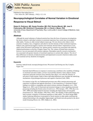
- 10. FIGURE 1. Ratings of negative emotive stimuli versus Wisconsin Card Sorting Test perseverative errors. Robinson et al. Page 10 J Nerv Ment Dis. Author manuscript; available in PMC 2007 November 30. NIH-PAAuthorManuscriptNIH-PAAuthorManuscriptNIH-PAAuthorManuscript
- 11. NIH-PAAuthorManuscriptNIH-PAAuthorManuscriptNIH-PAAuthorManuscript Robinson et al. Page 11TABLE1 DimensionalAnalysesofBackgroundCharacteristics,PsychiatricAssessmentScores,andEmotionResponses NegativeEmotionSpearman’sPositiveEmotionSpearman’s SDor(%)rho/Fprho/Fp Demographics Age(mean)5416.30.0420.8380.3780.057 Sex(#)female1765.4%F2.120.163F2.430.137 Handedness(#)right1973.1%F0.800.464F0.250.779 Race(#)Caucasian2492.3%F0.370.550F0.090.771 SES(#)ClassIII–V1869.2%F1.380.281F1.840.176 Education(meany)15.13.4−0.0650.754−0.0620.764 MeanSD Psychiatricassessment SCID84.16.3−0.1460.476−0.1100.593 PSE3.22.3−0.0880.6680.0340.870 Ham-A6.34.3−0.1610.4310.0320.878 Ham-D4.33.4−0.1580.442−0.2410.236 SFE0.0660.075−0.0830.688−0.2870.155 STC2.71.20.0610.7670.0940.649 TAS-R46.811.8−0.0290.889−0.2910.149 Emotionresponses NRstoneutralstimulation0.80.90.6750.00020.6800.0001 NRstonegativestimulation6.12.3——0.6030.001 PRstoneutralstimulation3.92.30.4940.0100.5910.001 PRstopositivestimulation7.41.80.6030.001—— HAM-A,HamiltonAnxietyScale;HAM-D,HamiltonDepressionScale;NR,negativeresponse;PR,positiveresponse;PSE,PresentStateExamination;SCID,StructuredClinicalInterviewforDSM- IV;SES,socioeconomicstatus;SFE,SocialFunctioningExam;STC,SocialTiesChecklist;TAS-R,TorontoAlexithymiaScale—Revised. J Nerv Ment Dis. Author manuscript; available in PMC 2007 November 30.
- 12. NIH-PAAuthorManuscriptNIH-PAAuthorManuscriptNIH-PAAuthorManuscript Robinson et al. Page 12TABLE2 DescriptivesandSpearman'sCorrelationsofEmotionResponsesVersusNeuropsychologicalMeasures NegativeEmotionSpearman’sPositiveEmotionSpearman’s NeuropsychologicalTestsbyDomainMeanSDrhoprhop Handedness EdinburghHandednessInventory61.855.30.0650.758−0.1800.389 Attention WAIS-R:DigitSpanACSS10.11.890.0670.7140.0360.868 GeneralIntellectualFunctioning WAIS-R:VIQ105.213.4−0.2180.2960.0610.772 WAIS-R:PIQ115.615.3−0.0850.687−0.0510.808 WAIS-R:FSIQ110.615.2−0.1830.382−0.0140.949 Visuospatialandperceptual RCFT:copyrawscore29.75.6−0.2650.200−0.6570.000 RCFT:immediaterecallrawscore15.65.3−0.2160.300−0.3610.077 RCFT:delayedrecallrawscore14.75.8−0.2150.302−0.4460.025 Frontalexecutivefunction COWAT:totalsum(listgeneration—C/F/L)34.610.9−0.2400.247−0.0370.862 WCST:categories3.51.4−0.5800.002−0.1950.350 WCST:correct48.89.6−0.4680.018−0.1280.543 WCST:errors15.29.60.4510.0240.1220.562 WCST:perseverations8.26.00.5170.0080.1480.480 WCST:perseverativeerrors7.85.20.5150.0080.1460.485 WCST:nonperseverativeerrors7.44.90.3340.1020.0090.965 ACSS,Age-correctedscaledscores;COWAT,ControlledOralWordAssociationTest;FSIQ,Full-ScaleIntelligenceQuotient;PIQ,PerformanceIntelligenceQuotient;RCFT,ReyComplexFigure Test;VIQ,VerbalIntelligenceQuotient;WAIS-R,WechslerAdultIntelligenceScale—Revised;WCST,WisconsinCardSortingTest.SignificantSpearmanrhopvaluesaredenotedbyboldcase. J Nerv Ment Dis. Author manuscript; available in PMC 2007 November 30.
- 13. NIH-PAAuthorManuscriptNIH-PAAuthorManuscriptNIH-PAAuthorManuscript Robinson et al. Page 13TABLE3 RelationshipBetweenEmotionResponsesandFrontalLobeROIGreyMatterVolumes NegativeEmotionSpearman’sPositiveEmotionSpearman’s MeanSDrhoprhop Leftfrontalstructures SFG21.6024.4550.1970.3340.1120.587 MFG19.6863.7510.0980.6330.1490.468 IFG12.3623.0260.0960.640−0.0100.960 OFC17.4152.182−0.0450.8260.2090.306 rAC6.3662.804−0.2170.288−0.2540.211 cAC6.6821.456−0.2600.199−0.0150.942 SG2.5360.5920.0800.6990.3770.057 Rightfrontalstructures SFG23.5064.7830.0330.8740.1470.475 MFG20.3624.6640.1090.5960.2220.276 IFG12.5502.636−0.2330.252−0.2750.174 OFC17.1662.2940.1360.5090.1570.445 rAC6.7472.500−0.0330.872−0.3090.125 cAC7.1771.213−0.2310.256−0.1220.551 SG2.6590.7380.1510.4620.2660.189 cAC,Caudal-anteriorcingulate;IFG,inferiorfrontalgyrus;MFG,middlefrontalgyrus;OFC,orbitofrontalcortex;rAC,rostral-anteriorcingulate;ROI,regionofinterest;SFG,superiorfrontalgyrus; SG,straightgyrus. J Nerv Ment Dis. Author manuscript; available in PMC 2007 November 30.