
Recomendados
Mais conteúdo relacionado
Mais procurados
Mais procurados (20)
Destaque
Destaque (13)
Semelhante a Dr Deepak Chahar Hip Dislocation JCOT
Semelhante a Dr Deepak Chahar Hip Dislocation JCOT (20)
Dr Deepak Chahar Hip Dislocation JCOT
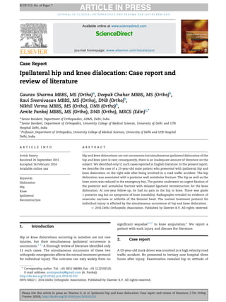
- 1. Case Report Ipsilateral hip and knee dislocation: Case report and review of literature Gaurav Sharma MBBS, MS (Ortho)a , Deepak Chahar MBBS, MS (Ortho)b , Ravi Sreenivasan MBBS, MS (Ortho), DNB (Ortho)b , Nikhil Verma MBBS, MS (Ortho), DNB (Ortho)b , Amite Pankaj MBBS, MS (Ortho), DNB (Ortho), MRCS (Edin)c,* a Senior Resident, Department of Orthopaedics, AIIMS, Delhi, India b Senior Resident, Department of Orthopedics, University College of Medical Sciences, University of Delhi and GTB Hospital Delhi, India c Professor, Department of Orthopedics, University College of Medical Sciences, University of Delhi and GTB Hospital Delhi, India 1. Introduction Hip or knee dislocations occurring in isolation are not rare injuries, but their simultaneous ipsilateral occurrence is uncommon.1–11 A thorough review of literature identified only 11 such cases. The simultaneous occurrence of these two orthopedic emergencies affects the normal treatment protocol for individual injury. The outcome can vary widely from no significant sequelae4,5,7 to knee amputation.6 We report a patient with such injury and discuss the literature. 2. Case report A 23-year-old truck driver was involved in a high velocity road traffic accident. He presented to tertiary care hospital three hours after injury. Examination revealed hip in attitude of j o u r n a l o f c l i n i c a l o r t h o p a e d i c s a n d t r a u m a x x x ( 2 0 1 6 ) x x x – x x x a r t i c l e i n f o Article history: Received 26 September 2015 Accepted 16 February 2016 Available online xxx Keywords: Dislocation Hip Knee Ipsilateral Reconstruction a b s t r a c t Hip and knee dislocations are not uncommon but simultaneous ipsilateral dislocation of the hip and knee joint is rare; consequently, there is an inadequate amount of literature on the subject. We identified only 11 such cases reported in English literature. In the present report, we describe the case of a 23-year-old male patient who presented with ipsilateral hip and knee dislocation on the right side after being involved in a road traffic accident. The hip dislocation was associated with a posterior wall acetabular fracture. The hip as well as the knee joints was reduced in the emergency bay. The patient underwent an urgent fixation of the posterior wall acetabular fracture with delayed ligament reconstruction for the knee dislocation. At one-year follow-up, he had no pain in the hip or knee. There was grade 1 posterior sag but no symptoms of knee instability. Radiographs revealed no evidence of avascular necrosis or arthritis of the femoral head. The normal treatment protocol for individual injury is affected by the simultaneous occurrence of hip and knee dislocation. # 2016 Delhi Orthopedic Association. Published by Elsevier B.V. All rights reserved. * Corresponding author. Tel.: +91 9811148080; fax: +91 1122592520. E-mail address: amitepankaj@gmail.com (A. Pankaj). JCOT-232; No. of Pages 7 Please cite this article in press as: Sharma G, et al. Ipsilateral hip and knee dislocation: Case report and review of literature, J Clin Orthop Trauma. (2016), http://dx.doi.org/10.1016/j.jcot.2016.02.012 Available online at www.sciencedirect.com ScienceDirect journal homepage: www.elsevier.com/locate/jcot http://dx.doi.org/10.1016/j.jcot.2016.02.012 0976-5662/# 2016 Delhi Orthopedic Association. Published by Elsevier B.V. All rights reserved.
- 2. flexion, adduction, and internal rotation with a posterior dislocation of the right knee. The posterior tibial and the dorsalis pedis artery were palpable. Dorsiflexion of the right ankle was absent along with reduced sensations along the lateral aspect of the leg and the dorsum of the foot, suggesting common peroneal nerve involvement. Radiographic evalua- tion revealed a posterior dislocation of the right hip with a posterior wall acetabular fracture (Fig. 1) and a posterior knee dislocation (Fig. 2). The patient underwent immediate closed reduction of the knee in the emergency bay under sedation followed by application of a posterior splint with the knee in 908 flexion. The hip joint was then reduced by giving traction over the distal part of thigh with the hip and knee in 90/90 flexed position and pelvis stabilized by second assistant. A third person stabilized the leg while traction was applied through the distal part of the thigh, with the hip reducing easily. Postreduction CT scan of the hip showed a concentrically reduced femoral head with a large posterior wall acetabular fracture (Fig. 3), while the postreduction MRI knee revealed disruption of the cruciates, medial collateral ligament (MCL), and the posterolateral corner (PLC) (type IV) (Fig. 4). Next morning the patient underwent osteosynthesis of the posterior wall acetabular fracture. He was operated in the lateral position using the Kocher Langenbeck approach. The posterior wall fragment was fixed with two lag screws and a buttress plate (Fig. 5). At the time of surgery, the knee was protected using a posterior splint. In order to reduce the surgical insult to the patient and to reduce the risk of arthrofibrosis, a delayed reconstruction was planned for the knee. It was placed in a hinged PCL brace initially locked in extension. Controlled range of motion exercises were started at three weeks. The patient was mobilized with crutches, nonweight bearing on the right lower extremity for the first eight weeks. At this time, the knee range of motion was 10–1208. Examination under anesthesia revealed a mildly positive Lachman test with a firm end point, a positive posterior drawer test with more than 15 mm translation, and a positive dial sign. Under fluoroscopy, valgus stress testing did not reveal instability, but varus stress testing at 08 and 308 showed more than 10 mm opening of lateral compartment of the knee. Ten weeks post injury, he underwent combined PCL and PLC reconstruction (Fig. 6). Fig. 1 – X-ray pelvis anteroposterior view showing fracture dislocation of hip. Note the large posterior wall fragment. Fig. 2 – X-ray knee anteroposterior and lateral views showing dislocation of knee. Note the fractures of the avulsion fracture of head of fibula and fracture of shaft of fibula. j o u r n a l o f c l i n i c a l o r t h o p a e d i c s a n d t r a u m a x x x ( 2 0 1 6 ) x x x – x x x2 JCOT-232; No. of Pages 7 Please cite this article in press as: Sharma G, et al. Ipsilateral hip and knee dislocation: Case report and review of literature, J Clin Orthop Trauma. (2016), http://dx.doi.org/10.1016/j.jcot.2016.02.012
- 3. Fig. 3 – Postreduction CT scan of the hip showed a concentrically reduced femoral head with a large posterior wall acetabular fracture. Fig. 4 – Postreduction MRI knee revealed disruption of the cruciates, medial collateral ligament (MCL), and the posterolateral corner (PLC) (type IV). Fig. 5 – Photograph and X-ray pelvis anteroposterior view illustrating the osteosynthesis of posterior acetabular wall fracture. j o u r n a l o f c l i n i c a l o r t h o p a e d i c s a n d t r a u m a x x x ( 2 0 1 6 ) x x x – x x x 3 JCOT-232; No. of Pages 7 Please cite this article in press as: Sharma G, et al. Ipsilateral hip and knee dislocation: Case report and review of literature, J Clin Orthop Trauma. (2016), http://dx.doi.org/10.1016/j.jcot.2016.02.012
- 4. Patient underwent PCL (arthroscopic single bundle with ipsilateral quadrupled hamstrings) and PLC reconstruction with Larson's procedure (contralateral semitendinosus graft). 3. Result At 18 months follow-up, the patient had no symptoms of knee instability, although he had mild discomfort in the knee. He had no pain in the hip and used no ambulatory aid. The CPN palsy had resolved completely. On examination, the right hip had a full painless range of motion. Knee examination revealed negative posterior drawer test as well as a negative dial test. Valgus and varus stress testing were negative. The ROM at the right knee was 10–1208 compared to 0–1408 on the left. Radiographs revealed a congruous hip without evidence of AVN or arthritis (Figs. 7 and 8). 4. Discussion Although injury to the ipsilateral knee is common in the setting of traumatic hip dislocation,12,13 the simultaneous occurrence of ipsilateral knee and hip dislocation seems to be a rare event.1–11 Table 1 lists the reported cases of ipsilateral hip and knee dislocation (twelve, including the present one). 4.1. Pattern of injury The right side was involved more often1,4–8,11 (eight times) as compared to the left3,4,9,10 (four times). All twelve cases of Fig. 6 – Photograph and line diagram illustrating the reconstruction of the posterolateral ligament complex. Fig. 7 – X-ray pelvis anteroposterior view revealed a congruous hip without evidence of AVN or arthritis. Fig. 8 – X-ray knee anteroposterior and lateral views revealed a congruous knee without evidence of arthritis. j o u r n a l o f c l i n i c a l o r t h o p a e d i c s a n d t r a u m a x x x ( 2 0 1 6 ) x x x – x x x4 JCOT-232; No. of Pages 7 Please cite this article in press as: Sharma G, et al. Ipsilateral hip and knee dislocation: Case report and review of literature, J Clin Orthop Trauma. (2016), http://dx.doi.org/10.1016/j.jcot.2016.02.012
- 5. Table 1 – Reported cases of ipsilateral hip and knee dislocation. Author name; year Side Hip dislocation Knee dislocation Associated injuries Treatment Result at final follow-up Hip Knee Hip Knee Malimson; 1984 Right P + PW Posterior closed; KD V Tarsometatarsal fracture dislocation, fracture body of sternum CR; Acetabular wall # treated conservatively Fractured LFC treated conservatively Painless 0–1008 ROM, knee stable Kreibich; 1989 Left P + FN Posterior closed; KD III L None OR with fixation of femoral neck # Primary repair of ligaments Intermittent pain, AVN Flexion up to 1058; no instability; Arthrosis+ Millea; 1991 Left P + FH Rotary closed Ipsilateral femoral shaft fracture, open bimalleolar fracture OR with fixation of femoral head # Primary repair/ fixation of ligaments Occasional discomfort of the hip Occasional discomfort of the knee Freedman; 1994 Right P Posterior closed; KD III L None CR Early ligament reconstruction Painless, no AVN 10–1058 ROM, mild Lachman+ Schierz; 2002 Right P + PW Posterior closed; KD III L None CR acetabular wall # treated conservatively PCL repaired, ACL planned for delayed reconstruction but patient refused Painless, no AVN 0–1258 ROM, Mild Lachman+ pivot shiftÀ Motsis; 2006 Right P + PW Posterior open; KD IV (C) None CR Acetabular wall # treated conservatively Through knee amputation Not mentioned Walking with crutches Dubois; 2006 Right P + PW Posterior closed; KD III L None CR using shanks' screw, Posterior wall treated conservatively Early ligament reconstruction Painless, no AVN 0–125 ROM, no instability Ali;2009 Right A Anterior KD I None CR Early ligament reconstruction Painless, no AVN Up to 120 ROM, mild instability Vaseenon; 2010 Left A Posterior closed; KD I Ipsilateral olecranon fracture CR Ligament reconstruction not discussed Not mentioned Not mentioned Sen; 2011 Left P + PW + FH Posterior closed; KD V Contralateral open leg fracture OR with fixation of femoral head and acetabular fragments Open reduction of tibial plateau fracture Painless, no AVN 0–105 ROM, no instability or arthrosis Waterman; 2011 Right P Posterior closed; KD III M (C) Ipsilateral tibiotalar dislocation, contralateral tibia fracture CR Patient refused ligament reconstruction Painless, no AVN 0–125, no instability Present case Right P + PW Posterior closed, KD IV None CR fixation of posterior wall fragment Delayed ligament reconstruction Painless, no AVN 0–120 ROM, mild Lachman+ P – posterior; PW – posterior wall; FH – femoral head; FN – femoral neck; (C) – vascular injury; CR – closed reduction; OR – open reduction; # – fracture; LFC – lateral femoral condyle. journalofclinicalorthopaedicsandtraumaxxx(2016)xxx–xxx5 JCOT-232;No.ofPages7 Pleasecitethisarticleinpressas:SharmaG,etal.Ipsilateralhipandkneedislocation:Casereportandreviewofliterature,JClinOrthop Trauma.(2016),http://dx.doi.org/10.1016/j.jcot.2016.02.012
- 6. ipsilateral hip and knee dislocation resulted from road traffic accidents. The hip dislocated posteriorly in ten cases, while two had anterior dislocations.8,9 The most common pattern was a posterior hip dislocation hip with a posterior wall acetabular fracture. The knee dislocated posteriorly in eleven of the twelve cases. Two of them were fracture dislocations, involving the tibial plateau,10 and lateral femoral condyle.1 Rest of them involved ligamentous disruptions (substance tears/avulsions). The most common pattern of knee dislocation involved a closed posterior ligamentous dislocation. 4.2. Associated injuries other than hip and knee Seven2,4–8 of the twelve patients, including the present one, had no other injury apart from the ipsilateral hip and knee dislocation. Three patients1,3,11 had ipsilateral foot and ankle injuries. This probably reflects the specific mechanism of injury involved, as well as transfer of most of the force to the knee, and through it, to the ipsilateral hip. 4.3. Nerve and vascular injuries Two of the patients had popliteal artery injury6,11 ; one had a successful arterial repair11 with grafting, whereas the other required an amputation.6 Three2,7 patients, including the present one, had common peroneal nerve injury, and all of them ultimately recovered. Cornwall and Radomisli14 reported a 10% incidence of nerve injury after traumatic hip dislocation. The peroneal component was most commonly involved. In cases of nerve injury associated with hip dislocation, explora- tion of the nerve is generally not recommended. The incidence of concomitant neurologic injury with knee dislocation is reported to be from 10% to 40%.15,16 The indications for peroneal neurolysis and cable grafting in the setting of knee dislocations are controversial. Patients who are undergoing PLC repair or reconstruction and have a peroneal nerve injury should be treated with at least a peroneal neurolysis.17 4.4. Technique of hip reduction Previous reports of ipsilateral hip and knee dislocation have described different techniques of reducing the hip. Freedman et al.4 reduced the hip with manual traction over the thigh. DuBois et al.7 described the use of Schanz pins placed in the femoral condyle as well as lateral aspect of the proximal femur to affect the reduction. Brian et al.11 described a technique where the patient's knee was flexed over the surgeon's shoulder and traction applied through the distal femur; gradual internal and external rotation completed the reduc- tion. Of note in most of the described techniques for reducing isolated hip dislocations, traction is applied through the proximal leg, either directly18 or using the surgeon's arms19 / knee as fulcrum.20 It makes sense that in case of an ipsilateral knee dislocation, traction be applied through the distal thigh instead of the proximal leg with the knee stabilized either by a third person or by the person attempting the reduction. We agree with Waterman et al.11 that an initial attempt of reducing the hip should be made in the trauma bay under sedation so as to reduce the time the hip remains dislocated. 4.5. Management of the knee dislocation Withrespecttothe timingofligamentreconstructioninisolated knee dislocations, recommendations have ranged from immo- bilization followed by delayed surgery21,22 to surgical treatment within three weeks after injury.23,24 The few published studies offering direct comparison of surgical timing have typically shown greater improvements in functional and clinical out- comes with early treatment.25,26 However, a recent systematic reviewofliteratureregardingtimingofsurgeryinmultiligament injured knees found residual anterior knee instability as well as more flexion deficits in acutely managed knees compared to delayed reconstructions.27 Additional treatment for joint stiffness was also more likely in association with acute treatment. One of the reasons for advocating early surgery in multiligament knee injuries is that the collaterals can be repaired; the repair becomes increasingly difficult two to three weeks after surgery. However, recent literature suggests that reconstructionofthePLCisbetterthanrepair,28,29 andtherefore, ifoneiscontemplating areconstruction,anearlysurgeryisnota necessity. Giannoudis et al.30 reported five cases of knee dislocation with ipsilateral femoral shaft fractures. They advocate a delayed knee ligament reconstruction in this setting. We opted for a delayed reconstruction to reduce the surgical insult to the patient as well as to reduce the chances of postoperative arthrofibrosis. All three4,7,8 of the twelve patients with ipsilateral hip and knee dislocation who underwent an acute reconstruction required an additional procedure for arthrofibrosis. Based on these findings, we recommend a delayed reconstruction of the multiligament injured knee in the setting of an ipsilateral hip and knee dislocation. 4.6. Rehabilitation The simultaneous occurrence of hip and knee dislocation precludes early weight bearing, which may be allowed after an isolated knee dislocation. The rehabilitation protocol however, also depends on the other associated injuries. 4.7. Outcome The outcome for ipsilateral hip and knee dislocation can vary widely from no significant sequelae4,5,7 to through the knee amputation.6 Surgeon-related factors that could improve outcome include emergent reduction of the knee joint, assessment of neurovascular injury, reduction of the hip joint as soon as possible, preferably in the emergency room with the knee stabilized, and management of the multiligament knee injury on an elective basis. 5. Conclusion The ipsilateral occurrence of knee and hip dislocation is a serious injury with important differences related to the technique of hip reduction, the timing of knee ligament reconstruction, and rehabilitation. The outcome is highly variable and remains guarded. j o u r n a l o f c l i n i c a l o r t h o p a e d i c s a n d t r a u m a x x x ( 2 0 1 6 ) x x x – x x x6 JCOT-232; No. of Pages 7 Please cite this article in press as: Sharma G, et al. Ipsilateral hip and knee dislocation: Case report and review of literature, J Clin Orthop Trauma. (2016), http://dx.doi.org/10.1016/j.jcot.2016.02.012
- 7. Conflicts of interest The authors have none to declare. r e f e r e n c e s 1. Malimson PD. Triple fracture-dislocation of the lower limb. Injury. 1984;16(1):11–12. 2. Kreibich DN, Moran CG, Pinder IM. Ipsilateral hip and knee dislocation. A case report. Acta Orthop Scand. 1990;61(1):90–91. 3. Millea TP, Romanelli RR, Segal LS, et al. Ipsilateral fracture- dislocation of the hip, knee and ankle: case report. J Trauma. 1991;31(3):416–419. 4. Freedman DM, Freedman EL, Shapiro MS. Ipsilateral hip and knee dislocation. J Orthop Trauma. 1994;8(2):177–180. 5. Schierz A, Hotz T, Kach K. Ipsilateral knee and hip joint dislocation. Unfallchirurg. 2002;105(7):660–663. 6. Motsis EK, Pakos EE, Zaharis K, et al. Concomitant ipsilateral traumatic dislocation of the hip and knee following high- energy trauma: a case report. J Orthop Surg (Hong Kong). 2006;14(3):322–324. 7. DuBois B, Montgomery Jr WH, Dunbar RP, et al. Simultaneous ipsilateral posterior knee and hip dislocations: case report, including a technique for closed reduction of the hip. J Orthop Trauma. 2006;20(3):216–219. 8. Ali C, Malkus T, Podskubka A. Ipsilateral traumatic dislocation of hip and knee joints. Case report. Acta Chir Orthop Traumatol Cech. 2009;76(4):329–334. 9. Vaseenon T, Wongtriratanachai P, Laohapoonrungsee A. Ipsilateral anterior hip dislocation and posterior knee subluxation: a case report. J Med Assoc Thai. 2010;93(1):128–131. 10. Sen RK, Tripathy SK, Krishnan V, Goyal T, Jagadeesh V. Ipsilateral fracture dislocations of the hip and knee joints with contralateral open fracture of the leg: a rare case and its management principles. Chin J Traumatol. 2011;14(June (3)):183–187. 11. Waterman BR, Banerjee R. Management of simultaneous ipsilateral dislocation of hip, knee, and ankle. Am J Orthop (Belle Mead NJ). 2011;40(June (6)):301–304. 12. Schmidt GL, Sciulli R, Altman GT. Knee injury in patients experiencing a high energy traumatic ipsilateral hip dislocation. J Bone Joint Surg Am. 2005;87(6):1200–1204. 13. Tabuenca J, Truan JR. Knee injuries in traumatic hip dislocation. Clin Orthop Relat Res. 2000;377:78–83. 14. Cornwall R, Radomisli TE. Nerve injury in traumatic dislocation of the hip. Clin Orthop Relat Res. 2000;377:84–91. 15. Harner CD, Waltrip R, Bennett GH, Francis KA, Cole B, Irrgang JJ. Surgical management of knee dislocation. J Bone Joint Surg Am. 2004;86:262–273. 16. Plancher. Siliski J. Long-term functional results and complications in patients with knee dislocations. J Knee Surg. 2008;21:261–268. 17. Goitz RJ, Tomaino MM. Management of peroneal nerve injuries associated with knee dislocations. Am J Orthop. 2003;32:14–16. 18. Allis OH. The Hip. Philadelphia: Dornan; 1895. 19. Lefkowitz M. A new method for reduction of hip dislocations. Orthopaed Rev. 1993;22:253–256. 20. Schafer SF, Anglen JO. The East Baltimore lift: a simple and effective method for reduction of posterior hip dislocation. J Orthop Trauma. 1999;13(1):56–57. 21. Fanelli GC, Giannotti BF, Edson CJ. Arthroscopically assisted combined anterior and posterior cruciate ligament reconstruction. Arthroscopy. 1996;12:5–14. 22. Shelbourne KD, Porter DA, Clingman JA, McCarroll JR, Rettig AC. Low-velocity knee dislocation. Orthop Rev. 1991;20:995–1004. 23. Noyes FR, Barber-Westin SD. Reconstruction of the anterior and posterior cruciate ligaments after knee dislocation. Use of early protected postoperative motion to decrease arthrofibrosis. Am J Sports Med. 1997;25:769–778. 24. Shapiro MS, Freedman EL. Allograft reconstruction of the anterior and posterior cruciate ligaments after traumatic knee dislocation. Am J Sports Med. 1995;23:580–587. 25. Harner CD, Waltrip RL, Bennett CH, Francis KA, Cole B, Irrgang JJ. Surgical management of knee dislocations. J Bone Joint Surg Am. 2004;86:262–273. 26. Tzurbakis M, Diamantopoulos A, Xenakis T, Georgoulis A. Surgical treatment of multiple knee ligament injuries in 44 patients: 2–8 years follow-up results. Knee Surg Sports Traumatol Arthrosc. 2006;14:739–749. 27. Mook WR, Miller MD, Diduch DR. Multiple-ligament knee injuries: a systematic review of the timing of operative intervention and postoperative rehabilitation. J Bone Joint Surg Am. 2009;91:2946–2957. 28. Stannard JP, Brown SL, Farris RC, McGwin Jr G, Volgas DA. The posterolateral corner of the knee: repair versus reconstruction. Am J Sports Med. 2005;33:881–888. 29. Levy BA, Dajani KA, Morgan JA, Shah JP, Dahm DL, Stuart MJ. Repair versus reconstruction of the fibular collateral ligament and posterolateral corner in the multiligament- injured knee. Am J Sports Med. 2010;38:804–809. 30. Giannoudis PV, Roberts CS, Parikh AR, Agarwal S, Hadjikoutidyer C, Macdonald dA.. Knee dislocation with ipsilateral femoral shaft fracture: a report of five cases. J Orthop Trauma. 2005;19(March (3)):205–210. j o u r n a l o f c l i n i c a l o r t h o p a e d i c s a n d t r a u m a x x x ( 2 0 1 6 ) x x x – x x x 7 JCOT-232; No. of Pages 7 Please cite this article in press as: Sharma G, et al. Ipsilateral hip and knee dislocation: Case report and review of literature, J Clin Orthop Trauma. (2016), http://dx.doi.org/10.1016/j.jcot.2016.02.012