SlideShare uma empresa Scribd logo
1 de 7
Baixar para ler offline
143
OBJECTIVE: To investigate the immunohistochemical
staining of hypoxia-inducible factor 1-alpha (HIF-1α)
and Ki-67 expression in the placenta of pregnant women
with placenta previa and placenta accreta.
STUDY DESIGN: Thirty placentas (10 normotensive,
10 placenta previa, and 10 placenta accreta) were
processed for routine histological tissue processing.
The biochemical parameters of patients were recorded.
Placentas were stained with hematoxylin-eosin and HIF-
1α and Ki-67 immunostaining.
RESULTS: Normal histology was observed in placentas
of normotensive pregnant women. Placenta previa
sections showed increased syncytial knots, intervillous
hemorrhage, fibrin accumulation, and hyalinization.
In placenta accreta sections, increased syncytial nodes,
vascular dilation/congestion, fibrin accumulation, and
hyalinization were observed. Normotensive placentas
showed no HIF-1α expression. In placenta previa tis­
sues, high HIF-1α expression was observed in vascular
endothelial cells, villous stromal cells, and syncytial
knots. High HIF-1α expression was recorded in villous
stromal cells and cytotrophoblast cells in placenta ac-
creta. In normotensive placental tissues, no Ki-67 ex-
pression was observed. In placenta previa sections,
high Ki-67 expression was observed mostly in root villi
stromal cells and some endothelial cells. High Ki-67
expression was observed mostly in villi stromal cells of
placenta accreta.
CONCLUSION: It is thought that HIF-1α is an im-
portant regulatory gene in the development of villus
in trophoblast invasion such as placenta accreta and
previa, while Ki-67 will play a key role in the develop­
ment of abnormal placenta with its stimulating effect
on inflammatory cell development and angiogenesis in
accreta and preeclampsia. (Anal Quant Cytopathol
Histpathol 2021;43:143–149)
Keywords:  female; hypoxia; hypoxia-inducible
factor 1, alpha subunit; placenta; placenta accreta;
placenta previa; placental lactogen; pregnancy,
physiology; preeclampsia.
The placenta is a temporary organ that nourishes
the fetus, regulates many metabolic activities, and
produces hormones necessary for the continuation
of pregnancy; its function ends at the end of
pregnancy.1 The placenta is implanted in any area
of the uterine wall; however, in some clinical
complications, it can prevent the fetal head from
descending during delivery if it is too low in the
uterus. Low-lying placentas can also cause unusual
bleeding during pregnancy or delivery.2
Analytical and Quantitative Cytopathology and Histopathology®
0884-6812/21/4303-0143/$18.00/0 © Science Printers and Publishers, Inc.
Analytical and Quantitative Cytopathology and Histopathology®
Immunohistochemical Analysis of Hypoxia-
Inducible Factor 1-Alpha and Ki-67 Expression
in Placenta Previa and Accreta
Serap Otçu, M.D., and Engin Deveci, Ph.D.
From the Department of Obstetrics and Gynecology, Health Sciences University, Diyarbakır Gazi Yaşargil Training and Research Hospi-
tal, Diyarbakır; and the Department of Histology and Embryology, Faculty of Medicine, Dicle University, Diyarbakır, Turkey.
Serap Otçu is Physician, Department of Obstetrics and Gynecology, Health Sciences University, Diyarbakır Gazi Yaşargil Training and
Research Hospital.
Engin Deveci is Professor and Head, Department of Histology and Embryology, Faculty of Medicine, Dicle University.
Address correspondence to: Engin Deveci, Ph.D., Department of Histology and Embryology, Faculty of Medicine, Dicle University,
21280 Diyarbakır, Turkey (engindeveci64@gmail.com)
Financial Disclosure:  The authors have no connection to any companies or products mentioned in this article.
As the embryo develops, substance exchange
between fetal and maternal blood circulation to-
wards the end of pregnancy can decrease due to
some abnormal structural changes such as thick­
ening of the basement membrane of fetal capil-
laries, increased fibrous tissue in the villous stro-
ma, and fibrinoid accumulation in the chorionic
plate and on root villi in the junction.3,4
Placenta previa occurs when the placenta at-
taches to the inside of the uterus, near or above the
cervical opening. It affects about 0.5% of pregnan­
cies. Risk factors include pregnancy in advanced
age, smoking, pre-aspiration and curettage, mul­
tiparity, previous cesarean surgery, labor induc-
tion, or termination of pregnancy. Incidence of
placenta previa is 1 in 300 deliveries.5,6 Placenta
previa can cause severe complications, such as
maternal placenta accreta, low blood pressure
or postpartum hemorrhage, and fetal growth re-
striction. The etiology of placenta previa is not
fully known, but there is a relationship between
abnormal endometrial vascularization due to scar­
ring or atrophy from previous trauma, surgery,
infection, and uterine scarring.7,8
Placenta accreta is the abnormal adhesion of the
placenta to the uterine cavity. It is characterized
by the absence of decidua basalis and by the
incomplete development of the fibrinoid layer.
Placenta accreta occurs in 5–10% of pregnancies
complicated with placenta previa. Its incidence
ranges from about 533 to 70,000 births.9 The best-
defined risk factors are previous uterine surgery
and placenta previa. Although risk factors for
placental attachment anomaly are well defined,
the correct etiology is largely unknown. Abnormal
or increased trophoblast invasion is seen. The
etiology of the placenta accreta is that the oxygen
imbalance in the uterine scar area causes this
con­
dition. The development of the embryo in a
relatively hypoxic environment causes prolifera-
tion or increased invasion of cytotrophoblasts.10,11
The aim of this study is to investigate HIF-1α
and Ki-67 expression in placenta previa and accreta
and to compare the expression differences.
Materials and Methods
Ethical approval was obtained from Dicle Univer­
sity Faculty of Medicine Non-Interventional Clin-
ical Research Ethics Committee (record no. 2020/
68). In our study, 10 normotensive, 10 placenta
previa, and 10 placenta accreta tissue samples
from pregnant women (regardless of age) were
obtained from Gynecology and Obstetrics Clinics,
Gazi Yasargil Training and Research Hospital.
All patients signed the informed patient consent
form. Biochemical parameters for each patient
were recorded. Placental tissues were processed
for routine paraffin wax embedding protocol.
Histological Tissue Processing
Placental tissues were fixed with zinc-formalin so-
lution (catalog no. Z2902, Sigma-Aldrich, St. Louis,
Missouri, USA) and washed under tap water for
5 minutes. Tissues were passed through ascend­
ing alcohol series for about 24 hours. Tissues were
washed with xylene 2×30 minutes and incubated
within paraffin wax. Sections of 5 µm were cut
with a microtome (catalog no. RM2265, Leica,
Wetzlar, Germany). Deparaffinized within xylene
for 2×30 minutes, sections were brought to dis­
tilled water. Some of the sections were stained with
routine hematoxylin and eosin, and the rest were
kept for immunohistochemical staining.
HIF-1α and Ki-67 Immunostaining
All placental tissues were brought to distilled
water. Hydrogen peroxide solution (catalog no.
TA-015-HP, Thermo Fisher, Fremont, California,
USA) was dropped on sections for 20 minutes.
After washing in PBS for 3×5 minutes, Ultra V
Block (catalog no. TA-015-UB, Thermo Fisher) was
applied to sections for 8 minutes. Sections were
incubated with primary antibodies anti-HIF-1α
(catalog no. ab216842, Abcam, Cambridge, Massa­
chusetts, USA) and anti-Ki-67 (catalog no. ab16667,
Abcam) at +4°C overnight. Sections were allowed
to warm at room temperature for 30–60 minutes.
Sections were washed with biotinylated secondary
antibody (catalog no. TP-015-BN, Thermo Fisher)
for 14 minutes. Streptavidin-peroxidase (catalog
no. TS-015-HR, Thermo Fisher) was dropped onto
sections for 15 minutes. Clearing with PBS, DAB
(catalog no. TA-001-HCX, Thermo Fisher) was
used as chromogen. Sections were counterstained
with Gill hematoxylin (catalog no. 105174, Sigma-
Aldrich) and mounted with Entellan (catalog no.
107961, Sigma-Aldrich). Slides were analyzed with
Zeiss Imager A2 Zen 3.0 software (Carl Zeiss,
Oberkochen, Germany) and photomicrographed.
Statistical Analysis
The data were recorded as arithmetic mean±
standard deviation with mean rank value. Statis­
tical analysis was done using the IBM SPSS 25.0
144 Analytical and Quantitative Cytopathology and Histopathology®
Otçu and Deveci
software (IBM SPSS Statistics for Windows, Version
25.0, released 2017. IBM Corp., Armonk, New York,
USA). One-way ANOVA with Tukey’s test was
used for multiple comparisons. P<0.05 was used as
the significance level.
Results
Statistical analysis of multiple comparisons be-
tween normotensive, placenta previa, and placen­
ta accreta is shown in Table I. No statistical dif-
ferences were recorded within group comparisons
for any parameters (ALT, AST, glucose, albumin,
total protein, urea, creatinine). Graphical illustra-
tions of all parameters are shown in Figures 1–2.
Placenta previa and accreta were stained with
hematoxylin-eosin and HIF-1α and Ki-67 immu­
nohistochemical staining. The staining is present­
ed in Figure 2. Normotensive placentas showed
normal placental histology (Figure 2A). In sections
of placenta previa, increased syncytial knots, in-
tervillous hemorrhage, fibrin accumulation, and
hyalinized regions were observed (Figure 2B).
Similar to placenta previa, placenta accreta show­
ed similar histopathology with increased syncytial
nodes, vascular dilation/congestion, fibrin accu­
mulation, and hyalinization (Figure 2C). In HIF-
1α immunohistochemical staining, no expression
was recorded in normotensive placental tissues.
In placenta previa tissues, high HIF-1α expres-
sion was observed in vascular endothelial cells,
villous stromal cells, and syncytial knots (Figure
2E). High HIF-1α expression was recorded in
villous stromal cells and cytotrophoblast cells in
placenta accreta (Figure 2F). No Ki-67 expression
was recorded in normotensive placental tissues
(Figure 2G). Placenta previa showed high Ki-67
expression mostly in root villi stromal cells and
some endothelial cells (Figure 2H). High Ki-67
expression was observed mostly in villi stromal
cells of placenta accreta (Figure 2I).
Discussion
In this study we investigated the tissues of placen­
ta previa and placenta accreta by biochemical and
immunohistochemical methods. Placenta previa is
a pregnancy complication in which placentation
is abnormally localized in the lower segment of
the uterus.12 It is one of the important causes of
maternal, fetal, and neonatal morbidity and mor­
tality characterized by third trimester bleeding. Al-
though its etiology is unknown, advanced mater­
nal age, multiparity, multiple pregnancy, previous
cesarean delivery, and smoking are risk factors,
causing placenta previa.6 Studies on histopathol­
ogy of placenta previa revealed fibrinoid necrosis,
polymorphonuclear cell infiltration, abnormal vas­
culatures, and dilated vessels. Biswas et al recorded
increased trophoblastic giant cells, hemorrhage,
absence of chorionic villi in the myometrium, and
inflammation in placenta previa tissues.13 Silver et
al also reported increased villous infarction with
fibrinoid and congested vessels in pathological
examination of placenta previa.14 In our study, we
observed increased syncytial knots, intervillous
hemorrhage, fibrin accumulation, and hyalinized
regions in placenta previa sections (Figure 1B).
Placenta accreta is defined as abnormally deep
adhesion of the placenta to the uterine muscles.
Its etiology is not fully known, but placenta previa
and previous cesarean deliveries are risk factors.
Complications include premature birth, maternal
bleeding, and morbidity.15 Histopathological anal­
ysis of placenta accreta showed chorionic villi
adjacent to myometrial fibers, fibrinoid formation,
intravascular chorionic villi, hemorrhage, and vas­
cular changes.14 In our study we observed in-
creased syncytial nodes, vascular dilation/conges­
tion, fibrin accumulation, and hyalinization in
placenta accreta tissues.
Perinatal studies have focused on the role of
oxygen and hypoxia during placentation and
pregnancy. Researchers have shown that oxygen
has vital functions to regulate trophoblast differ­
entiation.14 Hypoxia-inducible factor-1α (HIF-1α)
is a transcription regulator of genes in adaptive re-
sponse to hypoxic conditions. HIF-1α is involved
in activation of transcription of over 40 genes
such as erythropoietin, glucose transporters, gly-
colytic enzymes, and vascular endothelial growth
factor during hypoxia. HIF-1α regulates vessel
formation in the embryonic stage, tumor angio­
genesis, and ischemia.16 During placental forma­
tion, cellular hypoxia develops, and thus HIF-1α
is activated to induce trophoblast proliferation and
the formation of specific cell subtypes.17,18 Zamudio
et al investigated HIF-1α expression and its regu­
latory mechanisms in placentas from pregnancies
of women living at different altitudes. HIF-1α
were overexpressed in placentas of women living
at high altitudes.19 Ietta et al investigated the
role of HIF-1α expression during human placen­
tal development. Expression of HIF-1α was high
when oxygen tension was low but decreased when
placental oxygen tension increased. They also
Volume 43, Number 3/June 2021 145
HIF-1-α and Ki-67 in Placenta Previa and Accreta
showed that during early placentation, HIF-1α
localized in cytotrophoblasts.20 In our study we
found that placenta previa tissues showed high
HIF-1α expression in vascular endothelial cells,
146 Analytical and Quantitative Cytopathology and Histopathology®
Otçu and Deveci
Table I  Means and Standard Deviations of Age, ALT, AST, Glucose, Albumin, Total Protein, Urea, and Creatinine with Group Comparisons
				 One-way ANOVA
Parameter	 Group	 N	 Mean+SD	 (p value)
Age, years	 (1) Normotensive	 20	 33.35±4.72 	 (2): 0.868
				 (3): 0.082
	 (2) Placenta previa	 20	 32.55±5.04 	 (1): 0.868
				 (3): 0.221
	 (3) Placenta accreta	 20	 29.90±5.18 	 (1): 0.082
				 (2): 0.221
ALT, U/L	 (1) Normotensive	 20	 29.90±5.18 	 (2): 0.241
				 (3): 1.00
	 (2) Placenta previa	 20	 32.55±5.04 	 (1): 0.241
				 (3): 0.241
	 (3) Placenta accreta	 20	 29.90±5.18 	 (1): 1.00
				 (2): 0.241
AST, U/L	 (1) Normotensive	 20	 13.14±8.60 	 (2): 1.00
				 (3): 1.00
	 (2) Placenta previa	 20	 13.11±6.34 	 (1): 1.00
				 (3): 1.00
	 (3) Placenta accreta	 20	 13.14±8.60 	 (1): 1.00
				 (2): 1.00
Glucose, mg/dL	 (1) Normotensive	 20	 18.67±7.41 	 (2): 0.886
				 (3): 1.00
	 (2) Placenta previa	 20	 20.13±13.43	 (1): 0.886
				 (3): 0.886
	 (3) Placenta accreta	 20	 18.67±7.41 	 (1): 1.00
				 (2): 0.886
Albumin, g/L	 (1) Normotensive	 20	 86.80±25.99	 (2): 0.425
				 (3): 1.00
	 (2) Placenta previa	 20	 97.06±25.39	 (1): 0.425
				 (3): 0.425
	 (3) Placenta accreta	 20	 86.80±25.99	 (1): 1.00
				 (2): 0.425
Total protein, g/dL	 (1) Normotensive	 20	 21.84±16.42	 (2): 0.479
				 (3): 1.00
	 (2) Placenta previa	 20	 27.72±14.97	 (1): 0.479
				 (3): 0.479
	 (3) Placenta accreta	 20	 21.84±16.42	 (1): 1.00
				 (2): 0.479
Urea, mg/dL	 (1) Normotensive	 20	 38.38±27.08	 (2): 0.736
				 (3): 1.00
	 (2) Placenta previa	 20	 31.59±31.53	 (1): 0.736
				 (3): 0.479
	 (3) Placenta accreta	 20	 38.38±27.08	 (1): 1.00
				 (2): 0.736
Creatinine, mg/dL	 (1) Normotensive	 20	 16.43±4.91 	 (2): 0.337
				 (3): 1.00
	 (2) Placenta previa	 20	 19.19±8.03 	 (1): 0.337
				 (3): 0.337
	 (3) Placenta accreta	 20	 16.43±4.91 	 (1): 1.00
				 (2): 0.337
SD = standard deviation.
Volume 43, Number 3/June 2021 147
HIF-1-α and Ki-67 in Placenta Previa and Accreta
Figure 1 
Graphical illustration of
patient parameters regarding
placenta complication.
Figure 2 
Normotensive, placenta
previa, and placenta accreta
tissues with H-E staining (A–C)
and HIF-1α (D–F) and Ki-67
(G–I) immunostainings. (A)
Normal placental histology.
(B) Increased syncytial nodes
(black arrow), intervillous
hemorrhage (red star),
fibrinoid (black star), and
hyalinization (arrowhead). (C) Increased syncytial nodes (black arrow), vascular dilation/congestion (red star), villous fibrinoid (black
star), and hyalinization (arrowhead). (D) No HIF-1α in normotensive placental tissues. (E) High HIF-1α expression in vascular
endothelial cells (red arrow), villous stromal cells (arrowhead), and syncytial nodes (black arrow). (F) High HIF-1α expression in villous
stromal cells (arrowhead) and cytotrophoblast cells (black arrow). (G) No Ki-67 expression in normotensive placental tissues. (H) High
Ki-67 expression mostly in root villi stromal cells (arrow) and some endothelial cells (red arrow). (I) High Ki-67 expression in mostly villi
stromal cells (arrows).
villous stromal cells, and syncytial knots (Figure
1E). Placenta accreta tissues showed high HIF-1α
expression in villous stromal cells and cytotro­
phoblast cells in placenta accreta (Figure 1F).
Ki-67 is a cellular marker for proliferation which
functions in interphase and mitotic cells. Mea-
surement of Ki-67 expression level can be used for
the prognosis of many tumors.21 Ki-67 expression
index has been used in the normal placenta, hy-
datidiform moles, and choriocarcinoma. Aberrant
placenta formation may be related with abnor-
mal regulation of molecular changes and the
proliferation of trophoblast cells so that tropho­
blasts are able to migrate and invade the uterine
wall.21,22 Kaya et al recorded that Ki-67 expression
was increased in villous cytotrophoblasts of pre-
eclamptic placentas relative to controls. They
showed an increased number of villous cytotro­
phoblasts in preeclampsia as compared to in nor-
mal pregnancy by analyzing with Ki-67 immuno­
histochemical staining.23 Unek et al showed that
Ki-67 expression was increased in placental villi
of preeclampsia.24 Another preeclampsia study
recorded that the Ki-67 index was high in villous
trophoblasts of preeclamptic patients.25 A study
demonstrated that Ki-67 expression was high in
trophoblast columns of accreta subtypes.26 In our
study we found that placenta previa showed high
Ki-67 expression mostly in root villi stromal cells
and some endothelial cells (Figure 1H). We re-
corded high Ki-67 expression mostly in villi stro-
mal cells (Figure 1I).
In conclusion, it is thought that HIF-1α is an
important regulatory gene in the development
of villus in trophoblast invasion such as placenta
accreta and placenta previa, while Ki-67 will play
a key role in the development of abnormal placen­
ta with its stimulating effect on inflammatory cell
development and angiogenesis in accreta and pre­
eclampsia
References
 1. Handwerger S, Freemark M: The roles of placental growth
hormone and placental lactogen in the regulation of hu-
man fetal growth and development. J Pediatr Endocrinol
Metab 2000;13(4):343-356
 2. Yetter JF 3rd: Examination of the placenta. Am Fam Physi­
cian 1998;57(5):1045-1054
 3. Freemark M: The roles of growth hormone, prolactin and
placental lactogen in human fetal development. In Molecu-
lar and Cellular Pediatric Endocrinology. Edited by S Hand­
werger. Totowa, New Jersey, Human Press 1999, pp 57-84
 4. Handwerger S: Clinical counterpoint: The physiology of
placental lactogen in human pregnancy. [Review]. Endocr
Rev 1991;12:329-336
  5.  D’antonio F, Bhide A: Ultrasound in placental disorders. Best
Prac Res Clin Obstet Gynaecol 2014;28(3):429-442
  6.  McShane PM, Heyl PS, Epstein MF: Maternal and perinatal
morbidity resulting from placenta previa. Obstet Gynecol
1985;65:176-182
  7.  Crane JM, Van den Hof MC, Dodds L, Armson Ba, Liston R:
Maternal complications with placenta previa. Am J Perinatol
2000;17(2):101-105
 8. Ananth CV, Smulian JC, Vintzileos AM: The effect of
placenta previa on neonatal mortality: A population-based
study in the United States, 1989 during 1997. Am J Obstet
Gynecol 2003;188(5):1299-1304
 9. Oyelese Y, Smulian JC: Placenta previa, placenta accreta
and vasa previa. Obstet Gynecol 2006;107(4):927-941
10. Arcario T, Greene M, Ostheimer GW, Datta S, Nauity
JS: Risks of placenta previa/accreta in patients with pre-
vious caesarean deliveries. Anesthesiology 1988;69(Suppl
3A):A 659
11. Miller DA, Chollet JA, Goodwin TM: Clinical risk factors
for placenta previa placenta accreta. Am J Obstet Gynecol
1997;177:210-214
12. Lockwood CJ, Russo-Stieglitz K: Placenta previa: Epidemi­
ology, clinical feature, diagnosis, morbidity and mortality.
Available at https:/
/www.uptodate.com/contents/placenta-
previa-epidemiology-clinical-features-diagnosis-morbidity-
and-mortality. Accessed April 28, 2020
13. Biswas R, Sawhney H, Dass R, Saran RK, Vasishta K:
Histopathological study of placental bed biopsy in placenta
previa. Acta Obstet Gynecol Scand 1999;78(3):173-179
14. Silver R: Abnormal placentation: Placenta previa, vasa
previa, and placenta accreta. Obstet Gynecol 2015;126(3):
654-668
15.  Khong TY: The pathology of placenta accreta, a worldwide
epidemic. J Clin Pathol 2008;61:1243-1246
16. Soares MJ, Iqbal K, Kozai K: Hypoxia and placental de-
velopment. Birth Defects Res 2017;109(17):1309-1329
17. Lee JW, Ko J, Ju C, et al: Hypoxia signaling in human
diseases and therapeutic targets. Exp Mol Med 2019;51:1-13
18. Simmons DG, Cross JC: Determinants of trophoblast line­
age and cell subtype specification in the mouse placenta.
Dev Biol 2005;284:12-24
19. Zamudio S, Wu Y, Ietta F, Rolfo A, Cross A, Wheeler T,
Post M, Illsley NP, Caniggia I: Human placental hypoxia-
inducible factor-1alpha expression correlates with clinical
outcomes in chronic hypoxia in vivo. Am J Pathol 2007;
170(6):2171-2179
20. Ietta F, Wu Y, Winter J, Xu J, Wang J, Post M, Caniggia I:
Dynamic HIF1A regulation during human placental devel­
opment. Biol Reprod 2006;75(1):112-121
21.  Sun X, Kaufman PD: Ki-67: More than a proliferation mark­
er. Chromosoma 2018;127(2):175-186
22. Hilali N, Kocarslan S, Vural M, et al: Ki-67 proliferation
index in patients with placenta previa percreta in the third
trimester. Wien Klin Wochenschr 2015;127:98-102
23. Kaya B, Nayki U, Nayki C, Ulug P, Oner G, Gultekin E,
148 Analytical and Quantitative Cytopathology and Histopathology®
Otçu and Deveci
Yildirim Y: Proliferation of trophoblasts and Ki67 expres-
sion in preeclampsia. Arch Gynecol Obstet 2015;291(5):1041-
1046
24. Unek G, Oxmen A, Mendilcioglu I, Simsek M, Korgun ET:
The expression of cell cycle related proteins PCNA, Ki67,
p27 and p57 in normal and preeclamptic human placentas.
Tissue Cell 2014;46:198-205
25.  Arnholdt H, Meisel F, Fandrey K, Löhrs U: Proliferation of
villous trophoblast of the human placenta in normal and
abnormal pregnancies. Virchow Archiv B Cell Pathology
1991;60(6):365-372
26. Tantbirojn P, Crum CP, Parast MM: Pathophysiology of
placenta creta: The role of decidua and extravillous tro­
phoblast. Placenta 2008;29(7):639-645
Volume 43, Number 3/June 2021 149
HIF-1-α and Ki-67 in Placenta Previa and Accreta

Mais conteúdo relacionado

Mais procurados

Liver ischemia/reperfusion injury, a setting in which the functional mass is ...
Liver ischemia/reperfusion injury, a setting in which the functional mass is ...Liver ischemia/reperfusion injury, a setting in which the functional mass is ...
Liver ischemia/reperfusion injury, a setting in which the functional mass is ...Prof. Hesham N. Mustafa
 
TCT2012-veropaque-poster
TCT2012-veropaque-posterTCT2012-veropaque-poster
TCT2012-veropaque-posterSangita Biswas
 
Comparison of surgically_repaired_achilles_tendon_tears_using_platelet_rich_f...
Comparison of surgically_repaired_achilles_tendon_tears_using_platelet_rich_f...Comparison of surgically_repaired_achilles_tendon_tears_using_platelet_rich_f...
Comparison of surgically_repaired_achilles_tendon_tears_using_platelet_rich_f...Dr. Manuel Concepción
 
The effects of diethylstilbestrol administration on rat kidney. ultrastructur...
The effects of diethylstilbestrol administration on rat kidney. ultrastructur...The effects of diethylstilbestrol administration on rat kidney. ultrastructur...
The effects of diethylstilbestrol administration on rat kidney. ultrastructur...Prof. Hesham N. Mustafa
 
Techniques and Best Practices for Cardiomyocyte Isolation
Techniques and Best Practices for Cardiomyocyte IsolationTechniques and Best Practices for Cardiomyocyte Isolation
Techniques and Best Practices for Cardiomyocyte IsolationInsideScientific
 
Intraarticular Platelet-Rich Plasma
Intraarticular Platelet-Rich Plasma Intraarticular Platelet-Rich Plasma
Intraarticular Platelet-Rich Plasma Ade Wijaya
 
Effect of carvedilol on atrial remodeling in canine model of atrial fibrillation
Effect of carvedilol on atrial remodeling in canine model of atrial fibrillationEffect of carvedilol on atrial remodeling in canine model of atrial fibrillation
Effect of carvedilol on atrial remodeling in canine model of atrial fibrillationCardiovascular Diagnosis and Therapy (CDT)
 
Preclinical evaluation of drugs for osteoarthritis
Preclinical evaluation of drugs for osteoarthritisPreclinical evaluation of drugs for osteoarthritis
Preclinical evaluation of drugs for osteoarthritispeter donik
 
PRP ICL 16 - Case Presentation Isakos 2013
PRP ICL 16 - Case Presentation Isakos 2013PRP ICL 16 - Case Presentation Isakos 2013
PRP ICL 16 - Case Presentation Isakos 2013Smith & Nephew
 
Platelet Rich Plasma in Orthopaedics
Platelet Rich Plasma in OrthopaedicsPlatelet Rich Plasma in Orthopaedics
Platelet Rich Plasma in OrthopaedicsSrinath Gupta
 
Using Platelet Rich Plasma for Orthopedic Conditions
Using Platelet Rich Plasma for Orthopedic ConditionsUsing Platelet Rich Plasma for Orthopedic Conditions
Using Platelet Rich Plasma for Orthopedic Conditionsregenmedsr
 
XXIV Jornadas AEMB - Dr. Tahsin Beyzadeoglu
XXIV Jornadas AEMB - Dr. Tahsin BeyzadeogluXXIV Jornadas AEMB - Dr. Tahsin Beyzadeoglu
XXIV Jornadas AEMB - Dr. Tahsin BeyzadeogluBSJ Marketing
 

Mais procurados (20)

Effects of Nebivolol on Ischemia-Reperfusion Induced Testicular Injury in a R...
Effects of Nebivolol on Ischemia-Reperfusion Induced Testicular Injury in a R...Effects of Nebivolol on Ischemia-Reperfusion Induced Testicular Injury in a R...
Effects of Nebivolol on Ischemia-Reperfusion Induced Testicular Injury in a R...
 
Effects of Carvacrol on Experimental Testicular Torsion-Detorsion Model: Inve...
Effects of Carvacrol on Experimental Testicular Torsion-Detorsion Model: Inve...Effects of Carvacrol on Experimental Testicular Torsion-Detorsion Model: Inve...
Effects of Carvacrol on Experimental Testicular Torsion-Detorsion Model: Inve...
 
Liver ischemia/reperfusion injury, a setting in which the functional mass is ...
Liver ischemia/reperfusion injury, a setting in which the functional mass is ...Liver ischemia/reperfusion injury, a setting in which the functional mass is ...
Liver ischemia/reperfusion injury, a setting in which the functional mass is ...
 
TCT2012-veropaque-poster
TCT2012-veropaque-posterTCT2012-veropaque-poster
TCT2012-veropaque-poster
 
Comparison of surgically_repaired_achilles_tendon_tears_using_platelet_rich_f...
Comparison of surgically_repaired_achilles_tendon_tears_using_platelet_rich_f...Comparison of surgically_repaired_achilles_tendon_tears_using_platelet_rich_f...
Comparison of surgically_repaired_achilles_tendon_tears_using_platelet_rich_f...
 
The effects of diethylstilbestrol administration on rat kidney. ultrastructur...
The effects of diethylstilbestrol administration on rat kidney. ultrastructur...The effects of diethylstilbestrol administration on rat kidney. ultrastructur...
The effects of diethylstilbestrol administration on rat kidney. ultrastructur...
 
Effects of N-Acetylcysteine on Meteorin-like Protein and Asprosin in an Exper...
Effects of N-Acetylcysteine on Meteorin-like Protein and Asprosin in an Exper...Effects of N-Acetylcysteine on Meteorin-like Protein and Asprosin in an Exper...
Effects of N-Acetylcysteine on Meteorin-like Protein and Asprosin in an Exper...
 
The Effect of Losartan on Deformities Occurring in Brain Tissue Craniectomy
The Effect of Losartan on Deformities Occurring in Brain Tissue CraniectomyThe Effect of Losartan on Deformities Occurring in Brain Tissue Craniectomy
The Effect of Losartan on Deformities Occurring in Brain Tissue Craniectomy
 
Techniques and Best Practices for Cardiomyocyte Isolation
Techniques and Best Practices for Cardiomyocyte IsolationTechniques and Best Practices for Cardiomyocyte Isolation
Techniques and Best Practices for Cardiomyocyte Isolation
 
Intraarticular Platelet-Rich Plasma
Intraarticular Platelet-Rich Plasma Intraarticular Platelet-Rich Plasma
Intraarticular Platelet-Rich Plasma
 
Effect of carvedilol on atrial remodeling in canine model of atrial fibrillation
Effect of carvedilol on atrial remodeling in canine model of atrial fibrillationEffect of carvedilol on atrial remodeling in canine model of atrial fibrillation
Effect of carvedilol on atrial remodeling in canine model of atrial fibrillation
 
Preclinical evaluation of drugs for osteoarthritis
Preclinical evaluation of drugs for osteoarthritisPreclinical evaluation of drugs for osteoarthritis
Preclinical evaluation of drugs for osteoarthritis
 
PRP ICL 16 - Case Presentation Isakos 2013
PRP ICL 16 - Case Presentation Isakos 2013PRP ICL 16 - Case Presentation Isakos 2013
PRP ICL 16 - Case Presentation Isakos 2013
 
Platelet Rich Plasma in Orthopaedics
Platelet Rich Plasma in OrthopaedicsPlatelet Rich Plasma in Orthopaedics
Platelet Rich Plasma in Orthopaedics
 
JNB_Eszter_Tuboly
JNB_Eszter_TubolyJNB_Eszter_Tuboly
JNB_Eszter_Tuboly
 
( Isi, 2016) published paper effects of a combination therapy of sclerostin a...
( Isi, 2016) published paper effects of a combination therapy of sclerostin a...( Isi, 2016) published paper effects of a combination therapy of sclerostin a...
( Isi, 2016) published paper effects of a combination therapy of sclerostin a...
 
Nader
NaderNader
Nader
 
Autophagy Promotes the Expression of Vascular Endothelial Growth Factor in Hu...
Autophagy Promotes the Expression of Vascular Endothelial Growth Factor in Hu...Autophagy Promotes the Expression of Vascular Endothelial Growth Factor in Hu...
Autophagy Promotes the Expression of Vascular Endothelial Growth Factor in Hu...
 
Using Platelet Rich Plasma for Orthopedic Conditions
Using Platelet Rich Plasma for Orthopedic ConditionsUsing Platelet Rich Plasma for Orthopedic Conditions
Using Platelet Rich Plasma for Orthopedic Conditions
 
XXIV Jornadas AEMB - Dr. Tahsin Beyzadeoglu
XXIV Jornadas AEMB - Dr. Tahsin BeyzadeogluXXIV Jornadas AEMB - Dr. Tahsin Beyzadeoglu
XXIV Jornadas AEMB - Dr. Tahsin Beyzadeoglu
 

Semelhante a Immunohistochemical Analysis of Hypoxia-Inducible Factor 1-Alpha and Ki-67 Expression in Placenta Previa and Accreta

acidemia
acidemiaacidemia
acidemiajumaque
 
“A Study on Coagulation Profile in Pregnancy Induced Hypertension Cases”
“A Study on Coagulation Profile in Pregnancy Induced Hypertension Cases”“A Study on Coagulation Profile in Pregnancy Induced Hypertension Cases”
“A Study on Coagulation Profile in Pregnancy Induced Hypertension Cases”iosrjce
 
Intra-uterine fetal death and Post-term pregnancy
Intra-uterine fetal death and Post-term pregnancyIntra-uterine fetal death and Post-term pregnancy
Intra-uterine fetal death and Post-term pregnancyLipi Mondal
 
WHAT IS NEW IN ESHRE 2022 AND FIGO 2022 FOR GENERAL GYNAECOLOGIST
WHAT IS NEW IN ESHRE 2022 AND FIGO 2022 FOR GENERAL GYNAECOLOGISTWHAT IS NEW IN ESHRE 2022 AND FIGO 2022 FOR GENERAL GYNAECOLOGIST
WHAT IS NEW IN ESHRE 2022 AND FIGO 2022 FOR GENERAL GYNAECOLOGISTAboubakr Elnashar
 
Induction and augmentation of labour by dr jograjiya
Induction and augmentation of labour by dr jograjiyaInduction and augmentation of labour by dr jograjiya
Induction and augmentation of labour by dr jograjiyaJograjiya Gelabhai Raghubhai
 
BIOCHEMICAL FOETAL MEASURES PPT.pptx
BIOCHEMICAL FOETAL MEASURES PPT.pptxBIOCHEMICAL FOETAL MEASURES PPT.pptx
BIOCHEMICAL FOETAL MEASURES PPT.pptxsunnyalvakharshandi
 
! Final artic ro- imf&gd_modif
! Final   artic ro- imf&gd_modif! Final   artic ro- imf&gd_modif
! Final artic ro- imf&gd_modifilie rodica
 
Prognostic Value of Serum Amyloid A Protein in Egyptian Infants with Hypoxic ...
Prognostic Value of Serum Amyloid A Protein in Egyptian Infants with Hypoxic ...Prognostic Value of Serum Amyloid A Protein in Egyptian Infants with Hypoxic ...
Prognostic Value of Serum Amyloid A Protein in Egyptian Infants with Hypoxic ...Healthcare and Medical Sciences
 
anaesthesia for fetal surgery.pptx
anaesthesia for fetal surgery.pptxanaesthesia for fetal surgery.pptx
anaesthesia for fetal surgery.pptxAkhilaBuddha
 
Choriocarcinoma 1
Choriocarcinoma 1Choriocarcinoma 1
Choriocarcinoma 1danilfatah
 
Seminar prenatal genetic screening
Seminar prenatal genetic screeningSeminar prenatal genetic screening
Seminar prenatal genetic screeningSreelasya Kakarla
 
Hp mar06 rqectopic
Hp mar06 rqectopicHp mar06 rqectopic
Hp mar06 rqectopicAbeer Ahmed
 
Hydroceph in 3 rd trimester itp
Hydroceph in 3 rd trimester itpHydroceph in 3 rd trimester itp
Hydroceph in 3 rd trimester itpRashmi Parikh
 

Semelhante a Immunohistochemical Analysis of Hypoxia-Inducible Factor 1-Alpha and Ki-67 Expression in Placenta Previa and Accreta (20)

Morphometric and Immunohistochemical Examination of Changes in Umbilical Cord...
Morphometric and Immunohistochemical Examination of Changes in Umbilical Cord...Morphometric and Immunohistochemical Examination of Changes in Umbilical Cord...
Morphometric and Immunohistochemical Examination of Changes in Umbilical Cord...
 
acidemia
acidemiaacidemia
acidemia
 
ADAMTS-5 and Caspase-12 Protein Immunoexpression Levels in Normotensive and P...
ADAMTS-5 and Caspase-12 Protein Immunoexpression Levels in Normotensive and P...ADAMTS-5 and Caspase-12 Protein Immunoexpression Levels in Normotensive and P...
ADAMTS-5 and Caspase-12 Protein Immunoexpression Levels in Normotensive and P...
 
“A Study on Coagulation Profile in Pregnancy Induced Hypertension Cases”
“A Study on Coagulation Profile in Pregnancy Induced Hypertension Cases”“A Study on Coagulation Profile in Pregnancy Induced Hypertension Cases”
“A Study on Coagulation Profile in Pregnancy Induced Hypertension Cases”
 
Intra-uterine fetal death and Post-term pregnancy
Intra-uterine fetal death and Post-term pregnancyIntra-uterine fetal death and Post-term pregnancy
Intra-uterine fetal death and Post-term pregnancy
 
WHAT IS NEW IN ESHRE 2022 AND FIGO 2022 FOR GENERAL GYNAECOLOGIST
WHAT IS NEW IN ESHRE 2022 AND FIGO 2022 FOR GENERAL GYNAECOLOGISTWHAT IS NEW IN ESHRE 2022 AND FIGO 2022 FOR GENERAL GYNAECOLOGIST
WHAT IS NEW IN ESHRE 2022 AND FIGO 2022 FOR GENERAL GYNAECOLOGIST
 
Dr Madhusmita 15
Dr Madhusmita 15Dr Madhusmita 15
Dr Madhusmita 15
 
Pregnancy Outcomes of Oligohydramnios at Term diagnosed by Ultra Sonography (...
Pregnancy Outcomes of Oligohydramnios at Term diagnosed by Ultra Sonography (...Pregnancy Outcomes of Oligohydramnios at Term diagnosed by Ultra Sonography (...
Pregnancy Outcomes of Oligohydramnios at Term diagnosed by Ultra Sonography (...
 
Induction and augmentation of labour by dr jograjiya
Induction and augmentation of labour by dr jograjiyaInduction and augmentation of labour by dr jograjiya
Induction and augmentation of labour by dr jograjiya
 
BIOCHEMICAL FOETAL MEASURES PPT.pptx
BIOCHEMICAL FOETAL MEASURES PPT.pptxBIOCHEMICAL FOETAL MEASURES PPT.pptx
BIOCHEMICAL FOETAL MEASURES PPT.pptx
 
! Final artic ro- imf&gd_modif
! Final   artic ro- imf&gd_modif! Final   artic ro- imf&gd_modif
! Final artic ro- imf&gd_modif
 
Prognostic Value of Serum Amyloid A Protein in Egyptian Infants with Hypoxic ...
Prognostic Value of Serum Amyloid A Protein in Egyptian Infants with Hypoxic ...Prognostic Value of Serum Amyloid A Protein in Egyptian Infants with Hypoxic ...
Prognostic Value of Serum Amyloid A Protein in Egyptian Infants with Hypoxic ...
 
anaesthesia for fetal surgery.pptx
anaesthesia for fetal surgery.pptxanaesthesia for fetal surgery.pptx
anaesthesia for fetal surgery.pptx
 
Choriocarcinoma 1
Choriocarcinoma 1Choriocarcinoma 1
Choriocarcinoma 1
 
Techniques Of Art
Techniques Of ArtTechniques Of Art
Techniques Of Art
 
Seminar prenatal genetic screening
Seminar prenatal genetic screeningSeminar prenatal genetic screening
Seminar prenatal genetic screening
 
Hp mar06 rqectopic
Hp mar06 rqectopicHp mar06 rqectopic
Hp mar06 rqectopic
 
Immunohistochemical Distribution of ADAMTS-1, ADAMTS-4 Expression in Placenta...
Immunohistochemical Distribution of ADAMTS-1, ADAMTS-4 Expression in Placenta...Immunohistochemical Distribution of ADAMTS-1, ADAMTS-4 Expression in Placenta...
Immunohistochemical Distribution of ADAMTS-1, ADAMTS-4 Expression in Placenta...
 
Murray2005
Murray2005Murray2005
Murray2005
 
Hydroceph in 3 rd trimester itp
Hydroceph in 3 rd trimester itpHydroceph in 3 rd trimester itp
Hydroceph in 3 rd trimester itp
 

Mais de ANALYTICAL AND QUANTITATIVE CYTOPATHOLOGY AND HISTOPATHOLOGY

Mais de ANALYTICAL AND QUANTITATIVE CYTOPATHOLOGY AND HISTOPATHOLOGY (20)

Subcutaneous Epstein-Barr Virus–Positive Diffuse Large B Cell Lymphoma Follow...
Subcutaneous Epstein-Barr Virus–Positive Diffuse Large B Cell Lymphoma Follow...Subcutaneous Epstein-Barr Virus–Positive Diffuse Large B Cell Lymphoma Follow...
Subcutaneous Epstein-Barr Virus–Positive Diffuse Large B Cell Lymphoma Follow...
 
Association Between Telomerase Reverse Transcriptase Promoter Mutations and M...
Association Between Telomerase Reverse Transcriptase Promoter Mutations and M...Association Between Telomerase Reverse Transcriptase Promoter Mutations and M...
Association Between Telomerase Reverse Transcriptase Promoter Mutations and M...
 
Preparation and Properties of Chitosan-Based Thermosensitive Hydrogel and Its...
Preparation and Properties of Chitosan-Based Thermosensitive Hydrogel and Its...Preparation and Properties of Chitosan-Based Thermosensitive Hydrogel and Its...
Preparation and Properties of Chitosan-Based Thermosensitive Hydrogel and Its...
 
Evaluation of the Bond Strength of Resin-Modified Glass Ionomer Enhanced with...
Evaluation of the Bond Strength of Resin-Modified Glass Ionomer Enhanced with...Evaluation of the Bond Strength of Resin-Modified Glass Ionomer Enhanced with...
Evaluation of the Bond Strength of Resin-Modified Glass Ionomer Enhanced with...
 
Ultrasound Findings of Different Subtypes of Ovarian Borderline Tumors
Ultrasound Findings of Different Subtypes of Ovarian Borderline TumorsUltrasound Findings of Different Subtypes of Ovarian Borderline Tumors
Ultrasound Findings of Different Subtypes of Ovarian Borderline Tumors
 
Effect of Graft Application and Nebivolol Treatment on Tibial Bone Defect in ...
Effect of Graft Application and Nebivolol Treatment on Tibial Bone Defect in ...Effect of Graft Application and Nebivolol Treatment on Tibial Bone Defect in ...
Effect of Graft Application and Nebivolol Treatment on Tibial Bone Defect in ...
 
Cytogenetic Risk and Hemocyte Account for the Age-Related Poor Prognosis in A...
Cytogenetic Risk and Hemocyte Account for the Age-Related Poor Prognosis in A...Cytogenetic Risk and Hemocyte Account for the Age-Related Poor Prognosis in A...
Cytogenetic Risk and Hemocyte Account for the Age-Related Poor Prognosis in A...
 
Effect of Intracoronary Application of Nicorandil and Tirofiban on No-Reflow ...
Effect of Intracoronary Application of Nicorandil and Tirofiban on No-Reflow ...Effect of Intracoronary Application of Nicorandil and Tirofiban on No-Reflow ...
Effect of Intracoronary Application of Nicorandil and Tirofiban on No-Reflow ...
 
Kit-Positive Cells in the Murine Common Bile Duct
Kit-Positive Cells in the Murine Common Bile DuctKit-Positive Cells in the Murine Common Bile Duct
Kit-Positive Cells in the Murine Common Bile Duct
 
Effect of miR-21 on Oral Squamous Cell Carcinoma Cell Proliferation and Apopt...
Effect of miR-21 on Oral Squamous Cell Carcinoma Cell Proliferation and Apopt...Effect of miR-21 on Oral Squamous Cell Carcinoma Cell Proliferation and Apopt...
Effect of miR-21 on Oral Squamous Cell Carcinoma Cell Proliferation and Apopt...
 
Prolonged Simvastatin Treatment Provided a Decrease in Apoptotic, Inflammator...
Prolonged Simvastatin Treatment Provided a Decrease in Apoptotic, Inflammator...Prolonged Simvastatin Treatment Provided a Decrease in Apoptotic, Inflammator...
Prolonged Simvastatin Treatment Provided a Decrease in Apoptotic, Inflammator...
 
Silenced microRNA-135b-5p Inhibits Tongue Squamous Cell Carcinoma Proliferati...
Silenced microRNA-135b-5p Inhibits Tongue Squamous Cell Carcinoma Proliferati...Silenced microRNA-135b-5p Inhibits Tongue Squamous Cell Carcinoma Proliferati...
Silenced microRNA-135b-5p Inhibits Tongue Squamous Cell Carcinoma Proliferati...
 
Three-Dimensional Investigation of the Effects of Ectodermal Dysplasia on the...
Three-Dimensional Investigation of the Effects of Ectodermal Dysplasia on the...Three-Dimensional Investigation of the Effects of Ectodermal Dysplasia on the...
Three-Dimensional Investigation of the Effects of Ectodermal Dysplasia on the...
 
The Spectrum of Histopathological Patterns in Diabetic Kidney Disease in East...
The Spectrum of Histopathological Patterns in Diabetic Kidney Disease in East...The Spectrum of Histopathological Patterns in Diabetic Kidney Disease in East...
The Spectrum of Histopathological Patterns in Diabetic Kidney Disease in East...
 
Effect of Rutin on Angiotensin II–Induced Cardiomyocyte Hypertrophy and the I...
Effect of Rutin on Angiotensin II–Induced Cardiomyocyte Hypertrophy and the I...Effect of Rutin on Angiotensin II–Induced Cardiomyocyte Hypertrophy and the I...
Effect of Rutin on Angiotensin II–Induced Cardiomyocyte Hypertrophy and the I...
 
Cardiac Metastases from Solid Papillary Carcinoma of the Breast: Analysis of ...
Cardiac Metastases from Solid Papillary Carcinoma of the Breast: Analysis of ...Cardiac Metastases from Solid Papillary Carcinoma of the Breast: Analysis of ...
Cardiac Metastases from Solid Papillary Carcinoma of the Breast: Analysis of ...
 
Can Numeric Maturation Value Be Used as a Prognostic Indicator and Diagnostic...
Can Numeric Maturation Value Be Used as a Prognostic Indicator and Diagnostic...Can Numeric Maturation Value Be Used as a Prognostic Indicator and Diagnostic...
Can Numeric Maturation Value Be Used as a Prognostic Indicator and Diagnostic...
 
Olaparib Upregulates miR-630 Expression to Enhance the Chemotherapeutic Sensi...
Olaparib Upregulates miR-630 Expression to Enhance the Chemotherapeutic Sensi...Olaparib Upregulates miR-630 Expression to Enhance the Chemotherapeutic Sensi...
Olaparib Upregulates miR-630 Expression to Enhance the Chemotherapeutic Sensi...
 
3D Reconstruction of Peripheral Nerves Based on Calcium Chloride Enhanced Mic...
3D Reconstruction of Peripheral Nerves Based on Calcium Chloride Enhanced Mic...3D Reconstruction of Peripheral Nerves Based on Calcium Chloride Enhanced Mic...
3D Reconstruction of Peripheral Nerves Based on Calcium Chloride Enhanced Mic...
 
Expression of AFP, P-sel, and MMP-9 in Cirrhosis with Portal Vein Thrombosis
Expression of AFP, P-sel, and MMP-9 in Cirrhosis with Portal Vein ThrombosisExpression of AFP, P-sel, and MMP-9 in Cirrhosis with Portal Vein Thrombosis
Expression of AFP, P-sel, and MMP-9 in Cirrhosis with Portal Vein Thrombosis
 

Último

Bangalore Call Girls Marathahalli 📞 9907093804 High Profile Service 100% Safe
Bangalore Call Girls Marathahalli 📞 9907093804 High Profile Service 100% SafeBangalore Call Girls Marathahalli 📞 9907093804 High Profile Service 100% Safe
Bangalore Call Girls Marathahalli 📞 9907093804 High Profile Service 100% Safenarwatsonia7
 
Mumbai Call Girls Service 9910780858 Real Russian Girls Looking Models
Mumbai Call Girls Service 9910780858 Real Russian Girls Looking ModelsMumbai Call Girls Service 9910780858 Real Russian Girls Looking Models
Mumbai Call Girls Service 9910780858 Real Russian Girls Looking Modelssonalikaur4
 
High Profile Call Girls Jaipur Vani 8445551418 Independent Escort Service Jaipur
High Profile Call Girls Jaipur Vani 8445551418 Independent Escort Service JaipurHigh Profile Call Girls Jaipur Vani 8445551418 Independent Escort Service Jaipur
High Profile Call Girls Jaipur Vani 8445551418 Independent Escort Service Jaipurparulsinha
 
Call Girls Jp Nagar Just Call 7001305949 Top Class Call Girl Service Available
Call Girls Jp Nagar Just Call 7001305949 Top Class Call Girl Service AvailableCall Girls Jp Nagar Just Call 7001305949 Top Class Call Girl Service Available
Call Girls Jp Nagar Just Call 7001305949 Top Class Call Girl Service Availablenarwatsonia7
 
Call Girls Whitefield Just Call 7001305949 Top Class Call Girl Service Available
Call Girls Whitefield Just Call 7001305949 Top Class Call Girl Service AvailableCall Girls Whitefield Just Call 7001305949 Top Class Call Girl Service Available
Call Girls Whitefield Just Call 7001305949 Top Class Call Girl Service Availablenarwatsonia7
 
Call Girls Thane Just Call 9910780858 Get High Class Call Girls Service
Call Girls Thane Just Call 9910780858 Get High Class Call Girls ServiceCall Girls Thane Just Call 9910780858 Get High Class Call Girls Service
Call Girls Thane Just Call 9910780858 Get High Class Call Girls Servicesonalikaur4
 
call girls in munirka DELHI 🔝 >༒9540349809 🔝 genuine Escort Service 🔝✔️✔️
call girls in munirka  DELHI 🔝 >༒9540349809 🔝 genuine Escort Service 🔝✔️✔️call girls in munirka  DELHI 🔝 >༒9540349809 🔝 genuine Escort Service 🔝✔️✔️
call girls in munirka DELHI 🔝 >༒9540349809 🔝 genuine Escort Service 🔝✔️✔️saminamagar
 
Call Girls ITPL Just Call 7001305949 Top Class Call Girl Service Available
Call Girls ITPL Just Call 7001305949 Top Class Call Girl Service AvailableCall Girls ITPL Just Call 7001305949 Top Class Call Girl Service Available
Call Girls ITPL Just Call 7001305949 Top Class Call Girl Service Availablenarwatsonia7
 
Call Girls Hebbal Just Call 7001305949 Top Class Call Girl Service Available
Call Girls Hebbal Just Call 7001305949 Top Class Call Girl Service AvailableCall Girls Hebbal Just Call 7001305949 Top Class Call Girl Service Available
Call Girls Hebbal Just Call 7001305949 Top Class Call Girl Service Availablenarwatsonia7
 
call girls in green park DELHI 🔝 >༒9540349809 🔝 genuine Escort Service 🔝✔️✔️
call girls in green park  DELHI 🔝 >༒9540349809 🔝 genuine Escort Service 🔝✔️✔️call girls in green park  DELHI 🔝 >༒9540349809 🔝 genuine Escort Service 🔝✔️✔️
call girls in green park DELHI 🔝 >༒9540349809 🔝 genuine Escort Service 🔝✔️✔️saminamagar
 
Call Girl Koramangala | 7001305949 At Low Cost Cash Payment Booking
Call Girl Koramangala | 7001305949 At Low Cost Cash Payment BookingCall Girl Koramangala | 7001305949 At Low Cost Cash Payment Booking
Call Girl Koramangala | 7001305949 At Low Cost Cash Payment Bookingnarwatsonia7
 
Call Girl Bangalore Nandini 7001305949 Independent Escort Service Bangalore
Call Girl Bangalore Nandini 7001305949 Independent Escort Service BangaloreCall Girl Bangalore Nandini 7001305949 Independent Escort Service Bangalore
Call Girl Bangalore Nandini 7001305949 Independent Escort Service Bangalorenarwatsonia7
 
VIP Call Girls Mumbai Arpita 9910780858 Independent Escort Service Mumbai
VIP Call Girls Mumbai Arpita 9910780858 Independent Escort Service MumbaiVIP Call Girls Mumbai Arpita 9910780858 Independent Escort Service Mumbai
VIP Call Girls Mumbai Arpita 9910780858 Independent Escort Service Mumbaisonalikaur4
 
Russian Call Girls Chickpet - 7001305949 Booking and charges genuine rate for...
Russian Call Girls Chickpet - 7001305949 Booking and charges genuine rate for...Russian Call Girls Chickpet - 7001305949 Booking and charges genuine rate for...
Russian Call Girls Chickpet - 7001305949 Booking and charges genuine rate for...narwatsonia7
 
Call Girls Service Chennai Jiya 7001305949 Independent Escort Service Chennai
Call Girls Service Chennai Jiya 7001305949 Independent Escort Service ChennaiCall Girls Service Chennai Jiya 7001305949 Independent Escort Service Chennai
Call Girls Service Chennai Jiya 7001305949 Independent Escort Service ChennaiNehru place Escorts
 
Call Girl Lucknow Mallika 7001305949 Independent Escort Service Lucknow
Call Girl Lucknow Mallika 7001305949 Independent Escort Service LucknowCall Girl Lucknow Mallika 7001305949 Independent Escort Service Lucknow
Call Girl Lucknow Mallika 7001305949 Independent Escort Service Lucknownarwatsonia7
 
College Call Girls Vyasarpadi Whatsapp 7001305949 Independent Escort Service
College Call Girls Vyasarpadi Whatsapp 7001305949 Independent Escort ServiceCollege Call Girls Vyasarpadi Whatsapp 7001305949 Independent Escort Service
College Call Girls Vyasarpadi Whatsapp 7001305949 Independent Escort ServiceNehru place Escorts
 
Call Girls Hsr Layout Just Call 7001305949 Top Class Call Girl Service Available
Call Girls Hsr Layout Just Call 7001305949 Top Class Call Girl Service AvailableCall Girls Hsr Layout Just Call 7001305949 Top Class Call Girl Service Available
Call Girls Hsr Layout Just Call 7001305949 Top Class Call Girl Service Availablenarwatsonia7
 
Call Girls Frazer Town Just Call 7001305949 Top Class Call Girl Service Avail...
Call Girls Frazer Town Just Call 7001305949 Top Class Call Girl Service Avail...Call Girls Frazer Town Just Call 7001305949 Top Class Call Girl Service Avail...
Call Girls Frazer Town Just Call 7001305949 Top Class Call Girl Service Avail...narwatsonia7
 
Call Girls Jayanagar Just Call 7001305949 Top Class Call Girl Service Available
Call Girls Jayanagar Just Call 7001305949 Top Class Call Girl Service AvailableCall Girls Jayanagar Just Call 7001305949 Top Class Call Girl Service Available
Call Girls Jayanagar Just Call 7001305949 Top Class Call Girl Service Availablenarwatsonia7
 

Último (20)

Bangalore Call Girls Marathahalli 📞 9907093804 High Profile Service 100% Safe
Bangalore Call Girls Marathahalli 📞 9907093804 High Profile Service 100% SafeBangalore Call Girls Marathahalli 📞 9907093804 High Profile Service 100% Safe
Bangalore Call Girls Marathahalli 📞 9907093804 High Profile Service 100% Safe
 
Mumbai Call Girls Service 9910780858 Real Russian Girls Looking Models
Mumbai Call Girls Service 9910780858 Real Russian Girls Looking ModelsMumbai Call Girls Service 9910780858 Real Russian Girls Looking Models
Mumbai Call Girls Service 9910780858 Real Russian Girls Looking Models
 
High Profile Call Girls Jaipur Vani 8445551418 Independent Escort Service Jaipur
High Profile Call Girls Jaipur Vani 8445551418 Independent Escort Service JaipurHigh Profile Call Girls Jaipur Vani 8445551418 Independent Escort Service Jaipur
High Profile Call Girls Jaipur Vani 8445551418 Independent Escort Service Jaipur
 
Call Girls Jp Nagar Just Call 7001305949 Top Class Call Girl Service Available
Call Girls Jp Nagar Just Call 7001305949 Top Class Call Girl Service AvailableCall Girls Jp Nagar Just Call 7001305949 Top Class Call Girl Service Available
Call Girls Jp Nagar Just Call 7001305949 Top Class Call Girl Service Available
 
Call Girls Whitefield Just Call 7001305949 Top Class Call Girl Service Available
Call Girls Whitefield Just Call 7001305949 Top Class Call Girl Service AvailableCall Girls Whitefield Just Call 7001305949 Top Class Call Girl Service Available
Call Girls Whitefield Just Call 7001305949 Top Class Call Girl Service Available
 
Call Girls Thane Just Call 9910780858 Get High Class Call Girls Service
Call Girls Thane Just Call 9910780858 Get High Class Call Girls ServiceCall Girls Thane Just Call 9910780858 Get High Class Call Girls Service
Call Girls Thane Just Call 9910780858 Get High Class Call Girls Service
 
call girls in munirka DELHI 🔝 >༒9540349809 🔝 genuine Escort Service 🔝✔️✔️
call girls in munirka  DELHI 🔝 >༒9540349809 🔝 genuine Escort Service 🔝✔️✔️call girls in munirka  DELHI 🔝 >༒9540349809 🔝 genuine Escort Service 🔝✔️✔️
call girls in munirka DELHI 🔝 >༒9540349809 🔝 genuine Escort Service 🔝✔️✔️
 
Call Girls ITPL Just Call 7001305949 Top Class Call Girl Service Available
Call Girls ITPL Just Call 7001305949 Top Class Call Girl Service AvailableCall Girls ITPL Just Call 7001305949 Top Class Call Girl Service Available
Call Girls ITPL Just Call 7001305949 Top Class Call Girl Service Available
 
Call Girls Hebbal Just Call 7001305949 Top Class Call Girl Service Available
Call Girls Hebbal Just Call 7001305949 Top Class Call Girl Service AvailableCall Girls Hebbal Just Call 7001305949 Top Class Call Girl Service Available
Call Girls Hebbal Just Call 7001305949 Top Class Call Girl Service Available
 
call girls in green park DELHI 🔝 >༒9540349809 🔝 genuine Escort Service 🔝✔️✔️
call girls in green park  DELHI 🔝 >༒9540349809 🔝 genuine Escort Service 🔝✔️✔️call girls in green park  DELHI 🔝 >༒9540349809 🔝 genuine Escort Service 🔝✔️✔️
call girls in green park DELHI 🔝 >༒9540349809 🔝 genuine Escort Service 🔝✔️✔️
 
Call Girl Koramangala | 7001305949 At Low Cost Cash Payment Booking
Call Girl Koramangala | 7001305949 At Low Cost Cash Payment BookingCall Girl Koramangala | 7001305949 At Low Cost Cash Payment Booking
Call Girl Koramangala | 7001305949 At Low Cost Cash Payment Booking
 
Call Girl Bangalore Nandini 7001305949 Independent Escort Service Bangalore
Call Girl Bangalore Nandini 7001305949 Independent Escort Service BangaloreCall Girl Bangalore Nandini 7001305949 Independent Escort Service Bangalore
Call Girl Bangalore Nandini 7001305949 Independent Escort Service Bangalore
 
VIP Call Girls Mumbai Arpita 9910780858 Independent Escort Service Mumbai
VIP Call Girls Mumbai Arpita 9910780858 Independent Escort Service MumbaiVIP Call Girls Mumbai Arpita 9910780858 Independent Escort Service Mumbai
VIP Call Girls Mumbai Arpita 9910780858 Independent Escort Service Mumbai
 
Russian Call Girls Chickpet - 7001305949 Booking and charges genuine rate for...
Russian Call Girls Chickpet - 7001305949 Booking and charges genuine rate for...Russian Call Girls Chickpet - 7001305949 Booking and charges genuine rate for...
Russian Call Girls Chickpet - 7001305949 Booking and charges genuine rate for...
 
Call Girls Service Chennai Jiya 7001305949 Independent Escort Service Chennai
Call Girls Service Chennai Jiya 7001305949 Independent Escort Service ChennaiCall Girls Service Chennai Jiya 7001305949 Independent Escort Service Chennai
Call Girls Service Chennai Jiya 7001305949 Independent Escort Service Chennai
 
Call Girl Lucknow Mallika 7001305949 Independent Escort Service Lucknow
Call Girl Lucknow Mallika 7001305949 Independent Escort Service LucknowCall Girl Lucknow Mallika 7001305949 Independent Escort Service Lucknow
Call Girl Lucknow Mallika 7001305949 Independent Escort Service Lucknow
 
College Call Girls Vyasarpadi Whatsapp 7001305949 Independent Escort Service
College Call Girls Vyasarpadi Whatsapp 7001305949 Independent Escort ServiceCollege Call Girls Vyasarpadi Whatsapp 7001305949 Independent Escort Service
College Call Girls Vyasarpadi Whatsapp 7001305949 Independent Escort Service
 
Call Girls Hsr Layout Just Call 7001305949 Top Class Call Girl Service Available
Call Girls Hsr Layout Just Call 7001305949 Top Class Call Girl Service AvailableCall Girls Hsr Layout Just Call 7001305949 Top Class Call Girl Service Available
Call Girls Hsr Layout Just Call 7001305949 Top Class Call Girl Service Available
 
Call Girls Frazer Town Just Call 7001305949 Top Class Call Girl Service Avail...
Call Girls Frazer Town Just Call 7001305949 Top Class Call Girl Service Avail...Call Girls Frazer Town Just Call 7001305949 Top Class Call Girl Service Avail...
Call Girls Frazer Town Just Call 7001305949 Top Class Call Girl Service Avail...
 
Call Girls Jayanagar Just Call 7001305949 Top Class Call Girl Service Available
Call Girls Jayanagar Just Call 7001305949 Top Class Call Girl Service AvailableCall Girls Jayanagar Just Call 7001305949 Top Class Call Girl Service Available
Call Girls Jayanagar Just Call 7001305949 Top Class Call Girl Service Available
 

Immunohistochemical Analysis of Hypoxia-Inducible Factor 1-Alpha and Ki-67 Expression in Placenta Previa and Accreta

  • 1. 143 OBJECTIVE: To investigate the immunohistochemical staining of hypoxia-inducible factor 1-alpha (HIF-1α) and Ki-67 expression in the placenta of pregnant women with placenta previa and placenta accreta. STUDY DESIGN: Thirty placentas (10 normotensive, 10 placenta previa, and 10 placenta accreta) were processed for routine histological tissue processing. The biochemical parameters of patients were recorded. Placentas were stained with hematoxylin-eosin and HIF- 1α and Ki-67 immunostaining. RESULTS: Normal histology was observed in placentas of normotensive pregnant women. Placenta previa sections showed increased syncytial knots, intervillous hemorrhage, fibrin accumulation, and hyalinization. In placenta accreta sections, increased syncytial nodes, vascular dilation/congestion, fibrin accumulation, and hyalinization were observed. Normotensive placentas showed no HIF-1α expression. In placenta previa tis­ sues, high HIF-1α expression was observed in vascular endothelial cells, villous stromal cells, and syncytial knots. High HIF-1α expression was recorded in villous stromal cells and cytotrophoblast cells in placenta ac- creta. In normotensive placental tissues, no Ki-67 ex- pression was observed. In placenta previa sections, high Ki-67 expression was observed mostly in root villi stromal cells and some endothelial cells. High Ki-67 expression was observed mostly in villi stromal cells of placenta accreta. CONCLUSION: It is thought that HIF-1α is an im- portant regulatory gene in the development of villus in trophoblast invasion such as placenta accreta and previa, while Ki-67 will play a key role in the develop­ ment of abnormal placenta with its stimulating effect on inflammatory cell development and angiogenesis in accreta and preeclampsia. (Anal Quant Cytopathol Histpathol 2021;43:143–149) Keywords:  female; hypoxia; hypoxia-inducible factor 1, alpha subunit; placenta; placenta accreta; placenta previa; placental lactogen; pregnancy, physiology; preeclampsia. The placenta is a temporary organ that nourishes the fetus, regulates many metabolic activities, and produces hormones necessary for the continuation of pregnancy; its function ends at the end of pregnancy.1 The placenta is implanted in any area of the uterine wall; however, in some clinical complications, it can prevent the fetal head from descending during delivery if it is too low in the uterus. Low-lying placentas can also cause unusual bleeding during pregnancy or delivery.2 Analytical and Quantitative Cytopathology and Histopathology® 0884-6812/21/4303-0143/$18.00/0 © Science Printers and Publishers, Inc. Analytical and Quantitative Cytopathology and Histopathology® Immunohistochemical Analysis of Hypoxia- Inducible Factor 1-Alpha and Ki-67 Expression in Placenta Previa and Accreta Serap Otçu, M.D., and Engin Deveci, Ph.D. From the Department of Obstetrics and Gynecology, Health Sciences University, Diyarbakır Gazi Yaşargil Training and Research Hospi- tal, Diyarbakır; and the Department of Histology and Embryology, Faculty of Medicine, Dicle University, Diyarbakır, Turkey. Serap Otçu is Physician, Department of Obstetrics and Gynecology, Health Sciences University, Diyarbakır Gazi Yaşargil Training and Research Hospital. Engin Deveci is Professor and Head, Department of Histology and Embryology, Faculty of Medicine, Dicle University. Address correspondence to: Engin Deveci, Ph.D., Department of Histology and Embryology, Faculty of Medicine, Dicle University, 21280 Diyarbakır, Turkey (engindeveci64@gmail.com) Financial Disclosure:  The authors have no connection to any companies or products mentioned in this article.
  • 2. As the embryo develops, substance exchange between fetal and maternal blood circulation to- wards the end of pregnancy can decrease due to some abnormal structural changes such as thick­ ening of the basement membrane of fetal capil- laries, increased fibrous tissue in the villous stro- ma, and fibrinoid accumulation in the chorionic plate and on root villi in the junction.3,4 Placenta previa occurs when the placenta at- taches to the inside of the uterus, near or above the cervical opening. It affects about 0.5% of pregnan­ cies. Risk factors include pregnancy in advanced age, smoking, pre-aspiration and curettage, mul­ tiparity, previous cesarean surgery, labor induc- tion, or termination of pregnancy. Incidence of placenta previa is 1 in 300 deliveries.5,6 Placenta previa can cause severe complications, such as maternal placenta accreta, low blood pressure or postpartum hemorrhage, and fetal growth re- striction. The etiology of placenta previa is not fully known, but there is a relationship between abnormal endometrial vascularization due to scar­ ring or atrophy from previous trauma, surgery, infection, and uterine scarring.7,8 Placenta accreta is the abnormal adhesion of the placenta to the uterine cavity. It is characterized by the absence of decidua basalis and by the incomplete development of the fibrinoid layer. Placenta accreta occurs in 5–10% of pregnancies complicated with placenta previa. Its incidence ranges from about 533 to 70,000 births.9 The best- defined risk factors are previous uterine surgery and placenta previa. Although risk factors for placental attachment anomaly are well defined, the correct etiology is largely unknown. Abnormal or increased trophoblast invasion is seen. The etiology of the placenta accreta is that the oxygen imbalance in the uterine scar area causes this con­ dition. The development of the embryo in a relatively hypoxic environment causes prolifera- tion or increased invasion of cytotrophoblasts.10,11 The aim of this study is to investigate HIF-1α and Ki-67 expression in placenta previa and accreta and to compare the expression differences. Materials and Methods Ethical approval was obtained from Dicle Univer­ sity Faculty of Medicine Non-Interventional Clin- ical Research Ethics Committee (record no. 2020/ 68). In our study, 10 normotensive, 10 placenta previa, and 10 placenta accreta tissue samples from pregnant women (regardless of age) were obtained from Gynecology and Obstetrics Clinics, Gazi Yasargil Training and Research Hospital. All patients signed the informed patient consent form. Biochemical parameters for each patient were recorded. Placental tissues were processed for routine paraffin wax embedding protocol. Histological Tissue Processing Placental tissues were fixed with zinc-formalin so- lution (catalog no. Z2902, Sigma-Aldrich, St. Louis, Missouri, USA) and washed under tap water for 5 minutes. Tissues were passed through ascend­ ing alcohol series for about 24 hours. Tissues were washed with xylene 2×30 minutes and incubated within paraffin wax. Sections of 5 µm were cut with a microtome (catalog no. RM2265, Leica, Wetzlar, Germany). Deparaffinized within xylene for 2×30 minutes, sections were brought to dis­ tilled water. Some of the sections were stained with routine hematoxylin and eosin, and the rest were kept for immunohistochemical staining. HIF-1α and Ki-67 Immunostaining All placental tissues were brought to distilled water. Hydrogen peroxide solution (catalog no. TA-015-HP, Thermo Fisher, Fremont, California, USA) was dropped on sections for 20 minutes. After washing in PBS for 3×5 minutes, Ultra V Block (catalog no. TA-015-UB, Thermo Fisher) was applied to sections for 8 minutes. Sections were incubated with primary antibodies anti-HIF-1α (catalog no. ab216842, Abcam, Cambridge, Massa­ chusetts, USA) and anti-Ki-67 (catalog no. ab16667, Abcam) at +4°C overnight. Sections were allowed to warm at room temperature for 30–60 minutes. Sections were washed with biotinylated secondary antibody (catalog no. TP-015-BN, Thermo Fisher) for 14 minutes. Streptavidin-peroxidase (catalog no. TS-015-HR, Thermo Fisher) was dropped onto sections for 15 minutes. Clearing with PBS, DAB (catalog no. TA-001-HCX, Thermo Fisher) was used as chromogen. Sections were counterstained with Gill hematoxylin (catalog no. 105174, Sigma- Aldrich) and mounted with Entellan (catalog no. 107961, Sigma-Aldrich). Slides were analyzed with Zeiss Imager A2 Zen 3.0 software (Carl Zeiss, Oberkochen, Germany) and photomicrographed. Statistical Analysis The data were recorded as arithmetic mean± standard deviation with mean rank value. Statis­ tical analysis was done using the IBM SPSS 25.0 144 Analytical and Quantitative Cytopathology and Histopathology® Otçu and Deveci
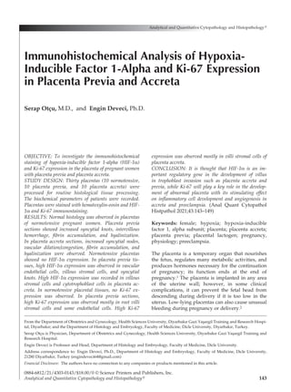
  • 3. software (IBM SPSS Statistics for Windows, Version 25.0, released 2017. IBM Corp., Armonk, New York, USA). One-way ANOVA with Tukey’s test was used for multiple comparisons. P<0.05 was used as the significance level. Results Statistical analysis of multiple comparisons be- tween normotensive, placenta previa, and placen­ ta accreta is shown in Table I. No statistical dif- ferences were recorded within group comparisons for any parameters (ALT, AST, glucose, albumin, total protein, urea, creatinine). Graphical illustra- tions of all parameters are shown in Figures 1–2. Placenta previa and accreta were stained with hematoxylin-eosin and HIF-1α and Ki-67 immu­ nohistochemical staining. The staining is present­ ed in Figure 2. Normotensive placentas showed normal placental histology (Figure 2A). In sections of placenta previa, increased syncytial knots, in- tervillous hemorrhage, fibrin accumulation, and hyalinized regions were observed (Figure 2B). Similar to placenta previa, placenta accreta show­ ed similar histopathology with increased syncytial nodes, vascular dilation/congestion, fibrin accu­ mulation, and hyalinization (Figure 2C). In HIF- 1α immunohistochemical staining, no expression was recorded in normotensive placental tissues. In placenta previa tissues, high HIF-1α expres- sion was observed in vascular endothelial cells, villous stromal cells, and syncytial knots (Figure 2E). High HIF-1α expression was recorded in villous stromal cells and cytotrophoblast cells in placenta accreta (Figure 2F). No Ki-67 expression was recorded in normotensive placental tissues (Figure 2G). Placenta previa showed high Ki-67 expression mostly in root villi stromal cells and some endothelial cells (Figure 2H). High Ki-67 expression was observed mostly in villi stromal cells of placenta accreta (Figure 2I). Discussion In this study we investigated the tissues of placen­ ta previa and placenta accreta by biochemical and immunohistochemical methods. Placenta previa is a pregnancy complication in which placentation is abnormally localized in the lower segment of the uterus.12 It is one of the important causes of maternal, fetal, and neonatal morbidity and mor­ tality characterized by third trimester bleeding. Al- though its etiology is unknown, advanced mater­ nal age, multiparity, multiple pregnancy, previous cesarean delivery, and smoking are risk factors, causing placenta previa.6 Studies on histopathol­ ogy of placenta previa revealed fibrinoid necrosis, polymorphonuclear cell infiltration, abnormal vas­ culatures, and dilated vessels. Biswas et al recorded increased trophoblastic giant cells, hemorrhage, absence of chorionic villi in the myometrium, and inflammation in placenta previa tissues.13 Silver et al also reported increased villous infarction with fibrinoid and congested vessels in pathological examination of placenta previa.14 In our study, we observed increased syncytial knots, intervillous hemorrhage, fibrin accumulation, and hyalinized regions in placenta previa sections (Figure 1B). Placenta accreta is defined as abnormally deep adhesion of the placenta to the uterine muscles. Its etiology is not fully known, but placenta previa and previous cesarean deliveries are risk factors. Complications include premature birth, maternal bleeding, and morbidity.15 Histopathological anal­ ysis of placenta accreta showed chorionic villi adjacent to myometrial fibers, fibrinoid formation, intravascular chorionic villi, hemorrhage, and vas­ cular changes.14 In our study we observed in- creased syncytial nodes, vascular dilation/conges­ tion, fibrin accumulation, and hyalinization in placenta accreta tissues. Perinatal studies have focused on the role of oxygen and hypoxia during placentation and pregnancy. Researchers have shown that oxygen has vital functions to regulate trophoblast differ­ entiation.14 Hypoxia-inducible factor-1α (HIF-1α) is a transcription regulator of genes in adaptive re- sponse to hypoxic conditions. HIF-1α is involved in activation of transcription of over 40 genes such as erythropoietin, glucose transporters, gly- colytic enzymes, and vascular endothelial growth factor during hypoxia. HIF-1α regulates vessel formation in the embryonic stage, tumor angio­ genesis, and ischemia.16 During placental forma­ tion, cellular hypoxia develops, and thus HIF-1α is activated to induce trophoblast proliferation and the formation of specific cell subtypes.17,18 Zamudio et al investigated HIF-1α expression and its regu­ latory mechanisms in placentas from pregnancies of women living at different altitudes. HIF-1α were overexpressed in placentas of women living at high altitudes.19 Ietta et al investigated the role of HIF-1α expression during human placen­ tal development. Expression of HIF-1α was high when oxygen tension was low but decreased when placental oxygen tension increased. They also Volume 43, Number 3/June 2021 145 HIF-1-α and Ki-67 in Placenta Previa and Accreta
  • 4. showed that during early placentation, HIF-1α localized in cytotrophoblasts.20 In our study we found that placenta previa tissues showed high HIF-1α expression in vascular endothelial cells, 146 Analytical and Quantitative Cytopathology and Histopathology® Otçu and Deveci Table I  Means and Standard Deviations of Age, ALT, AST, Glucose, Albumin, Total Protein, Urea, and Creatinine with Group Comparisons One-way ANOVA Parameter Group N Mean+SD (p value) Age, years (1) Normotensive 20 33.35±4.72  (2): 0.868 (3): 0.082 (2) Placenta previa 20 32.55±5.04  (1): 0.868 (3): 0.221 (3) Placenta accreta 20 29.90±5.18  (1): 0.082 (2): 0.221 ALT, U/L (1) Normotensive 20 29.90±5.18  (2): 0.241 (3): 1.00 (2) Placenta previa 20 32.55±5.04  (1): 0.241 (3): 0.241 (3) Placenta accreta 20 29.90±5.18  (1): 1.00 (2): 0.241 AST, U/L (1) Normotensive 20 13.14±8.60  (2): 1.00 (3): 1.00 (2) Placenta previa 20 13.11±6.34  (1): 1.00 (3): 1.00 (3) Placenta accreta 20 13.14±8.60  (1): 1.00 (2): 1.00 Glucose, mg/dL (1) Normotensive 20 18.67±7.41  (2): 0.886 (3): 1.00 (2) Placenta previa 20 20.13±13.43 (1): 0.886 (3): 0.886 (3) Placenta accreta 20 18.67±7.41  (1): 1.00 (2): 0.886 Albumin, g/L (1) Normotensive 20 86.80±25.99 (2): 0.425 (3): 1.00 (2) Placenta previa 20 97.06±25.39 (1): 0.425 (3): 0.425 (3) Placenta accreta 20 86.80±25.99 (1): 1.00 (2): 0.425 Total protein, g/dL (1) Normotensive 20 21.84±16.42 (2): 0.479 (3): 1.00 (2) Placenta previa 20 27.72±14.97 (1): 0.479 (3): 0.479 (3) Placenta accreta 20 21.84±16.42 (1): 1.00 (2): 0.479 Urea, mg/dL (1) Normotensive 20 38.38±27.08 (2): 0.736 (3): 1.00 (2) Placenta previa 20 31.59±31.53 (1): 0.736 (3): 0.479 (3) Placenta accreta 20 38.38±27.08 (1): 1.00 (2): 0.736 Creatinine, mg/dL (1) Normotensive 20 16.43±4.91  (2): 0.337 (3): 1.00 (2) Placenta previa 20 19.19±8.03  (1): 0.337 (3): 0.337 (3) Placenta accreta 20 16.43±4.91  (1): 1.00 (2): 0.337 SD = standard deviation.
  • 5. Volume 43, Number 3/June 2021 147 HIF-1-α and Ki-67 in Placenta Previa and Accreta Figure 1  Graphical illustration of patient parameters regarding placenta complication. Figure 2  Normotensive, placenta previa, and placenta accreta tissues with H-E staining (A–C) and HIF-1α (D–F) and Ki-67 (G–I) immunostainings. (A) Normal placental histology. (B) Increased syncytial nodes (black arrow), intervillous hemorrhage (red star), fibrinoid (black star), and hyalinization (arrowhead). (C) Increased syncytial nodes (black arrow), vascular dilation/congestion (red star), villous fibrinoid (black star), and hyalinization (arrowhead). (D) No HIF-1α in normotensive placental tissues. (E) High HIF-1α expression in vascular endothelial cells (red arrow), villous stromal cells (arrowhead), and syncytial nodes (black arrow). (F) High HIF-1α expression in villous stromal cells (arrowhead) and cytotrophoblast cells (black arrow). (G) No Ki-67 expression in normotensive placental tissues. (H) High Ki-67 expression mostly in root villi stromal cells (arrow) and some endothelial cells (red arrow). (I) High Ki-67 expression in mostly villi stromal cells (arrows).
  • 6. villous stromal cells, and syncytial knots (Figure 1E). Placenta accreta tissues showed high HIF-1α expression in villous stromal cells and cytotro­ phoblast cells in placenta accreta (Figure 1F). Ki-67 is a cellular marker for proliferation which functions in interphase and mitotic cells. Mea- surement of Ki-67 expression level can be used for the prognosis of many tumors.21 Ki-67 expression index has been used in the normal placenta, hy- datidiform moles, and choriocarcinoma. Aberrant placenta formation may be related with abnor- mal regulation of molecular changes and the proliferation of trophoblast cells so that tropho­ blasts are able to migrate and invade the uterine wall.21,22 Kaya et al recorded that Ki-67 expression was increased in villous cytotrophoblasts of pre- eclamptic placentas relative to controls. They showed an increased number of villous cytotro­ phoblasts in preeclampsia as compared to in nor- mal pregnancy by analyzing with Ki-67 immuno­ histochemical staining.23 Unek et al showed that Ki-67 expression was increased in placental villi of preeclampsia.24 Another preeclampsia study recorded that the Ki-67 index was high in villous trophoblasts of preeclamptic patients.25 A study demonstrated that Ki-67 expression was high in trophoblast columns of accreta subtypes.26 In our study we found that placenta previa showed high Ki-67 expression mostly in root villi stromal cells and some endothelial cells (Figure 1H). We re- corded high Ki-67 expression mostly in villi stro- mal cells (Figure 1I). In conclusion, it is thought that HIF-1α is an important regulatory gene in the development of villus in trophoblast invasion such as placenta accreta and placenta previa, while Ki-67 will play a key role in the development of abnormal placen­ ta with its stimulating effect on inflammatory cell development and angiogenesis in accreta and pre­ eclampsia References  1. Handwerger S, Freemark M: The roles of placental growth hormone and placental lactogen in the regulation of hu- man fetal growth and development. J Pediatr Endocrinol Metab 2000;13(4):343-356  2. Yetter JF 3rd: Examination of the placenta. Am Fam Physi­ cian 1998;57(5):1045-1054  3. Freemark M: The roles of growth hormone, prolactin and placental lactogen in human fetal development. In Molecu- lar and Cellular Pediatric Endocrinology. Edited by S Hand­ werger. Totowa, New Jersey, Human Press 1999, pp 57-84  4. Handwerger S: Clinical counterpoint: The physiology of placental lactogen in human pregnancy. [Review]. Endocr Rev 1991;12:329-336   5.  D’antonio F, Bhide A: Ultrasound in placental disorders. Best Prac Res Clin Obstet Gynaecol 2014;28(3):429-442   6.  McShane PM, Heyl PS, Epstein MF: Maternal and perinatal morbidity resulting from placenta previa. Obstet Gynecol 1985;65:176-182   7.  Crane JM, Van den Hof MC, Dodds L, Armson Ba, Liston R: Maternal complications with placenta previa. Am J Perinatol 2000;17(2):101-105  8. Ananth CV, Smulian JC, Vintzileos AM: The effect of placenta previa on neonatal mortality: A population-based study in the United States, 1989 during 1997. Am J Obstet Gynecol 2003;188(5):1299-1304  9. Oyelese Y, Smulian JC: Placenta previa, placenta accreta and vasa previa. Obstet Gynecol 2006;107(4):927-941 10. Arcario T, Greene M, Ostheimer GW, Datta S, Nauity JS: Risks of placenta previa/accreta in patients with pre- vious caesarean deliveries. Anesthesiology 1988;69(Suppl 3A):A 659 11. Miller DA, Chollet JA, Goodwin TM: Clinical risk factors for placenta previa placenta accreta. Am J Obstet Gynecol 1997;177:210-214 12. Lockwood CJ, Russo-Stieglitz K: Placenta previa: Epidemi­ ology, clinical feature, diagnosis, morbidity and mortality. Available at https:/ /www.uptodate.com/contents/placenta- previa-epidemiology-clinical-features-diagnosis-morbidity- and-mortality. Accessed April 28, 2020 13. Biswas R, Sawhney H, Dass R, Saran RK, Vasishta K: Histopathological study of placental bed biopsy in placenta previa. Acta Obstet Gynecol Scand 1999;78(3):173-179 14. Silver R: Abnormal placentation: Placenta previa, vasa previa, and placenta accreta. Obstet Gynecol 2015;126(3): 654-668 15.  Khong TY: The pathology of placenta accreta, a worldwide epidemic. J Clin Pathol 2008;61:1243-1246 16. Soares MJ, Iqbal K, Kozai K: Hypoxia and placental de- velopment. Birth Defects Res 2017;109(17):1309-1329 17. Lee JW, Ko J, Ju C, et al: Hypoxia signaling in human diseases and therapeutic targets. Exp Mol Med 2019;51:1-13 18. Simmons DG, Cross JC: Determinants of trophoblast line­ age and cell subtype specification in the mouse placenta. Dev Biol 2005;284:12-24 19. Zamudio S, Wu Y, Ietta F, Rolfo A, Cross A, Wheeler T, Post M, Illsley NP, Caniggia I: Human placental hypoxia- inducible factor-1alpha expression correlates with clinical outcomes in chronic hypoxia in vivo. Am J Pathol 2007; 170(6):2171-2179 20. Ietta F, Wu Y, Winter J, Xu J, Wang J, Post M, Caniggia I: Dynamic HIF1A regulation during human placental devel­ opment. Biol Reprod 2006;75(1):112-121 21.  Sun X, Kaufman PD: Ki-67: More than a proliferation mark­ er. Chromosoma 2018;127(2):175-186 22. Hilali N, Kocarslan S, Vural M, et al: Ki-67 proliferation index in patients with placenta previa percreta in the third trimester. Wien Klin Wochenschr 2015;127:98-102 23. Kaya B, Nayki U, Nayki C, Ulug P, Oner G, Gultekin E, 148 Analytical and Quantitative Cytopathology and Histopathology® Otçu and Deveci
  • 7. Yildirim Y: Proliferation of trophoblasts and Ki67 expres- sion in preeclampsia. Arch Gynecol Obstet 2015;291(5):1041- 1046 24. Unek G, Oxmen A, Mendilcioglu I, Simsek M, Korgun ET: The expression of cell cycle related proteins PCNA, Ki67, p27 and p57 in normal and preeclamptic human placentas. Tissue Cell 2014;46:198-205 25.  Arnholdt H, Meisel F, Fandrey K, Löhrs U: Proliferation of villous trophoblast of the human placenta in normal and abnormal pregnancies. Virchow Archiv B Cell Pathology 1991;60(6):365-372 26. Tantbirojn P, Crum CP, Parast MM: Pathophysiology of placenta creta: The role of decidua and extravillous tro­ phoblast. Placenta 2008;29(7):639-645 Volume 43, Number 3/June 2021 149 HIF-1-α and Ki-67 in Placenta Previa and Accreta