Using Carbon Nano-Tube Field Emitters to Miniaturize X-Ray Tubes
•
1 gostou•6,413 visualizações

Understand how carbon nanotube technology increases portability and image quality in medical imaging; and is more energy efficient.

Recomendados
Mais conteúdo relacionado
Mais procurados
Mais procurados (20)
Semelhante a Using Carbon Nano-Tube Field Emitters to Miniaturize X-Ray Tubes
Semelhante a Using Carbon Nano-Tube Field Emitters to Miniaturize X-Ray Tubes (20)
Mais de Carestream
Mais de Carestream (20)
Último
Último (20)
Using Carbon Nano-Tube Field Emitters to Miniaturize X-Ray Tubes
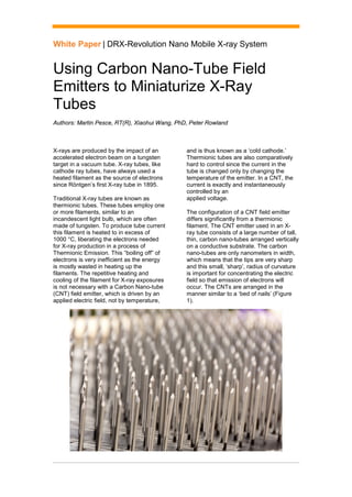
- 1. White Paper | DRX-Revolution Nano Mobile X-ray System Using Carbon Nano-Tube Field Emitters to Miniaturize X-Ray Tubes Authors: Martin Pesce, RT(R), Xiaohui Wang, PhD, Peter Rowland X-rays are produced by the impact of an accelerated electron beam on a tungsten target in a vacuum tube. X-ray tubes, like cathode ray tubes, have always used a heated filament as the source of electrons since Röntgen’s first X-ray tube in 1895. Traditional X-ray tubes are known as thermionic tubes. These tubes employ one or more filaments, similar to an incandescent light bulb, which are often made of tungsten. To produce tube current this filament is heated to in excess of 1000 °C, liberating the electrons needed for X-ray production in a process of Thermionic Emission. This “boiling off” of electrons is very inefficient as the energy is mostly wasted in heating up the filaments. The repetitive heating and cooling of the filament for X-ray exposures is not necessary with a Carbon Nano-tube (CNT) field emitter, which is driven by an applied electric field, not by temperature, and is thus known as a ‘cold cathode.’ Thermionic tubes are also comparatively hard to control since the current in the tube is changed only by changing the temperature of the emitter. In a CNT, the current is exactly and instantaneously controlled by an applied voltage. The configuration of a CNT field emitter differs significantly from a thermionic filament. The CNT emitter used in an X- ray tube consists of a large number of tall, thin, carbon nano-tubes arranged vertically on a conductive substrate. The carbon nano-tubes are only nanometers in width, which means that the tips are very sharp and this small, ‘sharp’, radius of curvature is important for concentrating the electric field so that emission of electrons will occur. The CNTs are arranged in the manner similar to a ‘bed of nails’ (Figure 1).
- 2. White Paper | DRX-Revolution Nano Mobile X-ray System 2 Figure 1: Illustration of the ‘bed of nails’ configuration of CNTs in an emitter A gate mesh structure, connected to an electrode inside the tube wall, is placed a small distance above the sharp tips of the CNTs. When the radiographer manipulates the two-position exposure switch to the prep position, a voltage gradient is applied externally between the grid mesh and the substrate. This voltage gradient creates a very strong electric field at the tips of the CNTs. Figure 2: Electrical field strength is concentrated at the sharp tip of a CNT. The electric field strength concentrated at the CNT tips forces field emission of negatively charged electrons to occur at the tips of the CNTs (Figure 2). This is very similar to the lighting rod effect. As no high temperature “boiling off” is needed, this ‘cold cathode’ translates to power- saving benefits in battery-operated X-ray units. Unlike a traditional tube, in which the electrons boil off in all directions, the CNT field emissions are attracted by the positively charged grid mesh. When the radiographer depresses the two-position exposure switch to the expose position, the electrons are emitted from the CNT tips and fly toward the grid mesh. The majority of electrons will pass through the mesh then to be accelerated by the anode high voltage, generating X-rays when the electrons impact the anode (Figure 3). Figure 3: The cold cathode using the CNT emitter replaces the filament normally associated with thermionic X-ray tubes. When voltage (V) is applied to the circuit, the electrons are concentrated at the tip of the CNT and attracted by the mesh. When voltage (V) is applied, the electrons are accelerated toward the anode to produce X-ray photons.
- 3. White Paper | DRX-Revolution Nano Mobile X-ray System 3 The amount of anode current can be very precisely controlled by varying the voltage applied between the grid mesh electrode and the cathode. Figure 4: The focus electrode focuses the electron beam to create a variable focal spot. In addition to room-temperature operation, the CNT cold cathode X-ray tube from Carestream Health has an electro-optic focus lens built inside to dynamically adjust the focal spot size. The lens is a collar-shaped focus electrode through which the electron beam passes. Changing the voltage applied to the collar electrode will alter the electron paths and change the amount of focusing of the beam. Thus with the addition of a focusing electrode, an X-ray tube using a CNT emitter can have a continuously variable focal spot size on the anode, controlled by the focus voltage applied at any instant (Figure 4). This feature can be used to great advantage to manage the heat load in the focal spot on a fixed-anode X-ray tube used for imaging. The X-ray cart control system automatically adjusts the focal spot size for each X-ray pulse to optimize the image quality according to the heat load of the exposure parameters chosen by the operator for each exam. By eliminating the traditional thermionic filament, rotating anode and stator, the CNT field emitter offers unique advantages over traditional tubes for mobile X-ray imaging. Size and weight are the most notable. The CNT X-ray tube weighs about 1 kg or 2 lb, compared to a traditional tube insert, which is 2 kg or 4.4 lb. However, the insert is then placed inside the housing, and the housing is filled with oil. The housing plus the tube and oil can weigh as much as 17 kg or 37.5 lb. When the collimator and lead shielding weight is combined, the CNT field emitter is approximately one fourth the weight of a traditional tube and collimator assembly (Figure 5).
- 4. White Paper | DRX-Revolution Nano Mobile X-ray System 4 Figure 5: (A) Thermionic Tube Housing Assembly weights. (B) CNT X-ray Tube Housing Assembly weights. With the miniaturization of the tube assembly, there is a domino effect that is translated throughout the design of a portable X-ray system. The tube head and collimator are considerably smaller and light, which means the size and operation of the tube arm and column can be decreased, reducing weight and size (Figure 6). A B
- 5. White Paper | DRX-Revolution Nano Mobile X-ray System carestream.com © Carestream Health, Inc., 2017. CARESTREAM is a trademark of Carestream Health. CAT 2000 219 04/17 Figure 1: (A) DRX-Revolution tube head and collimator assembly, ~ 56 cm (22 in.) in width and weighing 75 kg (165.3 lb). (B) DRX-Revolution Nano tube head and collimator assembly, ~32 cm (12.5 in.) in width and weighing 7 kg (15.4 lb). Although fairly new to medical imaging, CNT field emitter X-ray tubes offer a significant advancement to system design. CNT field emitters do not need to time to heat up a filament or spin up the rotating anode, so they are always in a near-ready state. They are smaller and weigh much less than a traditional tube, which allows for a more compact design and easier maneuverability. Finally, they are more energy efficient than a traditional tube, which reduces the need for large, heavy generators and the battery pack. This also helps to significantly decrease the overall size and weight of a portable X-ray system. A B